. 1997 Sep 15;17(18):6839-49.
doi: 10.1523/JNEUROSCI.17-18-06839.1997.

Serotonergic inhibition of the T-type and high voltage-activated Ca2+ currents in the primary sensory neurons of Xenopus larvae

Affiliations

Serotonergic inhibition of the T-type and high voltage-activated Ca2+ currents in the primary sensory neurons of Xenopus larvae

Q Q Sun et al. J Neurosci. .

Abstract

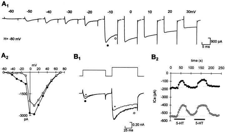
The primary sensory Rohon-Beard (R-B) neurons of Xenopus larvae are highly analogous to the C fibers of the mammalian pain pathway. We explored the actions of 5-HT by studying the modulation of Ca2+ currents. In approximately 80% of the acutely isolated R-B neurons, 5-HT inhibited the high voltage-activated (HVA) currents by 16% (n = 29) and the T-type currents by 24% (n = 41). The modulation of the T-type and the HVA currents was mimicked by selective 5-HT1A and 5-HT1D agonists: 8-OH-DPAT and L-694,247. The effects of the agonists were blocked by their respective 5-HT1A or 5-HT1D antagonists: p-MPPI and GR127935, suggesting that both 5-HT1A and 5-HT1D receptors were involved. Approximately 70% of the actions of 5-HT on HVA currents was occluded by omega-conotoxin-GVIA (N-type channel blocker), whereas the rest of the modulation ( approximately 30%) was occluded by <100 nM omega-agatoxin-TK (P/Q-type channel blocker). This suggests that 5-HT acts on N- and P/Q-type Ca2+ channels. Neither the modulation of the T-type nor that of the HVA currents was accompanied by changes in their voltage-dependent kinetics. Cell-attached patch-clamp recordings suggest that the modulation of the T-type channel occurs through a membrane-delimited second messenger. We have studied the functional consequences of the modulation of T-type Ca2+ channels and have found that these channels play a role in spike initiation in R-B neurons. Modulation of T-type channels by 5-HT therefore could modulate the sensitivity of this sensory pathway by increasing the thresholds of R-B neurons. This is a new and potentially important locus for modulation of sensory pathways in vertebrates.


Figures

Fig. 1.
Fig. 1.
Both the HVA and the T-type Ca2+ currents were reduced by 5-HT in acutely isolated R–B neurons. A, R–B neurons possess both the HVA and the T-type Ca2+ currents.A1, Whole-cell Ca2+ currents were recorded by using steps from a holding potential of −80 mV to test potentials between −60 and +30 mV in a stage 42 Xenopus R–B neuron.A2, I/V curve measured from the peak (showing the T-type and HVA currents) and end (showing only the HVA currents) of the Ca2+ currents (symbols correspond to measurements inA1).B1, T-type and HVA currents were elicited in the same neuron by test steps of −30 and +10 mV, respectively, from a holding potential of −90 mV. Both were reduced by 1 μm 5-HT (shown by asterisk); unlabeled traces are control and wash. B2, Courses of the effects of 5-HT showing that they were totally reversible on both currents in the same neuron (symbolscorrespond to measurements inB1).
Fig. 2.
Fig. 2.
Voltage-independent inhibition of the HVA Ca2+ currents by 5-HT.A1, Ca2+currents elicited by steps from a holding potential of −80 mV (asterisk = 1 μm 5-HT).A2, The inhibition of HVA currents by 5-HT (1 μm) was independent of membrane potential (n = 5);I5-HT, current in presence of 5-HT;ICon, current in the control.B1, Example showing that the inhibition of the HVA currents in RB neurons was not changed by applying a +120 mV prepulse (asterisk = trace elicited by test pulse with 120 mV prepulse in 1 μm5-HT). B2, Summary showing no significant difference between the mean inhibition of the HVA currents before and after applying a 120 mV prepulse in five R–B neurons.
Fig. 3.
Fig. 3.
The inhibition of HVA Ca2+currents by 5-HT is dose-dependent and mediated via 5-HT1Aand 5-HT1D receptors.A1, The selective 5-HT1D agonist L-694,247 (100 nm) inhibited HVA currents. The effects of L-694,247 (100 nm) were blocked by the selective 5-HT1D antagonist GR127935 (100 nm). A2, The selective 5-HT1A agonist 8-OH-DPAT (100 nm) also inhibited the HVA currents. The effects of 8-OH-DPAT (100 nm) were totally blocked by the selective 5-HT1A antagonist p-MPPI (100 nm). B, Summary of the antagonist effects on the agonists of 8-OH-DPAT and L-694,247 (*p < 0.05 vs agonists alone). C, The inhibition of HVA currents by 5-HT was dose-dependent (n = 7–39 for each dose). The smooth line is the best-fitting Hill equation.
Fig. 4.
Fig. 4.
The identity of the calcium channels inhibited by 5-HT in RB neurons. A1, Time courses of the effects of calcium channel blockers ω-agatoxin-TK (100 nm), ω-CgTx (1 μm), nifedipine (10 μm), and Cd2+ (100 μm) on the HVA currents. A2, Calcium currents elicited by steps to 10 mV from a holding potential of −50 mV during the application of the blockers (same neuron as inA1).A3, Summary of the effects of calcium channel blockers on the HVA currents in R–B neurons.B1, Recording showing that the inhibition of HVA currents by 5-HT (1 μm) was mostly occluded by ω-conotoxin (1 μm) in a R–B neuron.B2, Recording showing that the inhibition of HVA currents by 5-HT (1 μm) was partially occluded by ω-agatoxin-TK (100 nm), whereas the remaining inhibition was totally occluded by 1 μm ω-conotoxin (asterisk = HVA currents recorded in 5-HT on top of Ca2+ channel blockers).B3, Summary of the inhibition of the HVA currents by 5-HT alone and additional inhibition on top of calcium channel blockers (*p < 0.05 and **p < 0.01 vs 5-HT alone).
Fig. 5.
Fig. 5.
Inhibition of the T-type calcium currents by serotonergic agonists. A, Representative traces (asterisk represents recordings in 100 nmselective agonists; unlabeled traces are control and wash) and dose–response relations showing the block of T-type currents by selective serotonergic agonists: 5-HT (A1), 8-OH-DPAT (A2), and L-694,247 (A3); n = 7–41 for each point. The solid line is the best fit of the Hill equation.A4, The inhibition of T-type currents by L-694,247 (100 nm) and 8-OH-DPAT (100 nm) was additive. B, Summary of the mean inhibition of T-type currents by selective serotonergic agonists at maximum dose in R–B neurons: 5-HT (1 μm,n = 41), 8-OH-DPAT (100 nm,n = 23), L-694,247 (100 nm,n = 22), CGS-12066B (1 μm,n = 8), α-M-5-HT (1 μm,n = 7), and 5-CT (100 nm,n = 5) (**p < 0.01 vs control).
Fig. 6.
Fig. 6.
Effects of selective antagonists on the inhibition of T-type calcium currents by selective agonists inXenopus R–B neurons. Both p-MPPI (100 nm, A1) and GR127935 (100 nm, A2) partially blocked the effect of 5-HT (1 μm).A3, p-MPPI (100 nm) and GR127935 (100 nm) produced an additive block of the effect of 5-HT (1 μm). p-MPPI blocked the effect of 8-OH-DPAT (100 nm,A4), whereas GR127935 (100 nm) blocked the effect of L-694,247 (100 nm,A5).B1, Summary of the block by the selective antagonists on 8-OH-DPAT (100 nm) (**p < 0.01 vs 8-OH-DPAT).B2, Summary of the block by antagonists on L-694,247 (100 nm) (**p< 0.01 vs L-694,247). B3, Summary of the block by selective antagonists on 5-HT (1 μm) (**p < 0.01 vs 5-HT).
Fig. 7.
Fig. 7.
Effects of 5-HT on T-type unitary channel recordings. A1, Consecutive unitary T-type Ba2+ currents were elicited by steps from potentials equivalent to the membrane potential of approximately −90 to −30 mV (after allowing for the screening effect of high Ba2+ levels on membrane surface charge) in a cell-attached patch. All traces are leak-subtracted.A2, Average unitary T-type Ba2+ current record obtained by averaging of 50 consecutive recordings in the same patch. The solid lineis the best fit of the single exponential equation; time constant (τ) = 23.5 msec. B1, 5-HT (1 μm) did not modulate the average (50–100 consecutive traces) unitary T-type Ba2+ currents recorded in cell-attached patches. In Cell A there were no changes in the T-type current. In Cell B, however, the averaged currents were reduced, but this did not reverse on washing, suggesting that the reduction may result from a “run down” of channel activity. B2, Summary of the effects of 5-HT (1 μm) on T-type unitary channel recordings in six R–B neurons.
Fig. 8.
Fig. 8.
5-HT did not alter the kinetic characteristics of the whole-cell T-type calcium currents.A1, Leak-subtracted T-type tail currents (shown by arrow) were elicited by a series of test pulses (−80 to −20 mV in 5 mV steps) from a holding potential of −100 mV in control. A2, Summary showing that 5-HT (1 μm) had no effect on the activation of the T-type currents in five R–B neurons. The data were fit with the Boltzmann relation (solid line):I/Imax = {1 + exp[(V +V½)/K]}−1.Squares, Control (V½= −37.2 mV; K = −7.6); filled circles, 5-HT (1 μm) (V½ = −38.8 mV;K = −7.3).B1, The T-type currents, measured at the peak, undergo steady-state inactivation.B2, 5-HT (1 μm) had no effect on the steady-state inactivation of the T-type currents in four R–B neurons. The data were fit with the Boltzmann relation (solid line). Squares, Control (V½ = −74.7 mV;K = 5.2); filled circles, 5-HT (1 μm) (V½ = −75.2 mV;K = 5.4). C1,Top left, T-type currents recorded in control and 1 μm 5-HT (shown by asterisk). Top right, The T-type currents recorded in 5-HT were scaled up to the same peak magnitude as control to show that the two traces overlap almost completely. The time-dependent inactivation of the T-type currents was measured by fitting with a single exponential (solid line). C2, 5-HT (1 μm) had no effect on the time-dependent inactivation in three R–B neurons. Squares, Control;filled circles, 5-HT (1 μm).
Fig. 9.
Fig. 9.
Differential block of the T-type and HVA Ca2+ currents by Y3+ in R–B neurons. A, Time series measurements in a R–B neuron showing the dose–response for Y3+ on T-type currents (A1) and HVA currents (A2) recorded in the same neuron.A3, Example of the HVA and T-type currents recorded simultaneously in the same neuron, as shown inA1 andA2 (asterisks= 10 nm Y3+). B, Summary of the effects of Y3+ (10 nm) on the T-type currents and HVA currents (**p < 0.01 vs control).
Fig. 10.
Fig. 10.
Effects of the selective T-type Ca2+ channel blockers and 5-HT on R–B neuron firing. A1, Current-clamp recording in a R–B neuron, using steps of current injection to detect the threshold current that evoked an action potential.A2, Y3+ (10 nm; asterisk), which selectively blocked T-type currents, blocked the action potential evoked at threshold current injection. B1, Current-clamp recording from a R–B neuron showing that injection of repeated current pulses just above the threshold caused reliable firing. At resting membrane potentials more positive than −50 mV, neither Y3+ (10 nm) nor 5-HT (10 nm) had any effects on neuron firing.B2, At more negative resting membrane potentials (−90 mV), both Y3+ (10 nm) and 5-HT (10 nm) reversibly reduced the probability of firing in the neuron. C, Summary of the effects of Y3+ (10 nm) and 5-HT (10 nm) on the probability of R–B neuron firing in response to repetitive threshold current injection at a resting membrane potential of −90 mV, where the probability was measured as the number of pulses that evoked a spike/total number of pulses during control, drug treatment, and wash (**p < 0.01 vs control or wash).

Similar articles

Cited by

References

    1. Awouters F. The pharmacology of ketanserin, the first selective serotonin S2-antagonist. Drug Dev Res. 1985;6:263–300.
    1. Bayliss DA, Umemiya M, Berger AL. Inhibition of N- and P-type calcium currents and after-hyperpolarization in rat motoneurones by serotonin. J Physiol (Lond) 1995;485:635–647. - PMC - PubMed
    1. Beer MS, Stanton JA, Bevan Y, Chauhan NS, Middlemiss DN. An investigation of the 5-HT1D receptor binding affinity of 5-hydroxytryptamine, 5-carboxyamidotryptamine, and sumatriptan in the central nervous system of seven species. Eur J Pharmacol. 1992;213:193–197. - PubMed
    1. Beer MS, Stanton JA, Bevan Y, Heald A, Reeve AJ, Street LJ, Matassa VG. L-694,247: a potent 5-HT1D receptor agonist. Br J Pharmacol. 1993;110:1196–1200. - PMC - PubMed
    1. Berger AJ, Takahashi T. Serotonin enhances a low-voltage-activated calcium current in rat spinal motoneurons. J Neurosci. 1990;10:1922–1928. - PMC - PubMed

Publication types

LinkOut - more resources