. 1997 Sep 15;17(18):7007-16.
doi: 10.1523/JNEUROSCI.17-18-07007.1997.

Trk receptors function as rapid retrograde signal carriers in the adult nervous system

Affiliations

Trk receptors function as rapid retrograde signal carriers in the adult nervous system

A Bhattacharyya et al. J Neurosci. .

Abstract

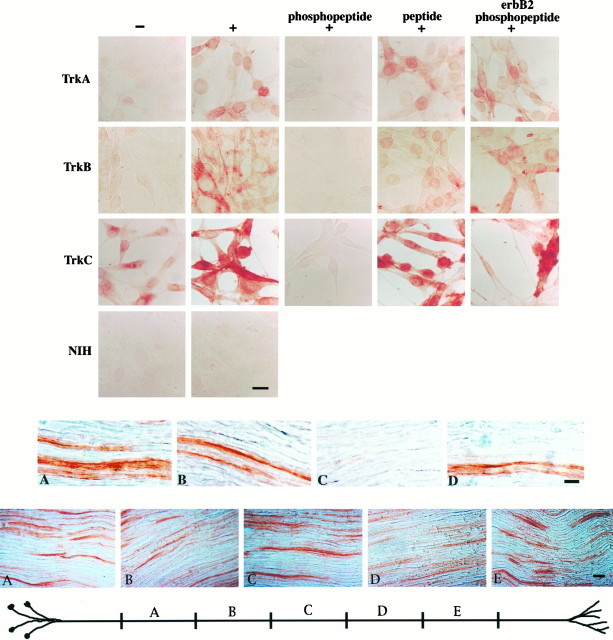
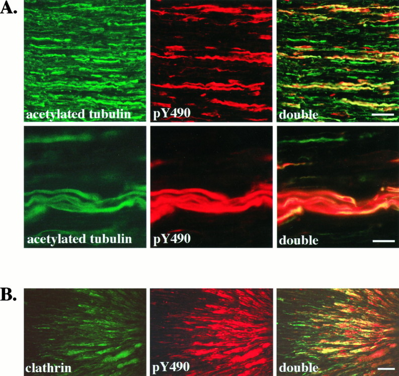
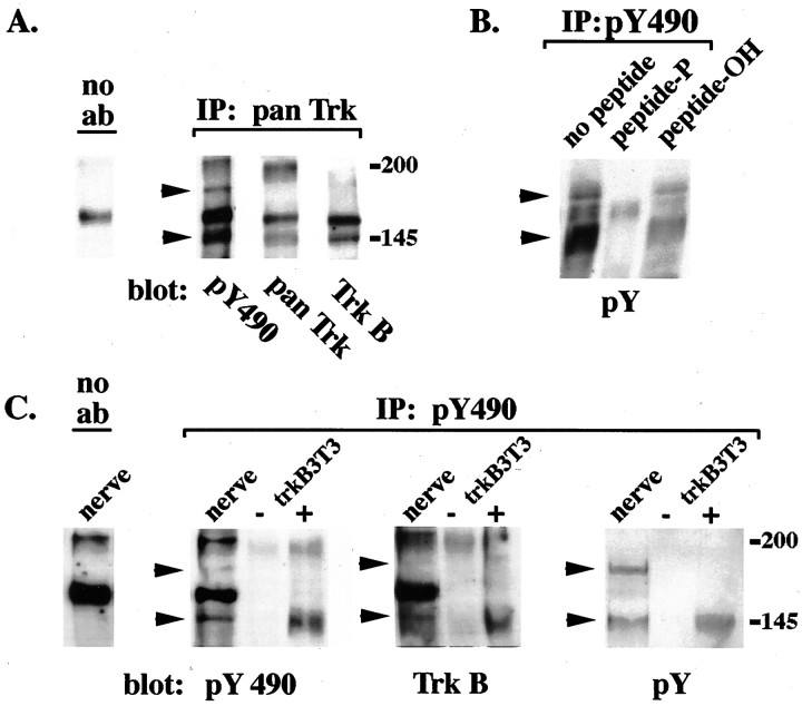
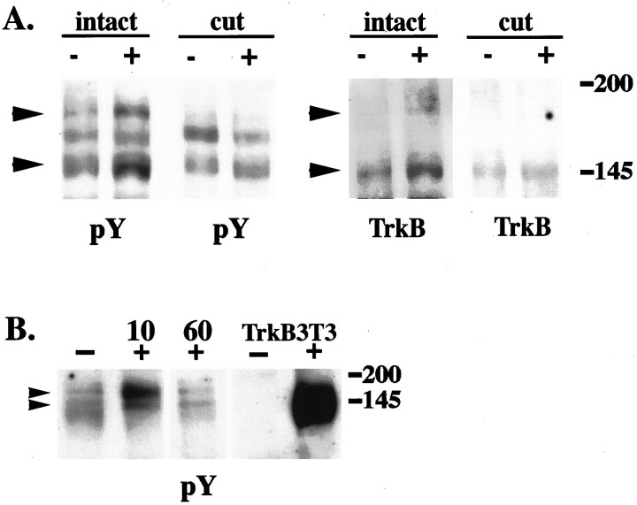
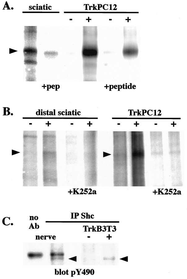
During development target-derived neurotrophins promote the survival of neurons. However, mature neurons no longer depend on the target for survival. Do target-derived neurotrophins retain retrograde signaling functions in mature neurons, and, if so, how are they executed? We addressed this question by using a phosphotyrosine-directed antibody to locate activated Trk receptors in adult rat sciatic nerve. We show that catalytically active Trk receptors are located within the axon of adult rat sciatic nerve and that they are distributed throughout the length of the axons. These catalytically active receptors are phosphorylated on tyrosine at a position that couples them to the signal-generating proteins Ras and PI3 kinase. Neurotrophin applied at sciatic nerve terminals increases both catalytic activity and phosphorylation state of Trk receptors at distant points within the axons. Trk activation initiated at the nerve terminals propagates through the axon toward the nerve cell body at an initial rate that exceeds that of conventional vesicular transport. However, our data suggest that this rapid signal is nevertheless vesicle-associated. Thus, in mature nerves, activated Trk receptors function as rapid retrograde signal carriers to execute remote responses to target-derived neurotrophins.


Figures

Fig. 1.
Fig. 1.
Top. Anti-pY490 detects activated TrkA, TrkB, and TrkC. TrkA-, TrkB-, and TrkC-expressing NIH3T3 cells were stimulated with neurotrophins and immunostained with the pY490 antibody, followed by avidin–biotin–HRP and DAB. TrkA, TrkB, and TrkC 3T3 cells stimulated with neurotrophin show increased pY490 immunostaining (+), as compared with control cells stimulated with vehicle alone (−). Immunostaining is abolished by preincubation of the antibody with its phosphopeptide immunogen (phosphopeptide). Preincubation of the antibody with the corresponding unphosphorylated peptide (peptide) or a phosphopeptide corresponding to the NPXY motif of the erbB2 receptor (erbB2 phosphopeptide) does not abolish the staining. No staining is observed in untransfected NIH3T3 cells (NIH). Scale bar, 10 μm.
Fig. 4.
Fig. 4.
Activated Trk receptors are confined to axons in the sciatic nerve and accumulate distal to a ligation.A, Longitudinal sections of adult rat sciatic nerve were immunostained with anti-acetylated α-tubulin (acetylated tubulin) and anti-pY490 (pY490), as described, and viewed with fluorescence optics. Double labeling is shown in sections viewed with both fluorescein and rhodamine filters (double). All axons in the sciatic nerve are labeled by the acetylated tubulin antibody. In contrast, fewer axons are stained by anti-pY490. Colocalization is apparent as yellow in the double panels, demonstrating that pY490 labels only a subset of axons. Another field viewed at higher magnification (second row) shows that the anti-pY490 staining colocalizes with the acetylated tubulin staining, verifying that the pY490 immunostaining is present in axons of the sciatic nerve. Scale bars: First row, 50 μm; second row, 25 μm.B, The sciatic nerve of an adult rat was ligated ∼2 cm from the gastrocnemius muscle to interrupt vesicular transport. At 24 hr after ligation the nerve was harvested, and longitudinal sections of the nerve were immunostained with an antibody to the coated vesicle protein, clathrin and anti-pY490. The portion of nerve distal to the ligation site is shown with the ligation site to theright. Clathrin immunostaining shows accumulation of clathrin in axons on the distal side of the ligation (clathrin). Phosphorylated Trks, as shown by pY490 immunostaining, also accumulate on the distal side of the ligation (pY490). Costaining shows that phosphorylated Trks colocalize with clathrin (double). Scale bar, 50 μm.
Fig. 5.
Fig. 5.
Anti-pY490 recognizes two forms of phosphorylated TrkB in the adult rat sciatic nerve. A, Extracts of adult rat sciatic nerve were immunoprecipitated with anti-Trk and immunoblotted with anti-pY490 (pY490), anti-Trk (pan Trk), or a TrkB-specific antibody (TrkB). Incubation of the samples with Protein A-Sepharose beads alone resulted in the binding of a nonspecific protein at ∼165 kDa (no ab). Anti-pY490 recognizes a diffuse band at 140–150 kDa as well as a distinct band at 180 kDa in the Trk immunoprecipitates. The broad band is recognized by anti-Trk, whereas anti-TrkB recognizes a subset within this band. The 180 kDa band is weakly recognized by anti-TrkB. B, Nerve extracts were immunoprecipitated by the pY490 antibody alone or preincubated with peptide and immunoblotted with anti-phosphotyrosine. Two proteins of 145 and 180 kDa are recognized by anti-phosphotyrosine (pY) in extracts immunoprecipitated with anti-pY490 alone (no peptide). Preincubation of the pY490 antibody with its phosphopeptide immunogen (peptide-P), but not preincubation of the antibody with the corresponding unphosphorylated peptide (peptide-OH), prevented immunoprecipitation of these two bands. C, Extracts of sciatic nerve (nerve) and unstimulated (−) or BDNF-stimulated (+) TrkB 3T3 cells were immunoprecipitated with pY490 antibody and immunoblotted with anti-pY490 (pY490), anti-TrkB (TrkB), and anti-phosphotyrosine (pY). Both the 145 and 180 kDa bands are immunoprecipitated from nerve extracts with the pY490 antibody. The TrkB antibody recognizes the 145 kDa band and weakly recognizes the 180 kDa band. Both bands are recognized by anti-phosphotyrosine.
Fig. 6.
Fig. 6.
TrkB receptors in sciatic nerve respond to target-derived BDNF. A, Cytochrome C (−) or BDNF (+) was injected into the gastrocnemius muscle of adult rats (intact). In cut nerve experiments (cut), sciatic nerves were cut near the gastrocnemius muscle before injection. Distal segments of sciatic nerves (1–3 cm from the injection site) were harvested 10 min after injection. Nerve extracts were immunoprecipitated with anti-pY490 and immunoblotted with anti-phosphotyrosine (pY) or anti-TrkB (TrkB). A nonspecific band is present in the blots at ∼165 kDa. Both the 145 and 180 kDa TrkB bands (arrows) are increased after BDNF injection (intact). When the nerve is cut before injection, no increase in these bands is observed with BDNF injection (cut). B, Cytochrome C (−) or BDNF (+) was injected into the gastrocnemius muscle of adult rats. Distal segments of sciatic nerves from these animals were harvested 10 and 60 min after injection. Nerve extracts and unstimulated (−) or BDNF-stimulated (+) TrkB 3T3 cells were immunoprecipitated with anti-pY490 and immunoblotted with anti-phosphotyrosine (P-tyr). There is an increase in both 145 and 180 kDa Trk bands within 10 min after BDNF injection. By 60 min after injection, the levels of Trk are equal to cytochrome C control (−).
Fig. 7.
Fig. 7.
Target-derived neurotrophin increases the catalytic activity of axonal Trk receptors. Extracts of normal whole sciatic nerves and TrkPC12 cells, either stimulated (+) or unstimulated (−) with NGF, were immunoprecipitated with anti-Trk or in the presence of peptide immunogen. An in vitro autophosphorylation assay was performed on the Trk immunoprecipitates. The immunoprecipitates were incubated with γ-32P-ATP. Thearrow indicates a broad band at 145 kDa, corresponding to autophosphorylation of Trk isoforms in the nerve. TrkA autophosphorylation is increased in TrkPC12 cells stimulated with NGF (+). The identities of the bands in these immunoprecipitates were confirmed by the peptide competition. B, Extracts of distal segments of cytochrome C-injected (−) or BDNF-injected (+) nerves and TrkPC12 cells, either control (−) or NGF-stimulated (+), were immunoprecipitated with anti-Trk. Note that the amount of protein used in each immunoprecipitation in this experiment is approximately one-half of that in A. The immunoprecipitates were incubated with γ-32P-ATP alone or in the presence of K252a (+K252a), a Trk kinase inhibitor. Thearrow indicates the 145 kDa species of Trk in the nerve. Trk autophosphorylation is increased in nerves injected with BDNF and in TrkPC12 cells stimulated with NGF. No kinase activity is apparent in extracts incubated with K252a. C, Extracts of normal rat sciatic nerves and TrkB 3T3 cells stimulated with BDNF (+) or unstimulated (−) were immunoprecipitated with anti-Shc and immunoblotted with anti-pY490. Precipitation with Protein A-Sepharose beads alone (no Ab) resulted in the binding of a nonspecific band at ∼165 kDa. A phosphorylated Trk band at 145 kDa is detected in the Shc immunoprecipitates, verifying that phospho-Trk is associated with Shc in nerve extracts.

References

    1. Acheson A, Conover J, Fandl J, DeChiara T, Russell M, Thadani A, Squinto S, Yancopoulos G, Lindsay R. A BDNF autocrine loop in adult sensory neurons prevents cell death. Nature. 1995;374:450–453. - PubMed
    1. Barbacid M. The Trk family of neurotrophin receptors. J Neurobiol. 1994;25:1386–1403. - PubMed
    1. Barde YA. Trophic factors and neuronal survival. Neuron. 1989;2:1525–1534. - PubMed
    1. Bothwell M. Keeping track of neurotrophin receptors. Cell. 1991;65:915–918. - PubMed
    1. Campenot RB. Development of sympathetic neurons in compartmentalized cultures. I. Local control of neurite growth by nerve growth factor. Dev Biol. 1982;93:1–12. - PubMed

Publication types

MeSH terms

LinkOut - more resources