. 1997 Sep 15;17(18):7037-44.
doi: 10.1523/JNEUROSCI.17-18-07037.1997.

Cell coupling and uncoupling in the ventricular zone of developing neocortex

Affiliations

Cell coupling and uncoupling in the ventricular zone of developing neocortex

K Bittman et al. J Neurosci. .

Abstract

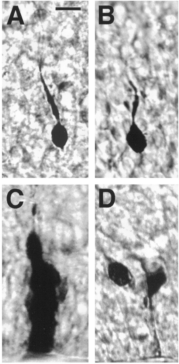
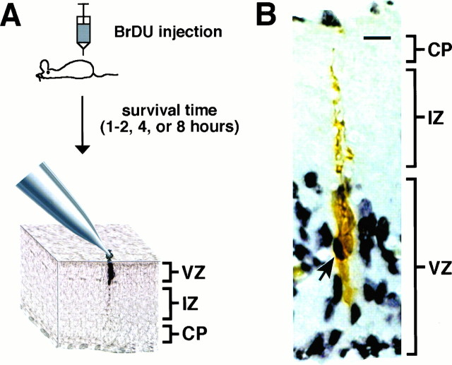
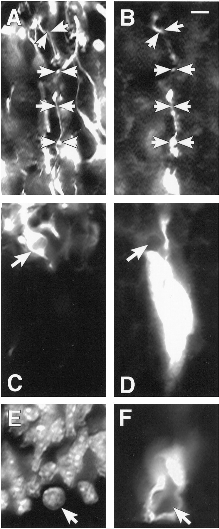
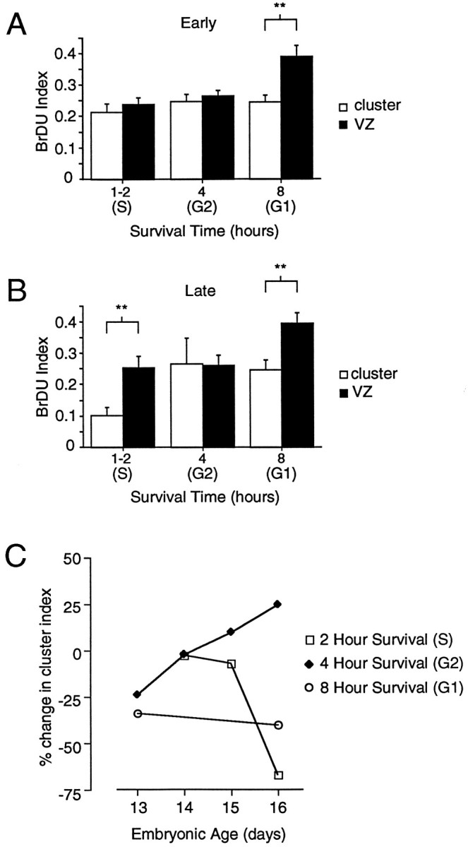
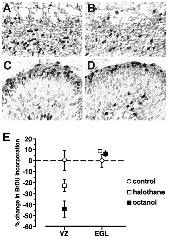
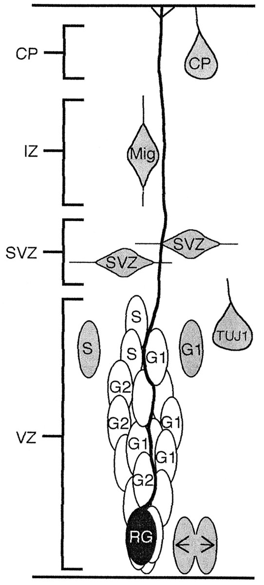
Cells within the ventricular zone (VZ) of developing neocortex are coupled together into clusters by gap junction channels. The specific role of clustering in cortical neurogenesis is unknown; however, clustering provides a means for spatially restricted local interactions between subsets of precursors and other cells within the VZ. In the present study, we have used a combination of 5-bromo-2'-deoxyuridine (BrDU) pulse labeling, intracellular biocytin labeling, and immunocytochemistry to determine when in the cell cycle VZ cells couple and uncouple from clusters and to determine what cell types within the VZ are coupled to clusters. Our results indicate that clusters contain radial glia and neural precursors but do not contain differentiating or migrating neurons. In early neurogenesis, all precursors in S and G2 phases of the cell cycle are coupled, and approximately half of the cells in G1 are coupled. In late neurogenesis, however, over half of the cells in both G1 and S phases are not coupled to VZ clusters, whereas all cells in G2 are coupled to clusters. Increased uncoupling in S phase during late neurogenesis may contribute to the greater percentage of VZ cells exiting the cell cycle at this time. Consistent with this hypothesis, we found that pharmacologically uncoupling VZ cells with octanol decreases the percentage of VZ cells that enter S phase. These results demonstrate that cell clustering in the VZ is restricted to neural precursors and radial glia, is dynamic through the cell cycle, and may play a role in regulating neurogenesis.


Figures

Fig. 1.
Fig. 1.
Only cells in the VZ of embryonic cortex form clusters of coupled cells. A, A cell in the IZ, a putative migrating neuron, not coupled to any other cells.B, A cell in the CP not coupled to any other cells.C, An entire cluster of cells in a 100 μm section labeled with biocytin. Cells are packed tightly, so individual cells are difficult to distinguish in a thick section. A single process exits from the top of the cluster. D, Two cells coupled at the top of the VZ. Scale bar, 10 μm.
Fig. 2.
Fig. 2.
Strategy for BrDU and biocytin double-labeling experiments. A, Pregnant mice at E13–E18 were injected with BrDU and allowed to survive for different times before the preparation of cortical explants and biocytin injection.B, Photomicrograph of tissue double-labeled for BrDU and biocytin. Black reaction product reveals BrDU staining, whereas brown product is biocytin. Thearrow indicates a cell within a cluster that is double labeled. Scale bar, 10 μm.
Fig. 3.
Fig. 3.
Cell types within and not within VZ clusters.A, RAT 401 nestin labeling of fibers in the IZ of E16 mouse cortex. Arrows mark the course of a glial fiber that is shown in B. B, A fiber extending into the IZ from a cluster filled in the VZ. The arrowsin A and B track the same fiber that is double-labeled for both RAT 401 and biocytin. C, TUJ labeling of the same field shown in D. Thearrow indicates a TUJ-labeled cell that is stained, and that cell is directly adjacent to the cluster labeled inD but is not coupled to the cluster. Also the fiber extending from the cluster does not stain with TUJ. D, A cluster of cells labeled with biocytin. The cluster is overexposed to show more clearly the borders of the cluster. E, A DAPI-stained view of the same field shown in F.F, The bottom of a cluster of cells at the ventricular surface. The arrow points to an M phase cell that is not coupled to the cluster. Scale bar, 10 μm.
Fig. 4.
Fig. 4.
Coupling changes throughout the cell cycle and the course of neurogenesis. A, A bar graph comparing the BrDU indices for cells in clusters with the indices for the VZ population. The BrDU index for a cluster and that for the VZ population were significantly different at the 8 hr survival time or during G1 (Tukey, **p < 0.01). Omnibus ANOVA comparing the VZ index with the cluster index across all times was significant at p < 0.01 (E13–E18).B, In late neurogenesis (E16–E18), there is a significant difference between the VZ and cluster indices at both 1–2 hr (primarily S phase cells) and 8 hr (G1 phase cells) (Tukey, **p < 0.01). C, A summary of the differences in BrDU indices between cells in clusters and cells in the VZ population. Unlike the analyses in A andB that show the means and SEM of the BrDU index determined separately for each cluster, the percentage differences reported in C are for the percentage of BrDU-labeled cells determined for cells in all clusters pooled together. The difference index indicates the fraction of BrDU cells in the population that cannot be accounted for in clusters in early and late neurogenesis for three phases of the cell cycle.
Fig. 5.
Fig. 5.
Pharmacological blockade of coupling decreases the number of cells in S phase. A, BrDU labeling in an explant of E16 dorsal cortex. The explant was incubated for 4 hr in ACSF, 95% O2/5% CO2, and then pulsed for 1 hr with 5 μm BrDU. B, BrDU labeling in an explant of cortex treated with 1 mm octanol.C, D, BrDU labeling in explants of cerebellum incubated for 4 hr and then pulsed with BrDU. The tissue inC was incubated in ACSF, and the tissue inD was incubated with ACSF and 1 mm octanol.E, Means and SEM showing the effects of octanol and halothane (1 mm) on the number of cells in S phase for explants of both neocortex and cerebellum. The uncoupling agents decreased the number of S phase cells in the VZ but not in the EGL, where cells are not coupled by gap junction channels.
Fig. 6.
Fig. 6.
Diagram summary depicting the composition of clusters in the VZ. Clusters are organized around radial glial (RG) fibers and contain cells in G1, S, and G2. Shaded cells are not members of VZ clusters; these include cells in M phase (arrows), postmitotic neurons (TUJ1), migrating neurons (Mig), SVZ cells, and some cells in S and G1phases of the cell cycle.

References

    1. Alvarez-Buylla A, Buskirk D, Nottebohm F. Monoclonal antibody reveals radial glia in adult avian brain. J Comp Neurol. 1987;264:159–170. - PubMed
    1. Antonopoulos J, Pappas I, Parnavelas J. Activation of the GABAA receptor inhibits the proliferative effects of bFGF in cortical progenitor cells. Eur J Neurosci. 1997;9:291–298. - PubMed
    1. Blanton M, LoTurco J, Kriegstein A. Whole cell recording from neuron in slices of reptilian and mammalian cerebral cortex. J Neurosci Methods. 1989;30:203–210. - PubMed
    1. Boulder Committee. Embryonic vertebrate central nervous system: revised terminology. Anat Rec. 1970;166:257–261. - PubMed
    1. Cai L, Hayes N, Nowakowski R. Synchrony of clonal cell proliferation and contiguity of clonally related cells: production of mosaicism in the ventricular zone of developing mouse neocortex. J Neurosci. 1997;17:2088–2100. - PMC - PubMed

Publication types

LinkOut - more resources