. 1997 Sep 8;138(5):1077-87.
doi: 10.1083/jcb.138.5.1077.

Transport and localization elements in myelin basic protein mRNA

Affiliations

Transport and localization elements in myelin basic protein mRNA

K Ainger et al. J Cell Biol. .

Abstract

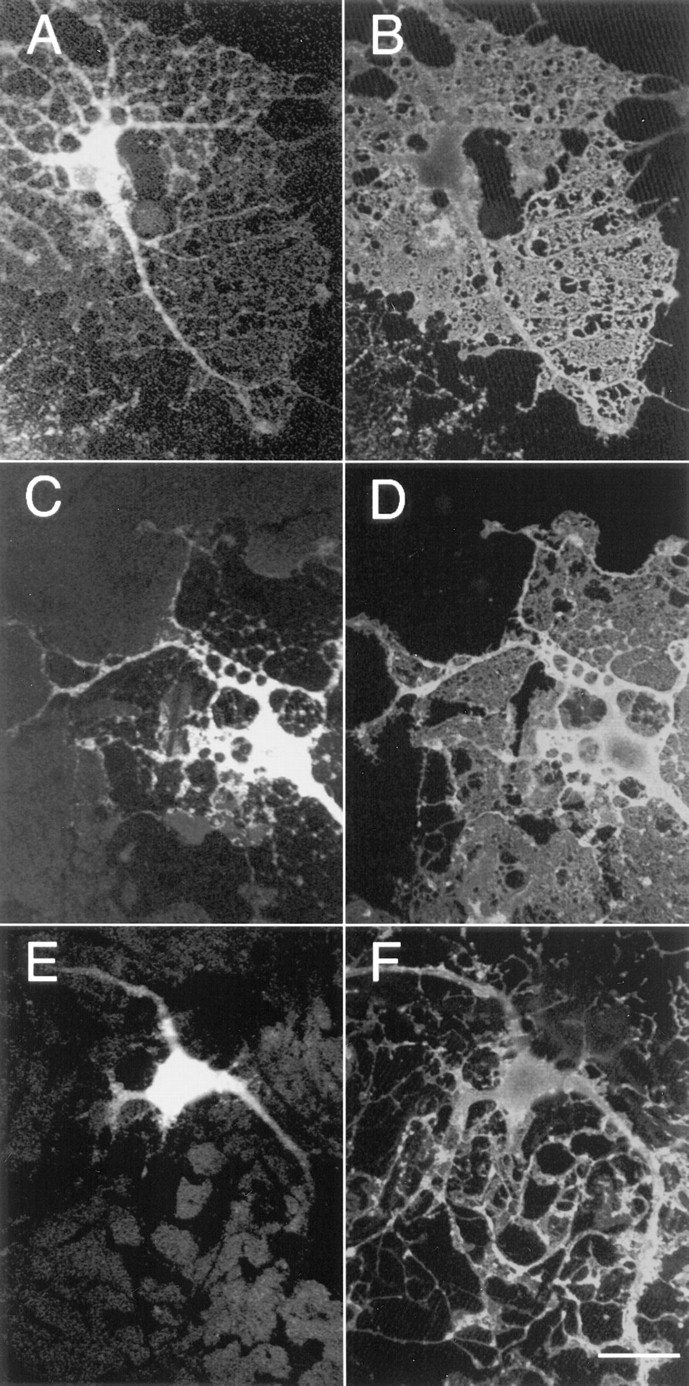
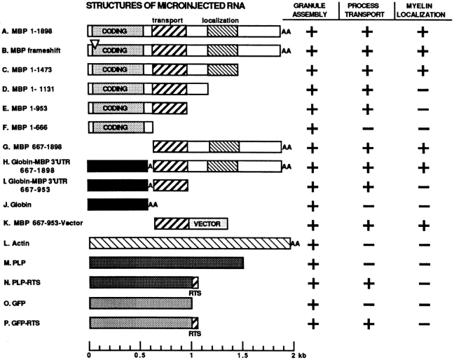
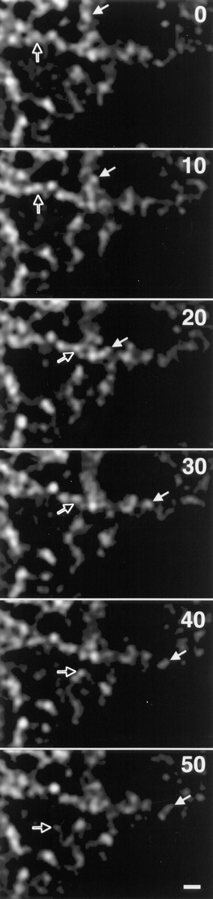
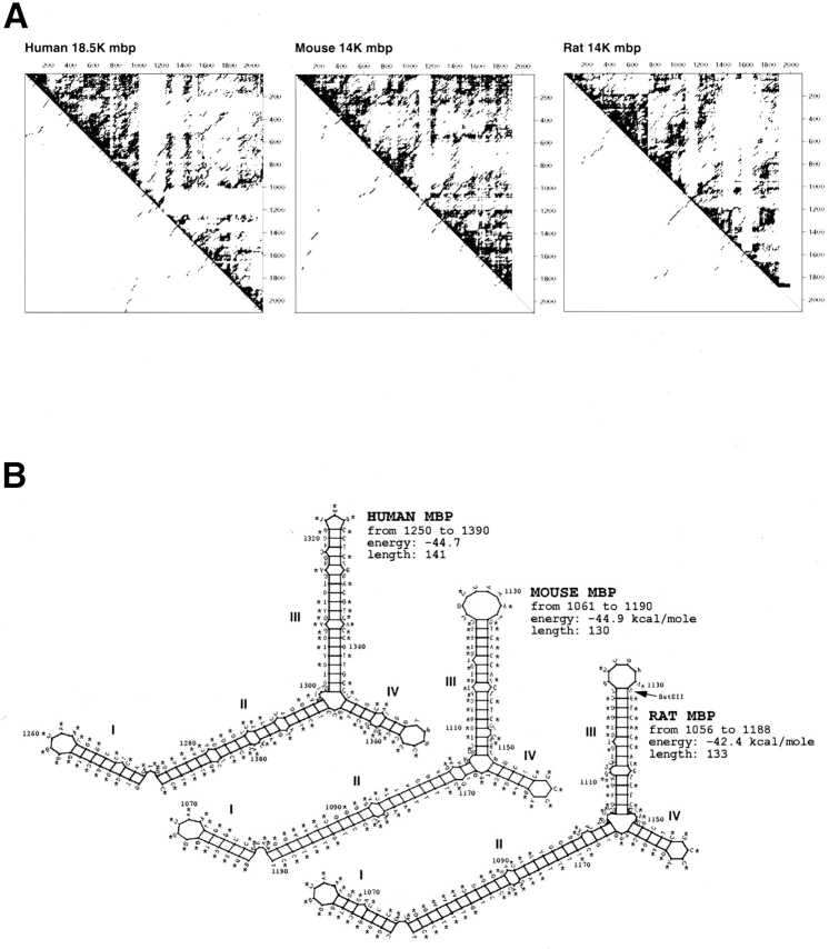
Myelin basic protein (MBP) mRNA is localized to myelin produced by oligodendrocytes of the central nervous system. MBP mRNA microinjected into oligodendrocytes in primary culture is assembled into granules in the perikaryon, transported along the processes, and localized to the myelin compartment. In this work, microinjection of various deleted and chimeric RNAs was used to delineate regions in MBP mRNA that are required for transport and localization in oligodendrocytes. The results indicate that transport requires a 21-nucleotide sequence, termed the RNA transport signal (RTS), in the 3' UTR of MBP mRNA. Homologous sequences are present in several other localized mRNAs, suggesting that the RTS represents a general transport signal in a variety of different cell types. Insertion of the RTS from MBP mRNA into nontransported mRNAs, causes the RNA to be transported to the oligodendrocyte processes. Localization of mRNA to the myelin compartment requires an additional element, termed the RNA localization region (RLR), contained between nucleotide 1,130 and 1, 473 in the 3' UTR of MBP mRNA. Computer analysis predicts that this region contains a stable secondary structure. If the coding region of the mRNA is deleted, the RLR is no longer required for localization, and the region between nucleotide 667 and 953, containing the RTS, is sufficient for both RNA transport and localization. Thus, localization of coding RNA is RLR dependent, and localization of noncoding RNA is RLR independent, suggesting that they are localized by different pathways.


Figures

Figure 2
Figure 2
Representative mouse oligodendrocytes microinjected and immunofluorescently labeled with both anti-digoxigenin (A, C, and E) and anti-MBP antibodies (B, D, and F). A and B show an oligodendrocyte injected with RNA of Fig. 1 A. The cell exhibits granule formation, transport along the processes, and localization to the myelin membranes. This pattern is representative of cells injected with RNAs of A, B, C, G, H, and K in Fig. 1. C and D show an oligodendrocyte injected with RNA of Fig. 1 D. The cell exhibits granule formation and transport along the processes but without localization to the myelin membrane. This pattern is representative of cells injected with RNAs of D, E, I, N and P in Fig. 1. E and F show an oligodendrocyte injected with RNA of Fig. 1 F. The cell exhibits granule formation but without transport along the processes or localization to the myelin membrane. This pattern is representative of cells injected with RNAs of F, J, L, M and O in Fig. 1. The extracellular staining in C and E is due to RNA leaking from the microinjection needle and adhering nonspecifically to the glass coverslip in regions not occluded by the cell. Bar, 25 μm.
Figure 1
Figure 1
Intracellular localization of microinjected RNAs. (A) Full length MBP. (B) Frameshifted full length MBP. (C) EcoRI-truncated MBP. (D) BstEII-truncated MBP. (E) PvuII-truncated MBP. (F) SalI-truncated MBP. (G) SalI-fragment of MBP 3′ UTR. (H) Chimeric construct of X. laevis β-globin with SalI fragment of MBP 3′ UTR. (I) Chimeric construct of X. laevis β-globin with SalI–PvuII fragment of MBP 3′ UTR. (J) X. laevis β-globin. (K) SalI–PvuII fragment of MBP 3′ UTR with vector-derived sequences. (L) Mouse β-actin. (M) Mouse PLP. (N) Mouse PLP-RTS. (O) GFP. (P) GFP-RTS. Plus (+) indicates that this distribution was observed in the majority of several hundred injected cells.
Figure 3
Figure 3
Time-lapse confocal imaging of RNA movement in oligodendrocyte microinjected with fluorescent PLP-RTS RNA. PLP-RTS RNA was fluorescently labeled and microinjected into oligodendrocytes. Time-lapse confocal images were collected at 10-s intervals. The region shown contains several oligodendrocyte processes. The cell body is out of the frame to the left. The majority of the labeled granules were immobile during the time interval shown. Two granules (indicated by open and closed arrows) exhibited sustained vectorial motion in an anterograde direction during the time course shown. Bar, 1 μm.
Figure 4
Figure 4
Secondary structure predictions for MBP mRNA. (A) Energy dot plots of predicted secondary structures in mRNAs encoding rat 14-kD, mouse 14-kD, and human 18.5-kD MBP. Suboptimal structures within 10 kcal/mole of the optimal structures are plotted above the diagonal. Sparsely populated vertical and horizontal regions indicate possible stable structures. Optimal structures are plotted below the diagonal. (B) Predicted secondary structures for rat 14 kD MBP mRNA from base 1,056 to 1,188, mouse 14-kD mRNA from 1,061 to 1,190, and human MBP 18.5-kD mRNA from base 1,250 to 1,390. Asterisks denote bases conserved in rat, mouse, and human MBP mRNAs. The BstEII site used to create the RNA in Fig. 1 D is indicated on the rat structure.

References

    1. Agarwal HC, Burton RM, Fishman MA, Mitchell RF, Prensky AL. Partial characterization of a new myelin component. J Neurochem. 1972;19:2083–2089. - PubMed
    1. Ainger K, Avossa D, Morgan F, Hill SJ, Barry C, Barbarese E, Carson JH. Transport and localization of exogenous myelin basic protein mRNA microinjected into oligodendrocytes. J Cell Biol. 1993;123:431–441. - PMC - PubMed
    1. Bacher G, Lütcke H, Jungnickel B, Rapoport TA, Dobberstein B. Regulation by the ribosome of the GTPase of the signal-recognition particle. Nature (Lond) 1996;381:248–251. - PubMed
    1. Barbarese E. Spatial distribution of myelin basic protein mRNA and polypeptide in quaking oligodendrocytes in culture. J Neurosci Res. 1991;29:271–281. - PubMed
    1. Barbarese E, Koppel DE, Deutscher MP, Smith CL, Ainger K, Morgan F, Carson JH. Protein translation components are colocalized in granules in oligodendrocytes. J Cell Sci. 1995;108:2781–2790. - PubMed

Publication types

MeSH terms