Comparative Study
. 1997 Sep 8;138(5):1149-57.
doi: 10.1083/jcb.138.5.1149.

ICAP-1, a novel beta1 integrin cytoplasmic domain-associated protein, binds to a conserved and functionally important NPXY sequence motif of beta1 integrin

Affiliations
Comparative Study

ICAP-1, a novel beta1 integrin cytoplasmic domain-associated protein, binds to a conserved and functionally important NPXY sequence motif of beta1 integrin

D D Chang et al. J Cell Biol. .

Abstract

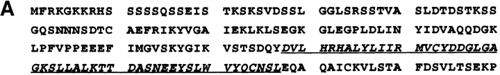
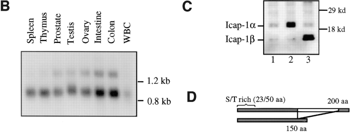
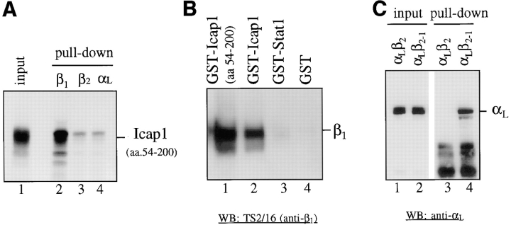
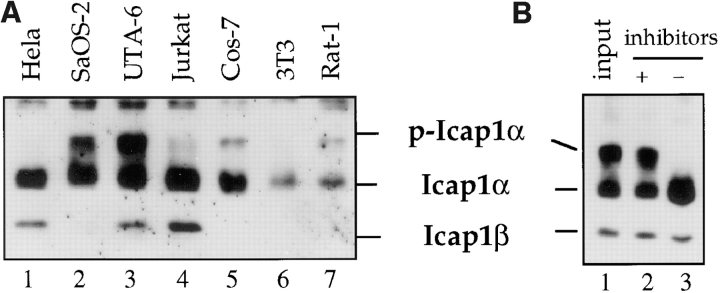
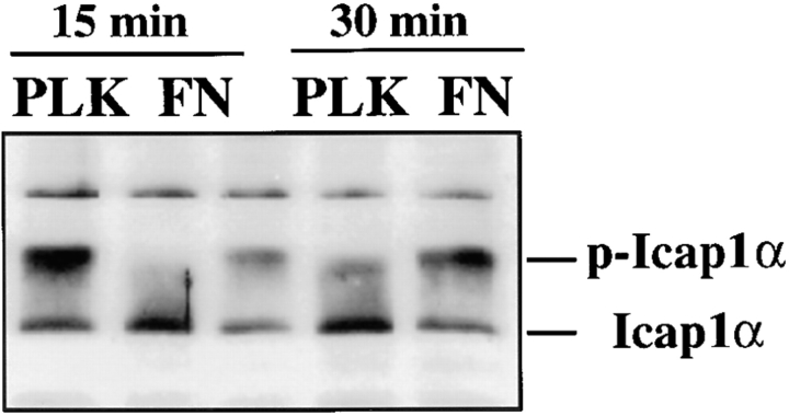
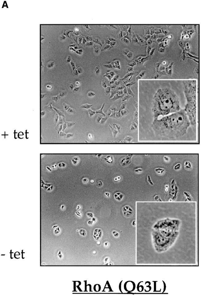
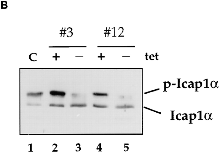
The cytoplasmic domains of integrins are essential for cell adhesion. We report identification of a novel protein, ICAP-1 (integrin cytoplasmic domain- associated protein-1), which binds to the 1 integrin cytoplasmic domain. The interaction between ICAP-1 and beta1 integrins is highly specific, as demonstrated by the lack of interaction between ICAP-1 and the cytoplasmic domains of other beta integrins, and requires a conserved and functionally important NPXY sequence motif found in the COOH-terminal region of the beta1 integrin cytoplasmic domain. Mutational studies reveal that Asn and Tyr of the NPXY motif and a Val residue located NH2-terminal to this motif are critical for the ICAP-1 binding. Two isoforms of ICAP-1, a 200-amino acid protein (ICAP-1alpha) and a shorter 150-amino acid protein (ICAP-1beta), derived from alternatively spliced mRNA, are expressed in most cells. ICAP-1alpha is a phosphoprotein and the extent of its phosphorylation is regulated by the cell-matrix interaction. First, an enhancement of ICAP-1alpha phosphorylation is observed when cells were plated on fibronectin-coated but not on nonspecific poly-L-lysine-coated surface. Second, the expression of a constitutively activated RhoA protein that disrupts the cell-matrix interaction results in dephosphorylation of ICAP-1alpha. The regulation of ICAP-1alpha phosphorylation by the cell-matrix interaction suggests an important role of ICAP-1 during integrin-dependent cell adhesion.


Figures

Figure 1
Figure 1
ICAP-1α is a novel serine/threonine-rich 20-kD protein. (A) Sequence of ICAP-1. The translated amino acid sequence of ICAP-1α is shown. 50 amino acids absent in ICAP-1β are indicated in underlined italics. The nucleotide sequence data are available from GenBank/EMBL/DDBJ under accession number AF012023. (B) Northern blot analysis of ICAP-1α. Northern blots of transcripts from various human tissues (Clontech) were hybridized with ICAP-1α cDNA probe. The 0.9-kb mRNA is expressed in all eight tissue samples. The minor 1.3-kb mRNA, derived from the use of the downstream polyadenylation site, is also expressed in varying amount in all eight samples. (C) Western blot analysis of ICAP-1. ICAP-1α and ICAP-1β were detected in the total cell lysate (5 μg) from 293T cells using polyclonal anti-ICAP-1 antibody (lane 1). The assignments of ICAP-1α and ICAP-1β were confirmed by transiently expressing ICAP-1α (lane 2) and ICAP-1β cDNA (lane 3) in 293T cells. (D) The full-length 200-aa ICAP-1α and an alternatively spliced ICAP-1β lacking internal 50 aa are shown in a schematic diagram.
Figure 1
Figure 1
ICAP-1α is a novel serine/threonine-rich 20-kD protein. (A) Sequence of ICAP-1. The translated amino acid sequence of ICAP-1α is shown. 50 amino acids absent in ICAP-1β are indicated in underlined italics. The nucleotide sequence data are available from GenBank/EMBL/DDBJ under accession number AF012023. (B) Northern blot analysis of ICAP-1α. Northern blots of transcripts from various human tissues (Clontech) were hybridized with ICAP-1α cDNA probe. The 0.9-kb mRNA is expressed in all eight tissue samples. The minor 1.3-kb mRNA, derived from the use of the downstream polyadenylation site, is also expressed in varying amount in all eight samples. (C) Western blot analysis of ICAP-1. ICAP-1α and ICAP-1β were detected in the total cell lysate (5 μg) from 293T cells using polyclonal anti-ICAP-1 antibody (lane 1). The assignments of ICAP-1α and ICAP-1β were confirmed by transiently expressing ICAP-1α (lane 2) and ICAP-1β cDNA (lane 3) in 293T cells. (D) The full-length 200-aa ICAP-1α and an alternatively spliced ICAP-1β lacking internal 50 aa are shown in a schematic diagram.
Figure 2
Figure 2
In vitro and in vivo interaction between ICAP-1α and β1 integrins. (A) Interaction between GST-β1 and ICAP-1α in vitro. ICAP-1α (aa 54–200) was synthesized in vitro using reticulocyte lysate (lane 1) and incubated with 2 μg of bacterially expressed GST fusion proteins containing the cytoplasmic domains of integrin β1 (lane 2), β2 (lane 3), and αL (lane 4). (B) Interaction between GST-ICAP1α and β1 integrins in vivo. ICAP-1α (aa 54– 200) (lane 1) and a full-length ICAP1α (lane 2) were expressed as GST fusion protein in 293T cells using eukaryotic expression vector pEBG. For controls, GST-Stat1 (lane 3) and GST (lane 4) were used. Coprecipitation of the endogenous β1 integrins with the GST fusion proteins were determined on a Western blot using TS2/16 (anti–human β1 integrin antibody). (C) Restricted binding specificity of ICAP1α. GST-ICAP1α was expressed in 293T cells along with expression constructs for αLβ2 or αLβ2-1 (chimeric β2 subunit that has the COOH-terminal 20 aa replaced with the COOH-terminal 21 aa of β1 integrin). Coprecipitation of transfected integrins with the GST-ICAP1α was determined on a Western blot using anti-αL antibody. Lanes 1 and 2, total lysates of cells expressing GST-ICAP1α and αLβ2 (lane 1) and αLβ2-1 (lane 2). Lanes 3 and 4, the same samples as in lanes 1 and 2, respectively, after GST “pull-down.”
Figure 3
Figure 3
ICAP-1α is a phosphoprotein. (A) Western blot analysis showing ICAP-1 expression. Total cell lysates (15 μg) from various cell lines were analyzed on a Western blot using anti-ICAP1 antibody. The positions of ICAP-1α and ICAP-1β as well as the slow migrating species corresponding to the phosphorylated ICAP-1α (p-ICAP1α) are indicated. (B) In vitro phosphatase treatment experiment. 15 μg of UTA-6 cell lysates were incubated at 30°C for 30 min. Lane 1, input; lane 2, incubation in the presence of sodium vanadate and calyculin A; lane 3, incubation at 30°C.
Figure 4
Figure 4
Regulation of ICAP-1α phosphorylation during cell adhesion. UTA-6 cells were trypsinized and replated on plates coated with poly-l-lysine (PLK) (lanes 1 and 3) or fibronectin (FN) (lanes 2 and 4). Adherent cells were lysed in 0.5% NP-40 at t = 15 min (lanes 1 and 2), and t = 30 min (lanes 3 and 4) and the extent of ICAP-1α phosphorylation was determined on a Western blot using anti-ICAP-1 antibody.
Figure 5
Figure 5
Control of ICAP-1α phosphorylation by RhoA protein. (A) Expression of activated RhoA interferes with cell spreading. The morphology of UTA-6 cells expressing RhoA(Q63L) under the control of tetracycline-inducible promoter is shown. In the absence of tetracycline, which induces the expression of RhoA (Q63L), cells remain rounded and fail to spread. (B) Expression of activated RhoA results in dephosphorylation of ICAP-1α. The extent of ICAP-1α phosphorylation in two independently derived UTA-6 cell lines expressing RhoA (Q63L) was assessed in Western blot analysis using anti-ICAP1 antibody. In parental cells (lane 1) or in the presence of tetracycline (lanes 2 and 4), a significant portion of ICAP-1α is phosphorylated. Induction of RhoA(Q63L) is associated with the loss of ICAP-1α phosphorylation is both cell lines (lanes 3 and 5).
Figure 5
Figure 5
Control of ICAP-1α phosphorylation by RhoA protein. (A) Expression of activated RhoA interferes with cell spreading. The morphology of UTA-6 cells expressing RhoA(Q63L) under the control of tetracycline-inducible promoter is shown. In the absence of tetracycline, which induces the expression of RhoA (Q63L), cells remain rounded and fail to spread. (B) Expression of activated RhoA results in dephosphorylation of ICAP-1α. The extent of ICAP-1α phosphorylation in two independently derived UTA-6 cell lines expressing RhoA (Q63L) was assessed in Western blot analysis using anti-ICAP1 antibody. In parental cells (lane 1) or in the presence of tetracycline (lanes 2 and 4), a significant portion of ICAP-1α is phosphorylated. Induction of RhoA(Q63L) is associated with the loss of ICAP-1α phosphorylation is both cell lines (lanes 3 and 5).

References

    1. Altieri DC, Edgington TS. The saturable high affinity association of factor X to ADP-stimulated monocytes defines a novel function of the Mac-1 receptor. J Biol Chem. 1988;263:7007–7015. - PubMed
    1. Ausubel, F.M., R. Brent, R.E. Kingston, D.D. Moore, J.G. Seidman, J.A. Smith, and K. Struhl. 1994. Current Protocols in Molecular Biology. John Wiley & Sons, Inc., New York.
    1. Balzac F, Belkin AM, Koteliansky VE, Balabanov YV, Altruda F, Silengo L, Tarone G. Expression and functional analysis of a cytoplasmic domain variant of the β1integrin subunit. J Cell Biol. 1993;121:171–178. - PMC - PubMed
    1. Bansal A, Geirasch LM. The NPXY internalization signal of the LDL receptor adopts a reverse-turn conformation. Cell. 1991;67:1195–1201. - PubMed
    1. Bennett JS, Vilaire G. Exposure of platelet fibrinogen receptors by ADP and epinephrine. J Clin Invest. 1979;64:1393–1401. - PMC - PubMed

Publication types

MeSH terms

Associated data