. 1997 Sep 8;138(5):1159-67.
doi: 10.1083/jcb.138.5.1159.

Integrin alpha2beta1 is a receptor for the cartilage matrix protein chondroadherin

Affiliations

Integrin alpha2beta1 is a receptor for the cartilage matrix protein chondroadherin

L Camper et al. J Cell Biol. .

Abstract

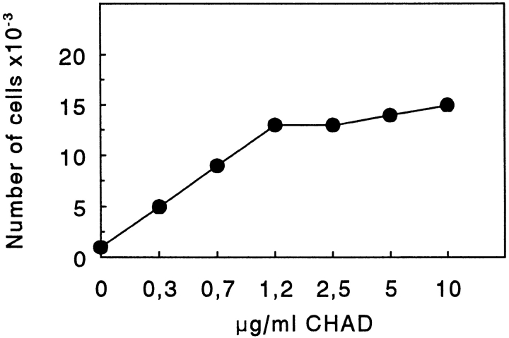
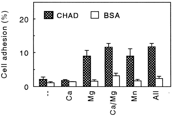
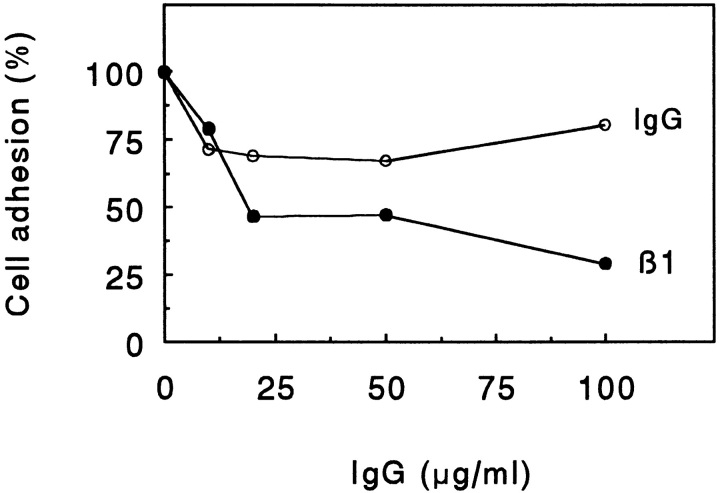
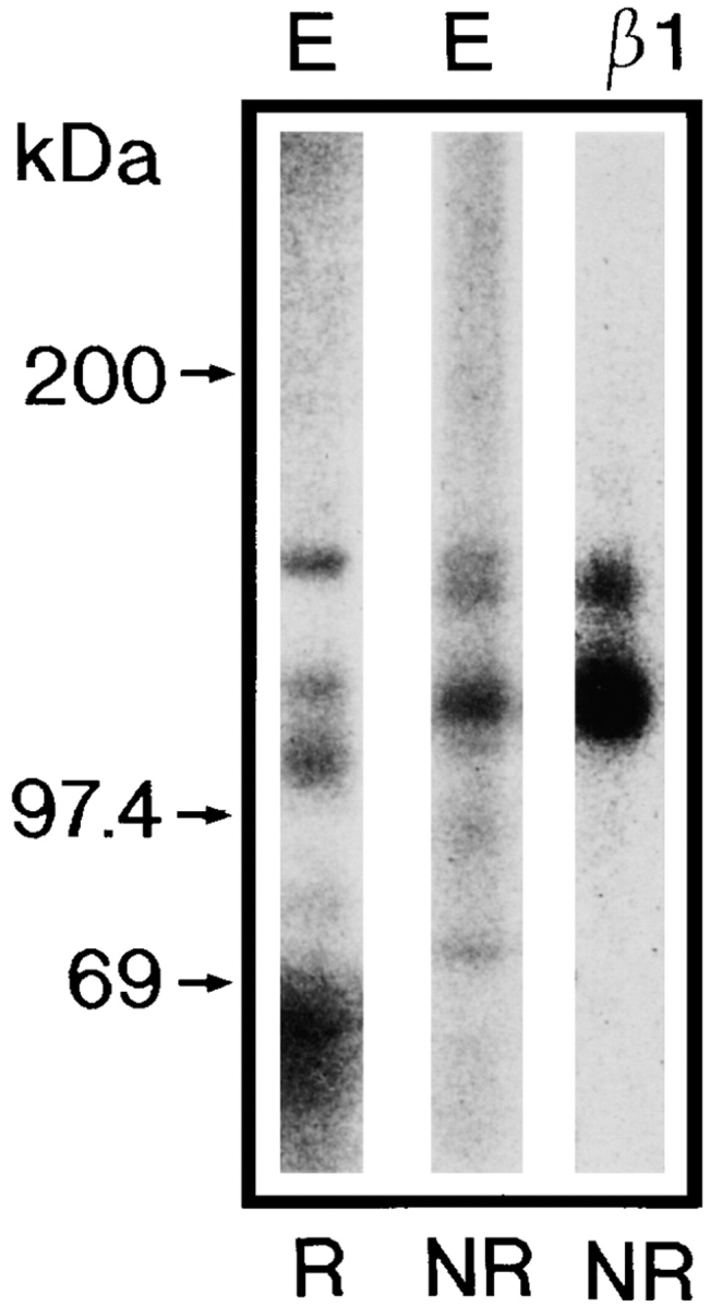
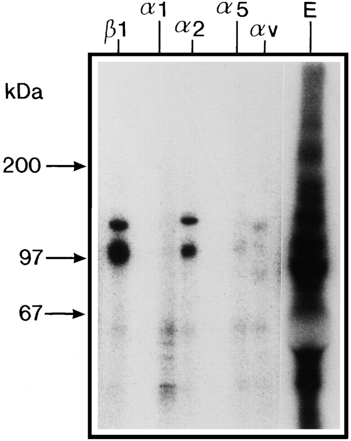
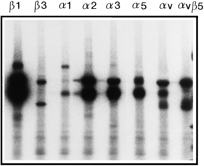
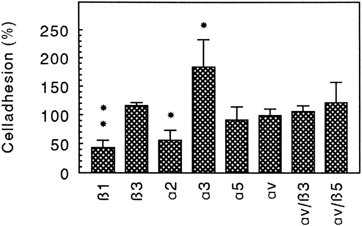
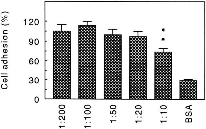
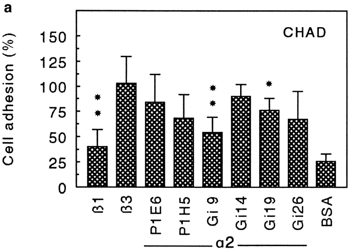
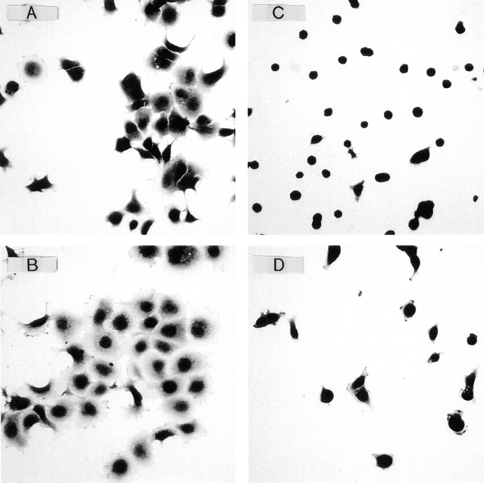
Chondroadherin (the 36-kD protein) is a leucine-rich, cartilage matrix protein known to mediate adhesion of isolated chondrocytes. In the present study we investigated cell surface proteins involved in the interaction of cells with chondroadherin in cell adhesion and by affinity purification. Adhesion of bovine articular chondrocytes to chondroadherin-coated dishes was dependent on Mg2+ or Mn2+ but not Ca2+. Adhesion was partially inhibited by an antibody recognizing beta1 integrin subunit. Chondroadherin-binding proteins from chondrocyte lysates were affinity purified on chondroadherin-Sepharose. The beta1 integrin antibody immunoprecipitated two proteins with molecular mass approximately 110 and 140 kD (nonreduced) from the EDTA-eluted material. These results indicate that a beta1 integrin on chondrocytes interacts with chondroadherin. To identify the alpha integrin subunit(s) involved in interaction of cells with the protein, we affinity purified chondroadherin-binding membrane proteins from human fibroblasts. Immunoprecipitation of the EDTA-eluted material from the affinity column identified alpha2beta1 as a chondroadherin-binding integrin. These results are in agreement with cell adhesion experiments where antibodies against the integrin subunit alpha2 partially inhibited adhesion of human fibroblast and human chondrocytes to chondroadherin. Since alpha2beta1 also is a receptor for collagen type II, we tested the ability of different antibodies against the alpha2 subunit to inhibit adhesion of T47D cells to collagen type II and chondroadherin. The results suggested that adhesion to collagen type II and chondroadherin involves similar or nearby sites on the alpha2beta1 integrin. Although alpha2beta1 is a receptor for both collagen type II and chondroadherin, only adhesion of cells to collagen type II was found to mediate spreading.


Figures

Figure 1
Figure 1
Adhesion of T47D cells to dishes coated with CHAD. Culture dishes (48 well) were coated with various concentrations of CHAD and blocked for nonspecific binding with BSA (0.25%). T47D cells (50,000/well) were allowed to adhere for 1 h at 37°C. Nonadherent cells were removed by washing, and adhesion was determined by analyzing lysosomal hexosaminidase. The results presented are the mean adhesion in duplicates from one of two experiments.
Figure 2
Figure 2
Divalent cation-dependent adhesion of chondrocytes to CHAD. Culture dishes (48 well) were coated with CHAD (5 μg/ ml) and blocked for nonspecific binding with BSA (0.25%). Bovine chondrocytes were allowed to adhere to the dishes for 1 h at 37°C in the absence of divalent cations or in the presence of Ca2+ (1 mM), Mg2+ (1 mM), or Mn2+ (50 μM). Nonadherent cells were removed by washing, and adhesion was determined by analyzing lysosomal hexosaminidase. Adhesion is expressed as a percentage of the total number of cells added to the dish. The numbers represent the mean adhesion from three wells ±SD from one of three experiments.
Figure 3
Figure 3
β1 integrin-dependent adhesion of chondrocytes to CHAD. Culture dishes (48 well) were coated overnight with chondroadherin (5 μg/ml) and blocked for nonspecific binding with BSA (0.25%). Bovine chondrocytes were allowed to adhere to the dishes for 1 h at 37°C in the presence of various concentrations of a polyclonal antibody against the rat β1 integrin subunit or control IgG. Nonadherent cells were removed by washing, and adhesion was determined by analyzing lysosomal hexosaminidase. The adhesion is expressed as a percentage of the control, and the numbers represent mean of duplicate adhesion from one of three experiments.
Figure 4
Figure 4
Affinity purification of CHAD-binding cell surface proteins. Bovine chondrocytes were 125I-labeled and lysed with 1% Triton X-100, 100 μg/ml aprotinin, 2 μg/ml leupeptin, 2 μg/ml pepstatin A, 1 mM PMSF, 1 mM MnCl2, and 1 mM MgCl2 in 10 mM Tris-HCl, pH 7.4. The lysate was passed over control agarose followed by CHAD agarose. Proteins with affinity for CHAD were eluted by EDTA (20 mM), passed over a desalting column (PD-10) equilibrated, and eluted with 0.3 M NaCl, 1% Triton X-100, 0.1% BSA 1 mM CaCl2, 1 mM MgCl2, 1 mM PMSF, in 50 mM Tris-HCl, pH 7.4. An aliquot of the protein peak was immunoprecipitated with the polyclonal rat β1 integrin antibody. Proteins in the eluate (E) and in the immunoprecipitate (β1) were separated by 4–12% SDS-PAGE under reducing (R) or nonreducing (NR) conditions.
Figure 5
Figure 5
Immunoprecipitation of CHAD-binding integrins from human fibroblasts. 125I-labeled A549 fibroblasts were lysed with 1% Triton X-100, 100 μg/ml aprotinin, 2 μg/ml leupeptin, 2 μg/ml pepstatin A, 1 mM PMSF, 1 mM MnCl2, and 1 mM MgCl2 in 10 mM Tris-HCl, pH 7.4. The lysate was passed over control agarose followed by CHAD agarose. Proteins with affinity for CHAD were eluted by EDTA (20 mM), passed over a desalting column (PD-10) equilibrated, and eluted with 0.3 M NaCl, 1% Triton X-100, 0.1% BSA, 1 mM CaCl2, 1 mM MgCl2, 1 mM PMSF in 50 mM Tris-HCl, pH 7.4. Aliquots of the protein peak were immunoprecipitated with monoclonal antibodies against the integrin subunits β1 (P4C10), α1 (TS2/7), α2 (P1E6), α5 (P1D6), and αv (VNR147). The immunoprecipitated proteins were separated by SDS-PAGE (4–12%) under nonreducing conditions and visualized by autoradiography.
Figure 6
Figure 6
Immunoprecipitation of integrins from human fibroblasts. 125I-labeled A549 fibroblasts were lysed with 1% Triton X-100, 100 μg/ml aprotinin, 2 μg/ml leupeptin, 2 μg/ml pepstatin A, 1 mM PMSF, 1 mM MnCl2, and 1 mM MgCl2 in 10 mM Tris-HCl, pH 7.4. Aliquots of the lysate were immunoprecipitated with monoclonal antibodies against the integrin subunits β1 (P4C10), β3 (RUU-PLF12), α1 (TS2/7), α2 (P1E6), α3 (P1B5), α5 (P1D6), αv (VNR147), and αvβ5 (P1F6). The immunoprecipitated proteins were separated by SDS-PAGE (4–12%) under nonreducing conditions and visualized by autoradiography.
Figure 7
Figure 7
Inhibition of fibroblast adhesion to CHAD by integrin antibodies. Culture dishes (48 well) were coated with CHAD (5 μg/ml) and blocked for nonspecific binding with BSA (0.25%). Human A549 fibroblasts were allowed to adhere to the dishes for 1 h at 37°C in the presence of monoclonal antibodies against the human integrin subunits β1 (P4C10), β3 (RUU-PLF12), α2 (Gi9), α3 (P1B5), α5 (P1D6), αv (VNR147), αvβ3 (LM609), and αvβ5 (P1F6). Nonadherent cells were removed by washing, and adhesion was determined by analyzing lysosomal hexosaminidase. The adhesion is expressed as a percentage of the control, and the numbers represent the mean of duplicate adhesion from three individual experiments ±SD. *P < 0.05; **P < 0.01; P β1 = 0.002; P α2 = 0.011; P α3 = 0.021.
Figure 8
Figure 8
Inhibition of human chondrocyte adhesion to CHAD by α2 integrin antibodies. Culture dishes (48 well) were coated with CHAD (5 μg/ml) and blocked for nonspecific binding with BSA (0.25%). Human chondrocytes were allowed to adhere to the dishes for 1 h at 37°C in the presence of various concentrations of the monoclonal antibody against the human integrin subunit α2 (Gi9). Nonadherent cells were removed by washing, and adhesion was determined by analyzing lysosomal hexosaminidase. The adhesion is expressed as a percentage of the control, and the numbers represent the mean adhesion ±SD from three wells in one of two experiments.
Figure 9
Figure 9
Inhibition of adhesion of T47D cells to CHAD (a) and to collagen type II (CII; b) by various α2 antibodies. Culture dishes (48 wells) were coated with 5 μg/ml of CHAD or CII and blocked for nonspecific binding with BSA (0.25%). T47D cells were allowed to adhere to the dishes for 1 h at 37°C in the absence or in the presence of monoclonal antibodies against the integrin subunits β1 (P4C10), β3 (RUU-PLF12), or various α2 antibodies. Nonadherent cells were removed by washing, and adhesion was determined by analyzing lysosomal hexosaminidase. The adhesion is expressed as a percentage of the control, and the numbers represent the mean of duplicate adhesion from three individual experiments ±SD. *P < 0.05; **P < 0.01; (a) Pβ1 = 0.004; PGi9 = 0.006; PGi19 = 0.026. (b) Pβ1 = 0.000; PP1E6 = 0.001; PP1H5 = 0.001; PGi9 = 0.000; PGi26 = 0.047.
Figure 9
Figure 9
Inhibition of adhesion of T47D cells to CHAD (a) and to collagen type II (CII; b) by various α2 antibodies. Culture dishes (48 wells) were coated with 5 μg/ml of CHAD or CII and blocked for nonspecific binding with BSA (0.25%). T47D cells were allowed to adhere to the dishes for 1 h at 37°C in the absence or in the presence of monoclonal antibodies against the integrin subunits β1 (P4C10), β3 (RUU-PLF12), or various α2 antibodies. Nonadherent cells were removed by washing, and adhesion was determined by analyzing lysosomal hexosaminidase. The adhesion is expressed as a percentage of the control, and the numbers represent the mean of duplicate adhesion from three individual experiments ±SD. *P < 0.05; **P < 0.01; (a) Pβ1 = 0.004; PGi9 = 0.006; PGi19 = 0.026. (b) Pβ1 = 0.000; PP1E6 = 0.001; PP1H5 = 0.001; PGi9 = 0.000; PGi26 = 0.047.
Figure 10
Figure 10
Spreading of T47D cells on collagen type II (CII) or CHAD. Chamber slides (eight chambers) were coated with 5 μg/ml of CII (A and B) or CHAD (C and D) and blocked for nonspecific binding with BSA (0.25%). Human T47D cells (20,000/ well) were plated onto the chambers and allowed to adhere and spread for 3 h at 37°C in the absence (A and C) or in the presence (B and D) of 10−8 M PMA. Nonadherent cells were removed by washing, and the adherent cells were fixed with 2% paraformaldehyde in PBS and stained with Meyer's hematoxilin and erythrosin. Spreading was visualized by light microscopy, and mean cell area (Table I) was calculated by image analyses using the Zeiss software KS400/ V2.00.

References

    1. Bengtsson E, Neames PJ, Heinegard D, Sommarin Y. The primary structure of a basic leucine-rich repeat protein, PRELP, found in connective tissue. J Biol Chem. 1995;270:25639–25644. - PubMed
    1. Blochberger TC, Vergnes J-P, Hempel J, Hassell JR. cDNA to chick lumican (corneal keratan sulfate proteoglycan) reveals homology to the small interstitial proteoglycan gene family and expression in muscle and intestine. J Biol Chem. 1992;267:347–352. - PubMed
    1. Carter W, Wayner E, Bouchard T, Kaur P. The role of integrins α2β1 in cell-cell and cell-substrate adhesion of human epidermal cells. J Cell Biol. 1990;110:1387–1404. - PMC - PubMed
    1. Chan BMC, Hemler ME. Multiple functional forms of integrin VLA-2 can be derived from a single α2 cDNA clone: interconversion of forms induced by an anti-β1 antibody. J Cell Biol. 1993;120:537–543. - PMC - PubMed
    1. Chen JD, Kim JP, Zhang K, Sarret Y, Wynn KC, Kramer RH, Woodley DT. Epidermal growth factor (EGF) promotes human keratinocyte locomotion on collagen by increasing the α2 integrin subunit. Exp Cell Res. 1993;209:216–223. - PubMed

Publication types

MeSH terms