Comparative Study
. 1997 Sep 8;138(5):1169-80.
doi: 10.1083/jcb.138.5.1169.

The kinetics of L-selectin tethers and the mechanics of selectin-mediated rolling

Affiliations
Comparative Study

The kinetics of L-selectin tethers and the mechanics of selectin-mediated rolling

R Alon et al. J Cell Biol. .

Abstract

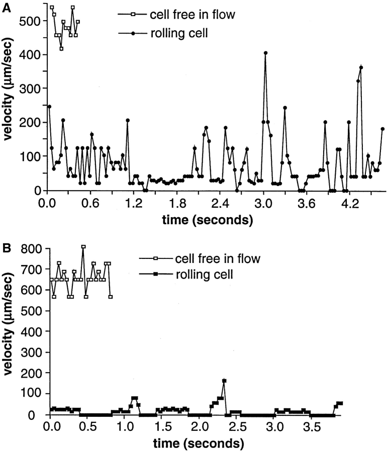
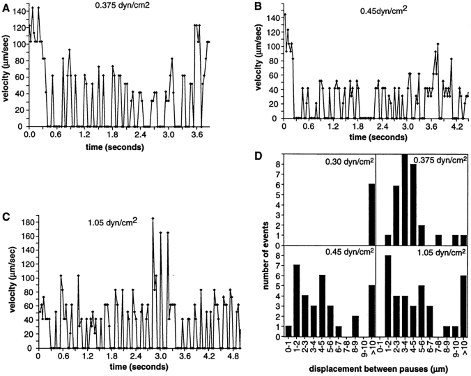
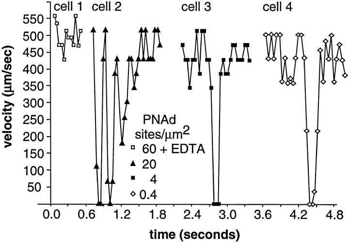
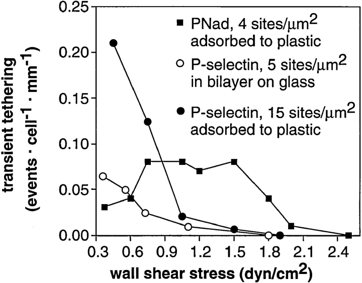
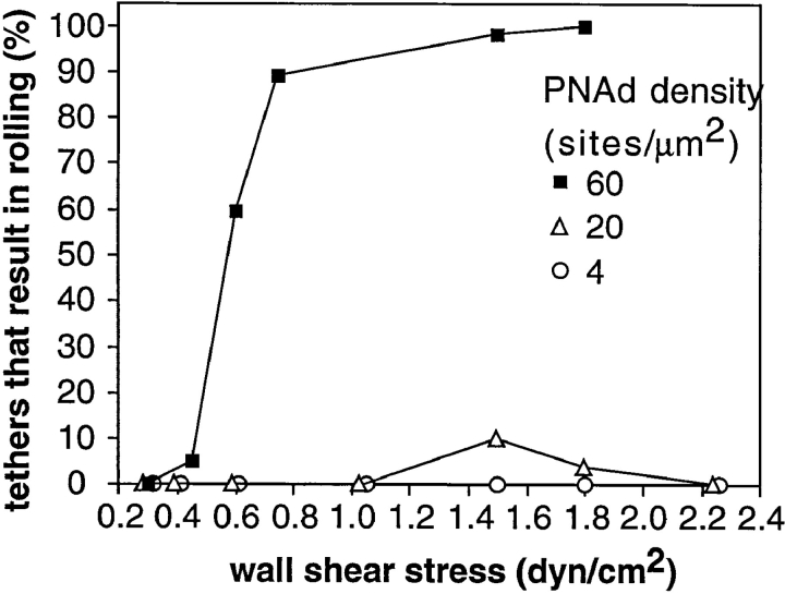
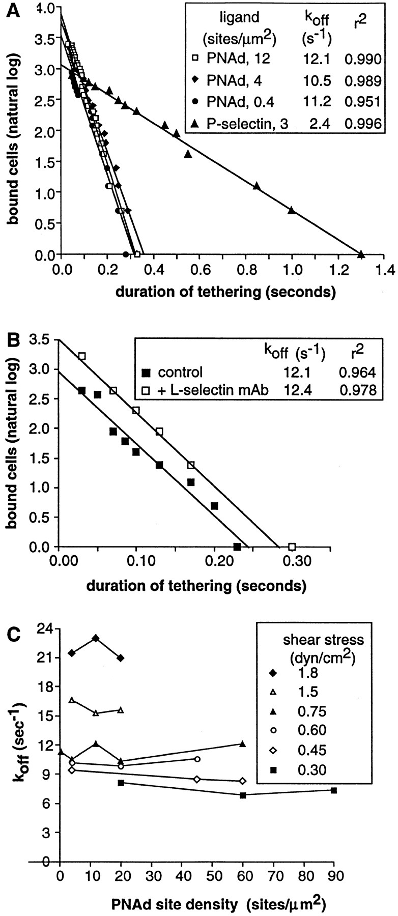
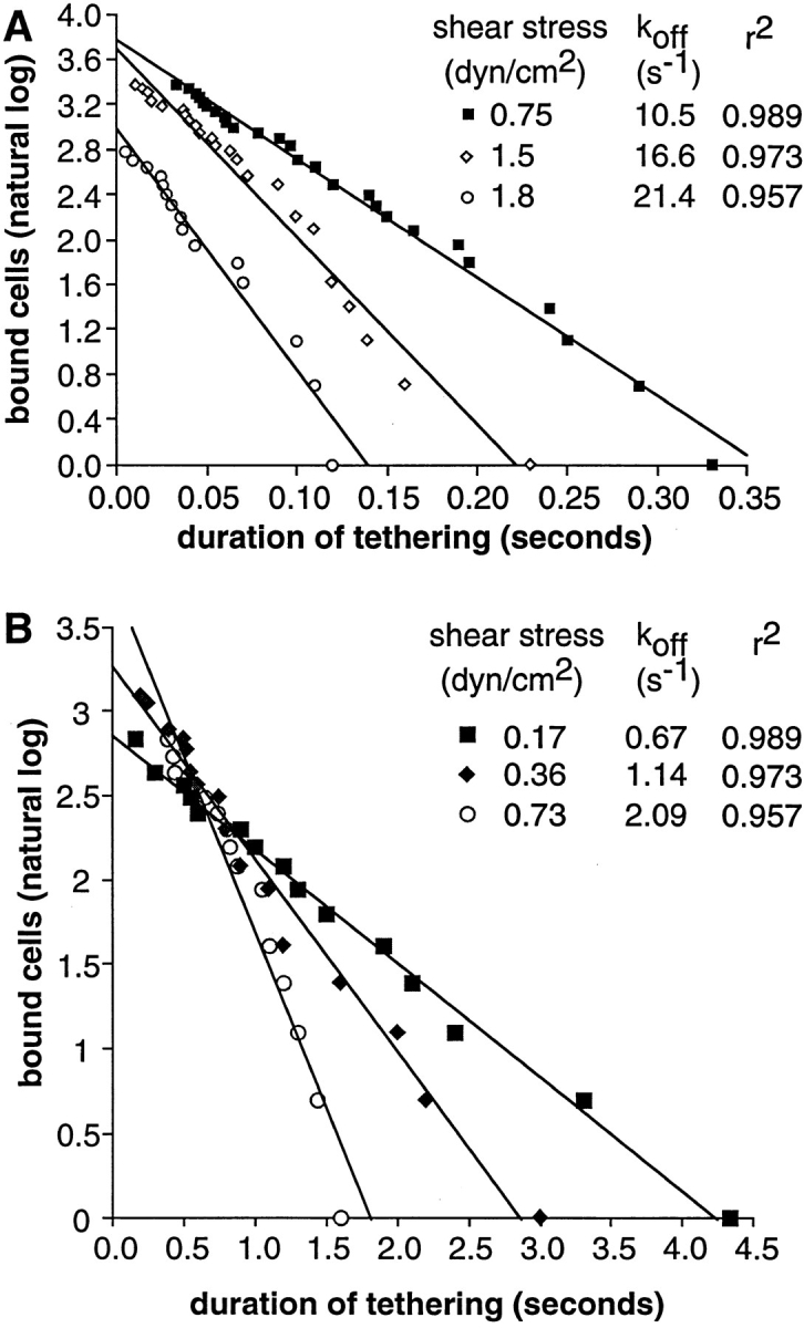
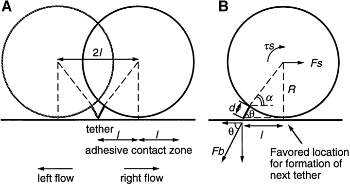
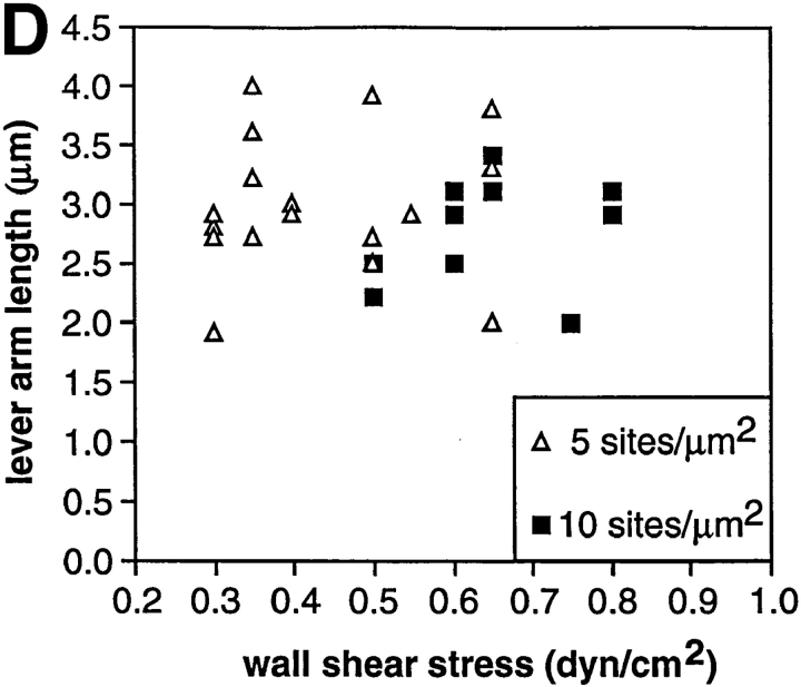
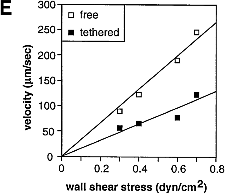
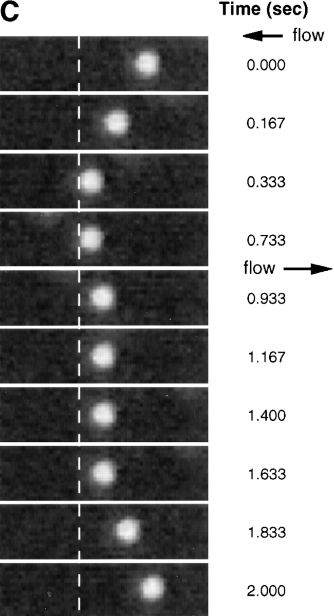
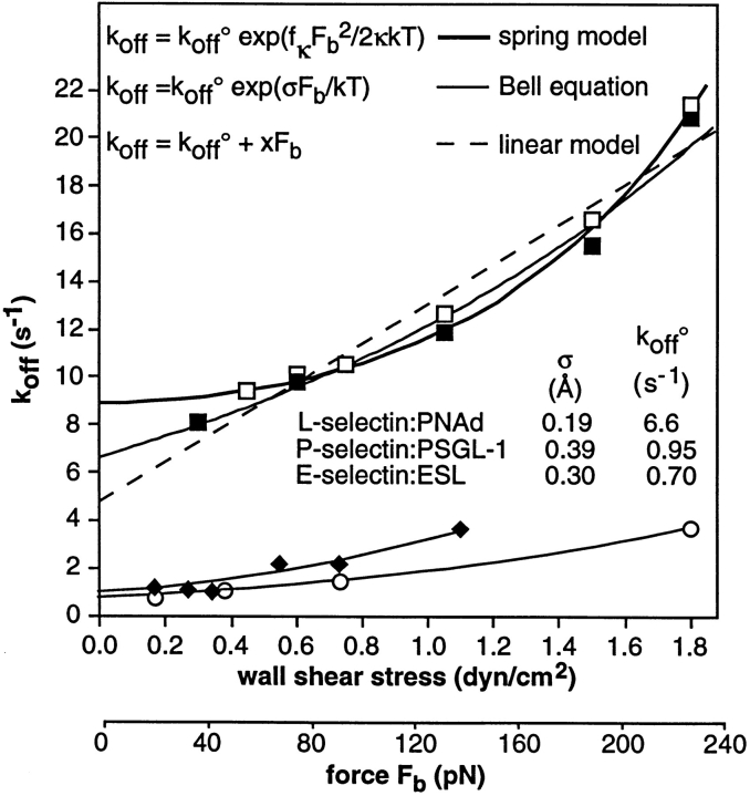
Two mechanisms have been proposed for regulating rolling velocities on selectins. These are (a) the intrinsic kinetics of bond dissociation, and (b) the reactive compliance, i.e., the susceptibility of the bond dissociation reaction to applied force. To determine which of these mechanisms explains the 7.5-11.5-fold faster rolling of leukocytes on L-selectin than on E- and P-selectins, we have compared the three selectins by examining the dissociation of transient tethers. We find that the intrinsic kinetics for tether bond dissociation are 7-10-fold more rapid for L-selectin than for E- and P-selectins, and are proportional to the rolling velocities through these selectins. The durations of pauses during rolling correspond to the duration of transient tethers on low density substrates. Moreover, applied force increases dissociation kinetics less for L-selectin than for E- and P-selectins, demonstrating that reactive compliance is not responsible for the faster rolling through L-selectin. Further measurements provide a biochemical and biophysical framework for understanding the molecular basis of rolling. Displacements of tethered cells during flow reversal, and measurements of the distance between successive pauses during rolling provide estimates of the length of a tether and the length of the adhesive contact zone, and suggest that rolling occurs with as few as two tethers per contact zone. Tether bond lifetime is an exponential function of the force on the bond, and the upper limit for the tether bond spring constant is of the same order of magnitude as the estimated elastic spring constant of the lectin-EGF unit. Shear uniquely enhances the rate of L-selectin transient tether formation, and conversion of tethers to rolling adhesions, providing further understanding of the shear threshold requirement for rolling through L-selectin.


Figures

Figure 1
Figure 1
The microkinetics of rolling of individual neutrophils interacting with the lowest densities of purified PNAd or P-selectin that can support rolling. (A) Neutrophils were perfused in a parallel wall flow chamber on PNAd at 60 sites per μm2 at a wall shear stress of 1.5 dyn/cm2. (B) Neutrophils were perfused on P-selectin at 30 sites per μm2 at 1.8 dyn/cm2. Velocities of cells free in flow adjacent to the wall are shown for comparison. Coordinates of cell centers were determined within ±0.68 μm and the velocity between each frame was calculated for individual cells.
Figure 2
Figure 2
The microkinetics of neutrophil rolling near the shear threshold. (A) The position of a neutrophil selected for continued interaction with a 100 sites per μm2 PNAd substrate at 0.375 dyn/ cm2 was determined to ±0.34 μm accuracy in each video frame and interframe velocities were calculated. (B) As in A, at 0.45 dyn/cm2. (C) As in A, at 1.05 dyn/cm2. Cells rolled for shorter distances at 0.375 dyn/cm2 than at 0.45 and 1.05 dyn/cm2; the cell in A rolled for 3.2 s. (D) The distance between pauses during rolling is modal near the shear threshold. The distance traveled between each pause (velocity = 0) was determined at each shear stress for two representative neutrophils that showed substantial interaction with the substrate after tethering within the field of view, in the same experiment as in A–C. Note that fewer events were seen at 0.3 dyn/cm2 because only transient tethers occurred.
Figure 3
Figure 3
The microkinetics of transient tethers on PNAd. Neutrophils were perfused at 1.5 dyn/cm2 on substrates with PNAd at the indicated density. Velocities of representative cells on each substrate are shown. Cell 1 in EDTA is shown to demonstrate the hydrodynamic velocity. Coordinates of cell centers were determined within one pixel accuracy (±0.9 μm) and the velocity between each frame was calculated for individual cells.
Figure 4
Figure 4
L-selectin forms tethers at higher shear forces than PSGL-1. The number of transient tethers was counted and divided by the distance cells were transported by flow across the field of view and by the number of cells that flowed across the field that were within the focal plane of the substrate. This yields the density of tethers. L-selectin blocking mAb DREG-56 added to neutrophils in the binding medium at 3 μg/ml, shortly before perfusion on the substrate, inhibited 95% of tethering events on PNAd. The density of leukocyte tethering to control substrates coated with HSA was <0.005 events cell−1 · mm−1. No tethering events were observed in the presence of 5 mM EDTA.
Figure 5
Figure 5
Conversion of tethering events to rolling is a function of wall shear stress and site density of PNAd. Tethers were considered to result in rolling when neutrophils moved for at least 2 s at a mean velocity of at least fivefold lower than the mean hydrodynamic velocity of untethered cells at a given shear stress.
Figure 6
Figure 6
The kinetics of dissociation of transiently tethered neutrophils from PNAd and its independence from PNAd or L-selectin densities. The k off values equal the negative slope of the lines through the dissociation data. (A) Kinetics at a wall shear stress of 0.75 dyn/cm2 on different low densities of PNAd. Data at the same wall shear stress on P-selectin (1) are shown for comparison. (B) Kinetics at a wall shear stress of 0.75 dyn/cm2 on higher density PNAd (60 sites per μm2) in the presence of subsaturating concentrations of control mAb AD38 to CD44 or mAb DREG-56 to L-selectin at 0.3 μg/ml. The L-selectin mAb reduced tethering frequency by 70% and abolished all rolling adhesions. (C) Dissociation rate constants at different wall shear stresses, as a function of PNAd site density. At low shear stresses, when both transiently tethered and continuously rolling cells could be observed, the few transient events that occurred had the same dissociation kinetics as those derived at low densities.
Figure 7
Figure 7
Effect of wall shear stress on the kinetics of neutrophil dissociation from PNAd and E-selectin. (A) PNAd at four sites per μm2. (B) E-selectin at seven sites per μm2.
Figure 8
Figure 8
The lever arm acting on a transient tether, the dimension of the adhesive contact zone, and the force on the tether bond. (A) Scheme for measurement of the lever arm by reversing the direction of flow. The lever arm, l, is the distance between the tether point and the projection of the center of the neutrophil on the substrate. (B) Estimation of forces on a neutrophil tethered in shear flow (drawn to scale). The force balance equations are F b cosθ = F s and F b lsinθ = τs + RF s, where F b is the force on the tether bond, F s is the force on the cell, and τs is the torque on the cell. The exact solution for F s for a motionless hard sphere in shear flow near a wall is F s = 6π·viscosity·R·h·shear·C where R is sphere radius, h is the distance from the center of the cell to the wall, and C is a numerical factor determined by integration that depends on h/R and ranges from 1 to 1.7 (17). With R = 4.25 μm for a neutrophil (48), variation of h–R from 0 to 0.5 μm has little effect on F s (±3%), and we have assumed h = R for a tethered cell. The assumption of hardness is good because no neutrophil deformation is visible microscopically within the range of shear used here. Roughness such as from microvilli is thought to increase F s only modestly; for a rough object, F s is intermediate between F s on a sphere contained in the object and F s on a sphere containing the object. We estimate uncertainty in R to measurement variation (48) and its effective increase by microvilli as ±0.25 μm. The calculated value of F s and its uncertainty are 59.8 ± 6.9 pN per dyn/cm2 wall shear stress. Both the uncertainty in R and in the lever arm measurement l = 3.06 ± 0.53 μm introduce uncertainties into the other calculated values. The uncertainties stemming from R and l were estimated separately for these values and their variances were added. We calculate θ = 62.3 ± 4.2°, d = 1.0 ± 0.32 μm, F b/F s = 2.15 ± 0.32, and F b = 124.4 ± 26.1 pN per dyn/cm2 wall shear stress. (C) Movement of a representative transiently tethered neutrophil on P-selectin at five sites per μm2 during reversal of flow at 0.3 dyn/cm2. The vertical dashed line marks the left boundary of the tethered cell before flow reversal. (D) Measurement of the lever arm. Each point is a measurement on an individual cell; the lever arm is defined as half the distance moved during flow reversal while cells were tethered to the indicated density of P-selectin. Shear stress was calibrated from the hydrodynamic velocity of cells in the same chamber using a syringe pump. (E) Velocities after flow reversal of untethered neutrophils and of neutrophils transiently tethered to P-selectin (five sites per μm2). The velocity of tethered cells was derived from a frame by frame analysis of the number of video frames required for a transiently tethered cell to move the distance of 2 l after flow reversal. Values are the average of two velocity determinations at each wall shear stress. Velocities of cells free in flow (i.e., untethered, traveling at the hydrodynamic velocity) were determined in the same video segments. Velocities of cells free in flow are the mean of four determinations. Similar velocity determination of cells free in flow was performed on an identical field using a syringe pump generating known wall shear stresses, and the shear stress was calibrated from these velocities. The movement of indicator nontethered cells in the same field of view was observed to mark flow reversal, and time was not counted until indicator cells reached full velocity (dead time was about one frame at the lowest shears).
Figure 8
Figure 8
The lever arm acting on a transient tether, the dimension of the adhesive contact zone, and the force on the tether bond. (A) Scheme for measurement of the lever arm by reversing the direction of flow. The lever arm, l, is the distance between the tether point and the projection of the center of the neutrophil on the substrate. (B) Estimation of forces on a neutrophil tethered in shear flow (drawn to scale). The force balance equations are F b cosθ = F s and F b lsinθ = τs + RF s, where F b is the force on the tether bond, F s is the force on the cell, and τs is the torque on the cell. The exact solution for F s for a motionless hard sphere in shear flow near a wall is F s = 6π·viscosity·R·h·shear·C where R is sphere radius, h is the distance from the center of the cell to the wall, and C is a numerical factor determined by integration that depends on h/R and ranges from 1 to 1.7 (17). With R = 4.25 μm for a neutrophil (48), variation of h–R from 0 to 0.5 μm has little effect on F s (±3%), and we have assumed h = R for a tethered cell. The assumption of hardness is good because no neutrophil deformation is visible microscopically within the range of shear used here. Roughness such as from microvilli is thought to increase F s only modestly; for a rough object, F s is intermediate between F s on a sphere contained in the object and F s on a sphere containing the object. We estimate uncertainty in R to measurement variation (48) and its effective increase by microvilli as ±0.25 μm. The calculated value of F s and its uncertainty are 59.8 ± 6.9 pN per dyn/cm2 wall shear stress. Both the uncertainty in R and in the lever arm measurement l = 3.06 ± 0.53 μm introduce uncertainties into the other calculated values. The uncertainties stemming from R and l were estimated separately for these values and their variances were added. We calculate θ = 62.3 ± 4.2°, d = 1.0 ± 0.32 μm, F b/F s = 2.15 ± 0.32, and F b = 124.4 ± 26.1 pN per dyn/cm2 wall shear stress. (C) Movement of a representative transiently tethered neutrophil on P-selectin at five sites per μm2 during reversal of flow at 0.3 dyn/cm2. The vertical dashed line marks the left boundary of the tethered cell before flow reversal. (D) Measurement of the lever arm. Each point is a measurement on an individual cell; the lever arm is defined as half the distance moved during flow reversal while cells were tethered to the indicated density of P-selectin. Shear stress was calibrated from the hydrodynamic velocity of cells in the same chamber using a syringe pump. (E) Velocities after flow reversal of untethered neutrophils and of neutrophils transiently tethered to P-selectin (five sites per μm2). The velocity of tethered cells was derived from a frame by frame analysis of the number of video frames required for a transiently tethered cell to move the distance of 2 l after flow reversal. Values are the average of two velocity determinations at each wall shear stress. Velocities of cells free in flow (i.e., untethered, traveling at the hydrodynamic velocity) were determined in the same video segments. Velocities of cells free in flow are the mean of four determinations. Similar velocity determination of cells free in flow was performed on an identical field using a syringe pump generating known wall shear stresses, and the shear stress was calibrated from these velocities. The movement of indicator nontethered cells in the same field of view was observed to mark flow reversal, and time was not counted until indicator cells reached full velocity (dead time was about one frame at the lowest shears).
Figure 9
Figure 9
Fit to theoretical predictions of the effect of F b on k off. k off was measured on PNAd at 4 and 20 sites per μm2 (▪ and □, respectively) or E-selectin (○); data on P-selectin (⋄) (1) are plotted against F b using the lever arm measured in the current paper. F b was calculated as described in the legend to Fig. 7 B. The thin lines are fit of all three selectin interactions to Bell's expression (4) k off = k off° exp (σF b/kT) where k off° is the unstressed k off, σ is the separation between receptor and ligand that weakens the bond, k is Boltzmann's constant, and T is the absolute temperature. The fits yield for PNAd: L-selectin k off° = 6.8 ± 0.2 s−1, σ = 0.20 ± 0.01 Å; for P-selectin k off° = 0.93 ± 0.15 s−1, σ = 0.40 ± 0.08 Å; for E-selectin k off° = 0.7 ± 0.05 s−1, σ = 0.31 ± 0.02 Å. The thick line is fit of the Hookean spring model, k off = k off° exp (f κ F b 2/2κkT), where κ is the spring constant for the bond; in the formalism of Dembo et al. (9) and Hammer and Apte (19), f κ is the fraction of the bond spring constant that is applied to bond dissociation; the remainder is applied to bond association. In other words, f κ is the fraction of bond strain that is devoted to bond dissociation, and is also known as the fractional spring slippage (9, 19). The fit yields for the PNAd: L-selectin k off° = 8.8 ± 0.2 s−1 and κ/f κ = 7.1 ± 0.4 N/m. Data was fit using the program Igor (WaveMetrics, Inc., Lake Oswego, OR). Values for σ and k off° shown in the figure are from the fit to Bell's equation.

References

    1. Alon R, Hammer DA, Springer TA. Lifetime of the P-selectin; carbohydrate bond and its response to tensile force in hydrodynamic flow. Nature (Lond) 1995;374:539. - PubMed
    1. Bargatze RF, Kurk S, Butcher EC, Jutila MA. Neutrophils roll on adherent neutrophils bound to cytokine-induced endothelial cells via L-selectin on the rolling cells. J Exp Med. 1994;180:1785–1792. - PMC - PubMed
    1. Bargatze RF, Jutila MA, Butcher EC. Distinct roles of L-selectin and integrins α4β7 and LFA-1 in lymphocyte homing to Peyer's patch-HEV in situ: the multistep model confirmed and refined. Immunity. 1995;3:99–108. - PubMed
    1. Bell GI. Models for the specific adhesion of cells to cells: a theoretical framework for adhesion mediated by reversible bonds between cell surface molecules. Science (Wash DC) 1978;200:618–627. - PubMed
    1. Berg EL, Robinson MK, Warnock RA, Butcher EC. The human peripheral lymph node vascular addressin is a ligand for LECAM-1, the peripheral lymph node homing receptor. J Cell Biol. 1991;114:343–349. - PMC - PubMed

Publication types