. 1997 Aug 15;11(16):2029-39.
doi: 10.1101/gad.11.16.2029.

Mice lacking the ski proto-oncogene have defects in neurulation, craniofacial, patterning, and skeletal muscle development

Affiliations

Mice lacking the ski proto-oncogene have defects in neurulation, craniofacial, patterning, and skeletal muscle development

M Berk et al. Genes Dev. .

Abstract

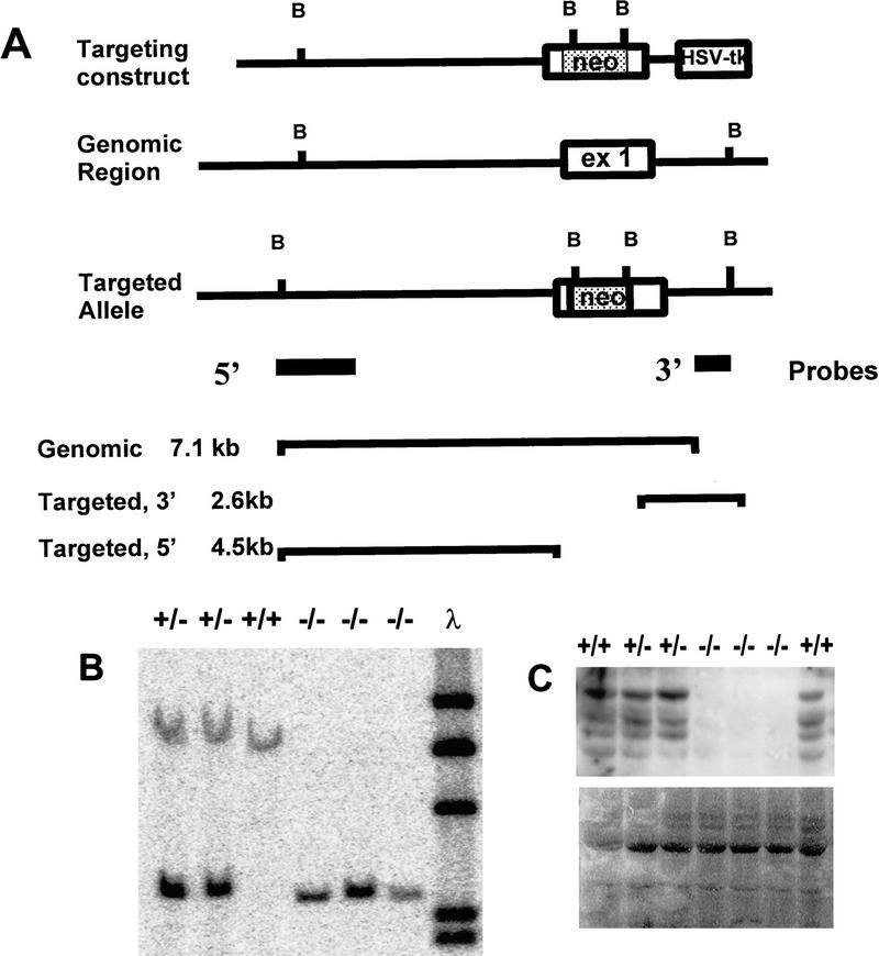
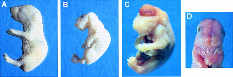
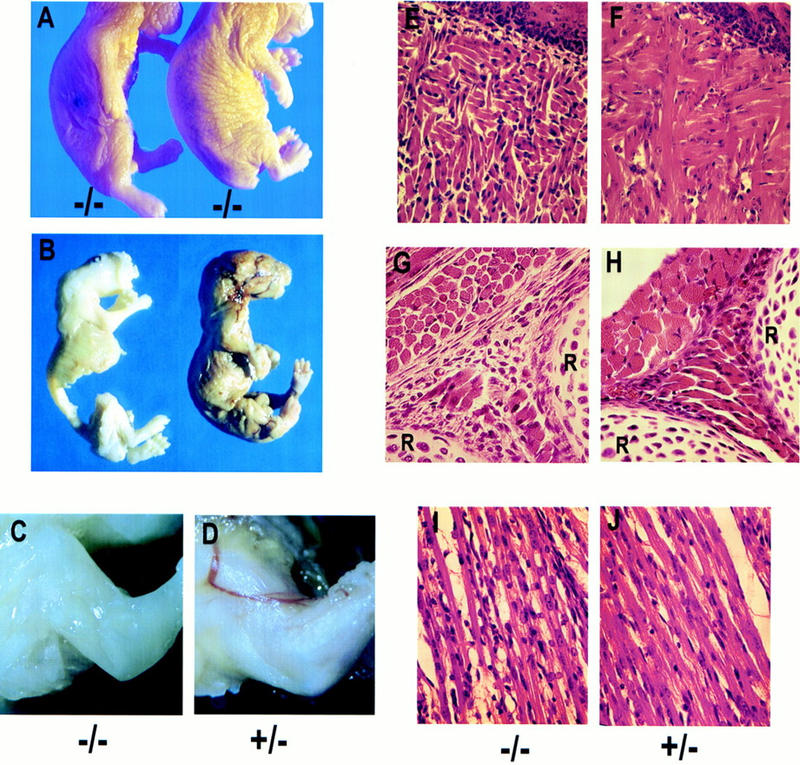
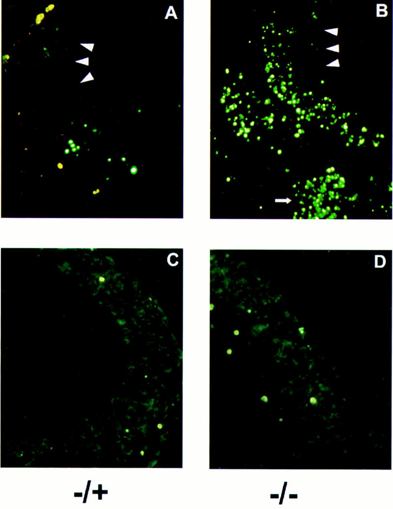
The c-ski proto-oncogene has been implicated in the control of cell growth and skeletal muscle differentiation. To determine its normal functions in vivo, we have disrupted the mouse c-ski gene. Our results show a novel role for ski in the morphogenesis of craniofacial structures and the central nervous system, and confirm its proposed function as a player in skeletal muscle development. Homozygous mutant mice show perinatal lethality resulting from exencephaly, a defect caused by failed closure of the cranial neural tube during neurulation. The timing of the neural tube defect in ski -/- embryos coincides with excessive apoptosis in the cranial neuroepithelium, as well as in the cranial mesenchyme. Homozygous ski mutants also exhibit a dramatic reduction in skeletal muscle mass, consistent with a defect in expansion of a myogenic precursor population. Nestin is an intermediate filament expressed in highly proliferative neuroepithelial stem cells and in myogenic precursors. Interestingly, we find decreased nestin expression in both the cranial neural tube and the somites of ski -/- embryos, compared with their normal littermates, but no reduction of nestin in the caudal neural tube. These results are consistent with a model in which ski activities are required for the successful expansion of a subset of precursors in the neuroepithelial or skeletal muscle lineages.


Figures

Figure 1
Figure 1
Targeted disruption of the ski gene in mice. (A) Diagram of the targeting vector and map of the targeted allele. (neo) pMC1 neo-expression cassette lacking a polyadenylation signal; (HSV-tk) pMC1-tk expression cassette used for negative selection. Positions of critical BamHI sites, location of the probes, and sizes of diagnostic fragments are indicated. (B) Southern analysis of genomic DNA isolated from offspring of heterozygote matings. DNA was digested with BamHI; blots were probed with the 3′ probe shown in Fig. 1A, as well as with labeled λ DNA, and exposed with a Molecular Dynamics PhosphorImager screen. (C) Western blot probed with G8 monoclonal antibody to Ski. The largest band corresponds to full-length ski; smaller forms may be degradation products. (Bottom) The same membrane stained with Sudan black to confirm equal loading and transfer of samples.
Figure 2
Figure 2
Morphological analysis of wild-type and ski −/− neonates. (A) Lateral view of a wild-type pup. (B) Lateral view of ski −/−, exencephalic pup. Note abnormal curvature of the back, neck, and head; the abnormal square jaw, flaccid limbs, and skinny forelimbs. (C) ski-deficient newborn delivered by C-section, showing the highly vascularized brain mass; this region is normally absent because it is sheared off during birth. Eyes are present and closed but not visible because this mutant is albino; most ski-deficient mice were born with open eyes, but some had normally shut eyelids. An abdominal cut was made to facilitate fixation. (D) ski −/− mouse with a cranial vault and brain, showing frontonasal clefting. Approximately 10%–15% of ski −/− mice had this phenotype (see Tables 1 and 2).
Figure 6
Figure 6
Reduced skeletal muscle mass in ski-deficient newborns. (A) Comparison of two ski −/−, exencephalic newborns with different degrees of muscle deficiency. Note the loose skin along the fore- and hindlimbs of the mouse on the right, which is not emaciated but skinny. (B) Comparison of wild-type and ski-deficient newborns after removal of the skin in preparation for skeletal staining. The ski −/− mouse (left) did not appear emaciated but merely skinny before the skin was removed. (C,D) Close-up comparison of the forelimbs from a ski-deficient pup and its normal heterozygous littermate, with the skin removed after fixation. Mutants appear white because of bleeding from the exposed, angiomatous brain mass; bleeding occurs throughout development but is most severe at birth, when the brain mass is sheared off. (E–J) Histological analysis of muscle groups in the tongue, intercostal region, and forelimb. Sagittal sections; sections from ski −/− newborns are on the left. (E,F) Sections through the tongue, with the epithelial surface on the upper right corner of each section. Note the shorter fibers, and empty space between fibers. (G,H) Sections through the intercostal muscles, with ribs (R) at bottom and right edges; note the reduced diameter of the muscle fibers in cross-section (upper left) and the disorganization and reduced number of fibers in the area between the two ribs (extreme right and bottom). (I,J) Sections through a forelimb muscle; note the reduced diameter of fibers in I, and increased numbers of nuclei not associated with fibers.
Figure 7
Figure 7
Skeletal abnormalities in ski-deficient mice. In each pair, the ski −/− sample is on the left. (A,B) Skeletal preparations of a ski −/− mouse A and a wild-type littermate B. The mouse in A is missing the frontal, parietal, interparietal, and supraoccipital bones, but the nasal bone is present. Arrowheads indicate the first and second cervical vertebrae (C1 and C2), which are fused in the mutant. Arrow indicates the mandible, which is wider and shorter in the mutant than in the normal littermate. (C,D) Comparison of the basal bones of the skull shows that the supraoccipital bone (s) and the presphenoid bone (ps) are completely absent in the ski-deficient skeleton. bo, basioccipital; bs, basisphenoid. (E) Comparison of the basioccipital bones. Note the anterior arch of the atlas, which is fused to the top of the mutant bone (left). (F) Malformation of the basisphenoid bone.
Figure 3
Figure 3
Morphological analysis of E9.5 embryos. Left (A,C,E,G) show a ski −/− embryo, right (B,D,F) a normal +/− littermate. (A) Lateral view showing open neural folds starting at the midbrain and extending rostrally and impaired development of the forebrain. Except for the head, size of the mutant and normal embryos is comparable. (B) Normal heterozygous littermate of embryo shown in A. (C,D) Dorsal views showing the open and everted neural folds of the ski −/− embryo. The neural tube of the normal embryo in D is completely closed; the discontinuity is a photographic artifact. (E,F) Frontal sections through the plane of the paper of embryos shown in C and D, stained with hematoxylin and eosin. The arrowhead shows the ventricular surface of the neuroepithelium, which in the ski −/− embryo becomes exposed to the exterior. Arrows indicate the cranial mesenchyme. (G) Frontal section, at higher magnification, showing the caudal neural tube of the ski −/− embryo in E. (nt) Neural tube (caudal); (n) notochord; (s) somite. Tears in the neuroepithelium are sectioning artifacts.
Figure 4
Figure 4
Excessive apoptosis in ski-deficient embryos at neurulation. Frontal frozen sections were prepared from embryos at E9.5, and analyzed with the TUNEL assay. (A,B) Ventricular edges of the neuroepithelium are indicated by arrowheads. Red blood cells are stained bright yellow; apoptotic cells that have incorporated labeled dUTP fluoresce green. DAPI staining (not shown) revealed that cells labeled with dUTP also showed condensed chromatin characteristic of apoptosis. The small arrow at the bottom of B shows abundant apoptosis in the craniofacial mesenchyme. In the neuroepithelium, apoptosis is concentrated along the mantle layer. (C,D) Frontal sections of embryos at E10.5, processed as described above. The amount of apoptosis is comparable in the −/− mutant and its heterozygous littermate at this stage.
Figure 5
Figure 5
Expression of nestin and β-III tubulin in heterozygous and ski-deficient E9.5 embryos. Frontal sections correspond to those shown in Fig. 3 (E–G), except that the caudal neural tube (C,D) is rotated sideways to show a larger area; large arrowheads in A, B, E, and F point to the ventricular surface of the neuroepithelium. Frozen sections were prepared and labeled with monoclonal antibodies to nestin or β-III tubulin, followed by FITC-labeled second antibody. In each pair, the ski −/− sample is on the left. (A,B) Expression of nestin in the cranial neural tube is evident at the outer edge of the mantle zone, where differentiated cells accumulate. Nestin-positive cells along the future mantle zone (away from the ventricular zone) are less abundant in the mutant A than in the +/− embryo shown in B. In addition, the −/− sample shows some staining of cells in the intermediate and exposed ventricular zone, which is not seen in the normal heterozygote and may indicate premature differentiation. (C,D) Expression of nestin in the somitic myotome and caudal neural tube. Small arrowheads point to the somitic myotomes; note nestin expression in myotome of the ski −/− embryo C is drastically reduced compared with the heterozygous embryo D, although expression in the caudal and closed neural tube is comparable in both sections. Note, however, the reduced size of the spinal cord in C. (E,F) Expression of β-III tubulin is comparable in ski-deficient E and heterozygous F embryos at E9.5.

References

    1. Chen ZF, Behringer RR. Twist is required in head mesenchyme cranial neural tube morphogenesis. Genes & Dev. 1995;9:686–699. - PubMed
    1. Colmenares C, Stavnezer E. The ski oncogene induces muscle differentiation in quail embryo cells. Cell. 1989;59:293–303. - PubMed
    1. Colmenares C, Sutrave P, Hughes SH, Stavnezer E. Activation of the c-ski oncogene by overexpression. J Virol. 1991a;65:4929–4935. - PMC - PubMed
    1. Colmenares C, Teumer JK, Stavnezer E. Transformation-defective v-ski induces MyoD and myogenin expression but not myotube formation. Mol Cell Biol. 1991b;11:1167–1170. - PMC - PubMed
    1. Copp AJ. Neural tube defects. Trends Neurosci. 1993;16:381–383. - PubMed

Publication types

MeSH terms