. 1997 Aug 15;11(16):2137-51.
doi: 10.1101/gad.11.16.2137.

Drosophila P-element transposase is a novel site-specific endonuclease

Affiliations

Drosophila P-element transposase is a novel site-specific endonuclease

E L Beall et al. Genes Dev. .

Abstract

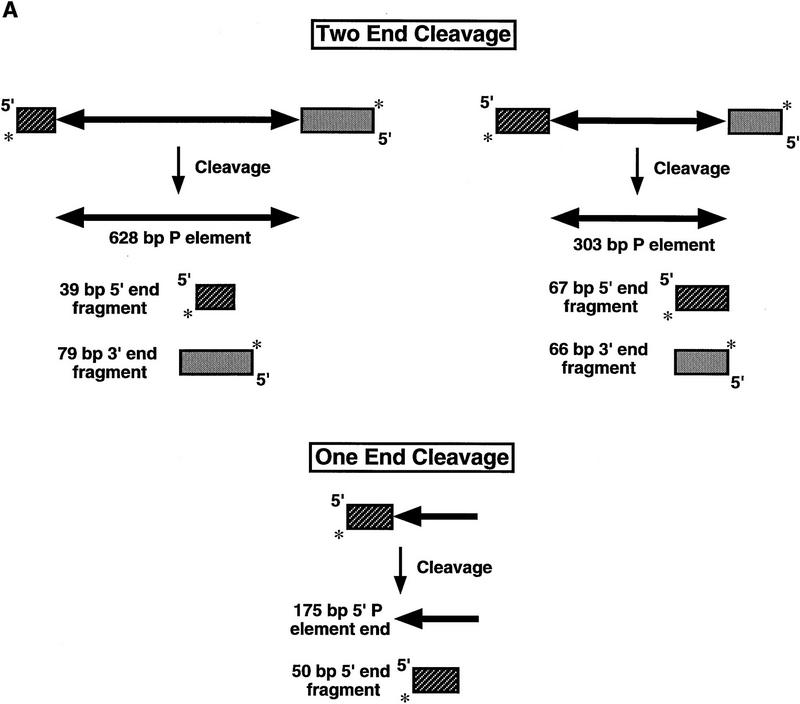
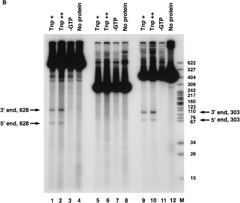
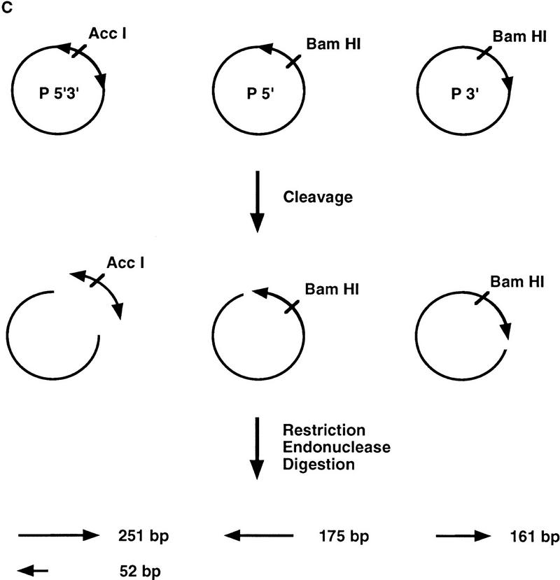
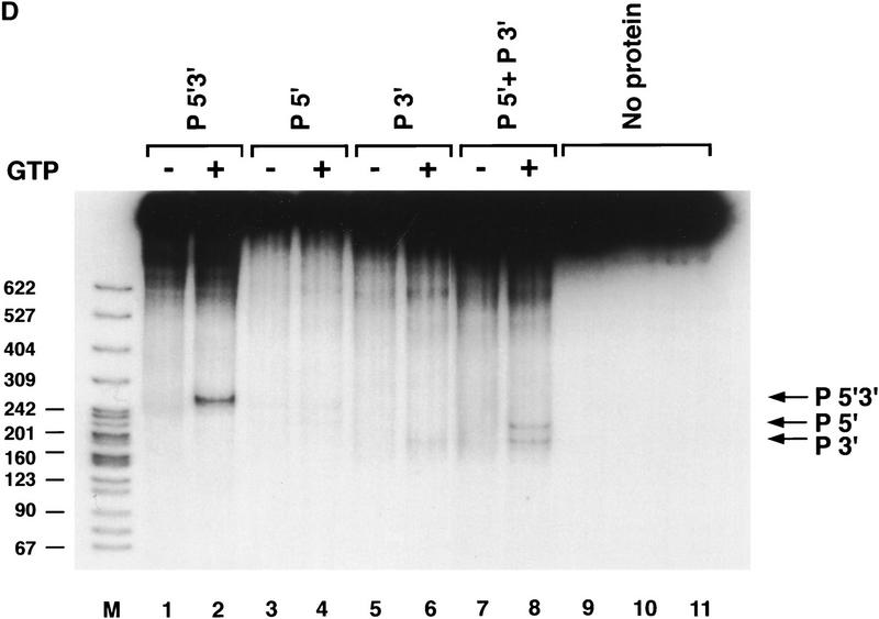
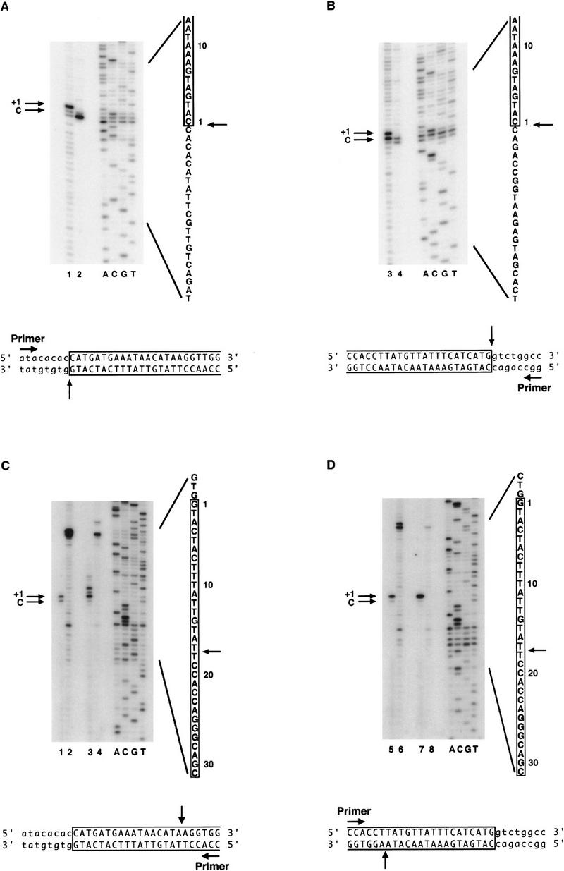
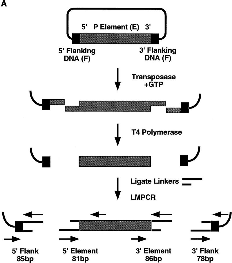
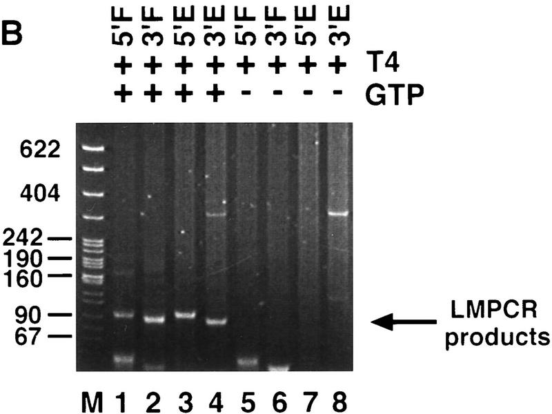
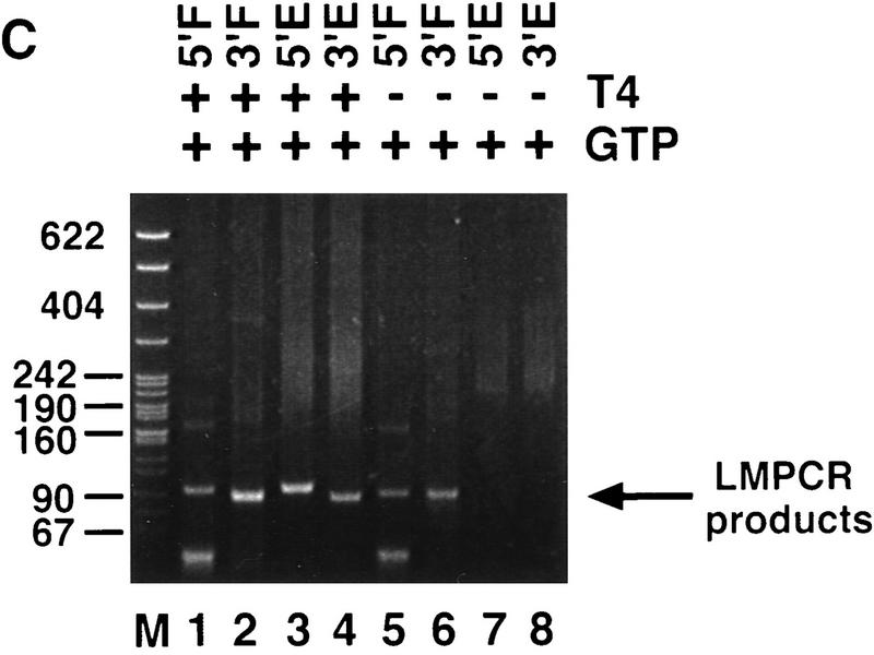
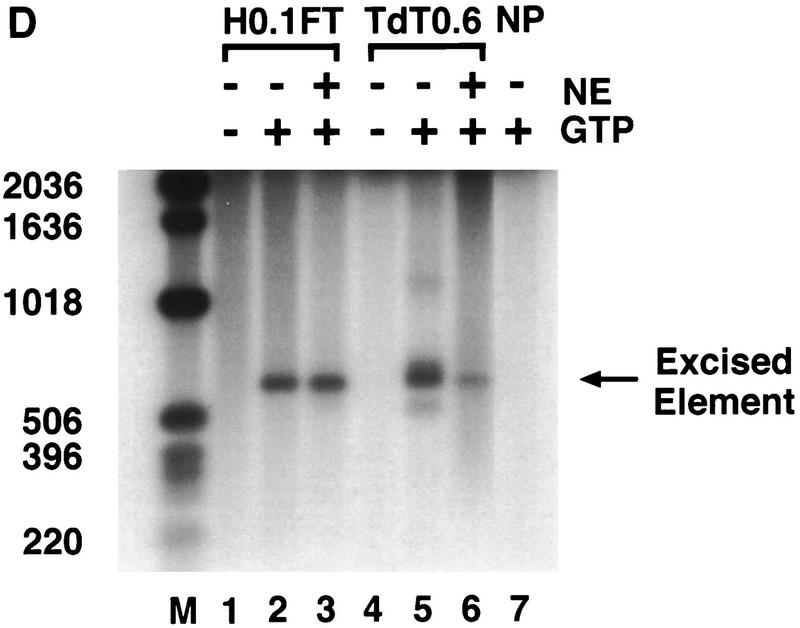
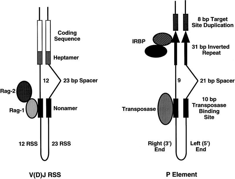
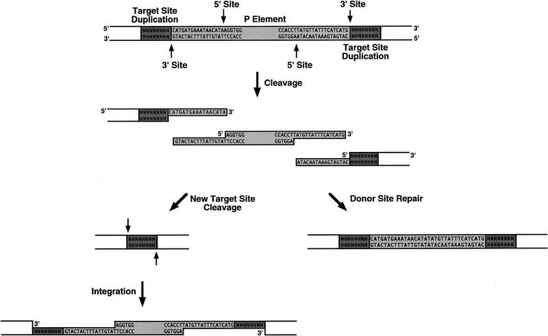
We developed in vitro assays to study the first step of the P-element transposition reaction: donor DNA cleavage. We found that P-element transposase required both 5' and 3' P-element termini for efficient DNA cleavage to occur, suggesting that a synaptic complex forms prior to cleavage. Transposase made a staggered cleavage at the P-element termini that is novel for all known site-specific endonucleases: the 3' cleavage site is at the end of the P-element, whereas the 5' cleavage site is 17 bp within the P-element 31-bp inverted repeats. The P-element termini were protected from exonucleolytic degradation following the cleavage reaction, suggesting that a stable protein complex remains bound to the element termini after cleavage. These data are consistent with a cut-and-paste mechanism for P-element transposition and may explain why P elements predominantly excise imprecisely in vivo.


Figures

Figure 1
Figure 1
Transposase requires both 5′ and 3′ P-element termini during cleavage. (A) Schematic diagram of the assay used to study the first step in the P-element transposition reaction: donor DNA cleavage. Restriction endonuclease fragments carrying internally deleted P elements of 303 or 628 bp in length (as indicated by arrows), along with varying lengths of flanking DNA (hatched and shaded boxes), were radiolabeled at the 3′ end as indicated by an asterisk. These substrates contain both P-element termini that include the 31-bp terminal inverted repeats, the internal transposase binding sites, and the internal 11-bp inverted repeat enhancer elements. A DNA fragment containing a single 5′ P-element end was also radiolabeled and tested as a cleavage substrate for transposase. The predicted sizes for each cleavage product are as indicated. (B) Partially purified transposase (H0.1FT) was tested for double-stranded DNA cleavage activity as diagrammed in A. Reactions were performed at 27°C for 2 hr, and products analyzed by native PAGE and autoradiography. Shown is an autoradiograph from an experiment in which three different substrates were tested for cleavage activity: the 628-bp P element-containing fragment (lanes 1–4), the 175-bp single left (5′) P-element end-containing fragment (lanes 5–8), and the 303-bp P-element-containing fragment (lanes 9–12). For each substrate, two different amounts of the H0.1FT were tested for activity (either ∼2 μg, or 4 μg total protein, as indicated by + or ++). GTP-dependent cleavage products are indicated by arrows. (M) Radiolabeled pBR322 MspI molecular weight markers. (C) Schematic diagram of the assay used to test whether P-element transposase requires both 5′ and 3′ P-element termini for DNA cleavage to occur. Plasmid substrates containing the 303-bp P element (P 5′3′, left), the left end only (P5′, middle), or the right end only (P3′, right) are as diagrammed. The predicted sizes for each product following transposase-mediated cleavage and restricion endonuclease digestion with the indicated enzymes are outlined at the bottom. (D) Partially purified transposase (H0.1FT) was tested for cleavage activity as diagrammed in Fig. 1C. Reactions were performed at 27°C for 2 hr, and products analyzed by DNA blot hybridization with a radiolabeled P-element probe following native PAGE. Shown is an autoradiograph from an experiment in which substrates containing both P-element termini (P 5′3′, lanes 1,2; only the 251-bp AccI fragment is shown), each terminus alone (P 5′, lanes 3,4; P 3′, lanes 5,6), or a mixture containing equimolar amounts of both P 5′ and P 3′ (lanes 7 and 8) were tested for cleavage by transposase. For each substrate, ∼2 μg of the H0.1FT was tested for activity. GTP-dependent cleavage products are indicated by arrows. (M) radiolabeled pBR322 MspI molecular weight markers.
Figure 1
Figure 1
Transposase requires both 5′ and 3′ P-element termini during cleavage. (A) Schematic diagram of the assay used to study the first step in the P-element transposition reaction: donor DNA cleavage. Restriction endonuclease fragments carrying internally deleted P elements of 303 or 628 bp in length (as indicated by arrows), along with varying lengths of flanking DNA (hatched and shaded boxes), were radiolabeled at the 3′ end as indicated by an asterisk. These substrates contain both P-element termini that include the 31-bp terminal inverted repeats, the internal transposase binding sites, and the internal 11-bp inverted repeat enhancer elements. A DNA fragment containing a single 5′ P-element end was also radiolabeled and tested as a cleavage substrate for transposase. The predicted sizes for each cleavage product are as indicated. (B) Partially purified transposase (H0.1FT) was tested for double-stranded DNA cleavage activity as diagrammed in A. Reactions were performed at 27°C for 2 hr, and products analyzed by native PAGE and autoradiography. Shown is an autoradiograph from an experiment in which three different substrates were tested for cleavage activity: the 628-bp P element-containing fragment (lanes 1–4), the 175-bp single left (5′) P-element end-containing fragment (lanes 5–8), and the 303-bp P-element-containing fragment (lanes 9–12). For each substrate, two different amounts of the H0.1FT were tested for activity (either ∼2 μg, or 4 μg total protein, as indicated by + or ++). GTP-dependent cleavage products are indicated by arrows. (M) Radiolabeled pBR322 MspI molecular weight markers. (C) Schematic diagram of the assay used to test whether P-element transposase requires both 5′ and 3′ P-element termini for DNA cleavage to occur. Plasmid substrates containing the 303-bp P element (P 5′3′, left), the left end only (P5′, middle), or the right end only (P3′, right) are as diagrammed. The predicted sizes for each product following transposase-mediated cleavage and restricion endonuclease digestion with the indicated enzymes are outlined at the bottom. (D) Partially purified transposase (H0.1FT) was tested for cleavage activity as diagrammed in Fig. 1C. Reactions were performed at 27°C for 2 hr, and products analyzed by DNA blot hybridization with a radiolabeled P-element probe following native PAGE. Shown is an autoradiograph from an experiment in which substrates containing both P-element termini (P 5′3′, lanes 1,2; only the 251-bp AccI fragment is shown), each terminus alone (P 5′, lanes 3,4; P 3′, lanes 5,6), or a mixture containing equimolar amounts of both P 5′ and P 3′ (lanes 7 and 8) were tested for cleavage by transposase. For each substrate, ∼2 μg of the H0.1FT was tested for activity. GTP-dependent cleavage products are indicated by arrows. (M) radiolabeled pBR322 MspI molecular weight markers.
Figure 2
Figure 2
Transposase makes 17-bp staggered cleavages at the P-element termini. Large-scale cleavage reactions were performed with partially purified transposase and plasmid substrates containing either the 628-bp or 303-bp P elements. Reaction products corresponding to both the cleaved plasmid vector and the excised element were isolated from agarose gel slices. A PCR-based primer extension analysis was performed on each product in order to determine the transposase cleavage sites. Shown are autoradiographs of sequencing gels that display primer extension products from reactions in which nontemplated addition of a single nucleotide by Taq polymerase occurs (as indicated by +1 in each panel). The authentic cleavage sites are indicated by C. Sequencing reactions (ACGT) are shown as markers. The relevant sequence is indicated, with P-element-derived sequences boxed and numbered from the terminal P-element nucleotide, and the cleavage sites indicated by arrows. Schematic diagrams of the direction for primer extension and the cleavage positions for each strand are indicated below each panel. (A,B) Extension products to determine the 3′ cleavage site at the left (5′) P-element end (A) or the 3′ cleavage site at the right (3′) P-element end (B). Products were analyzed for the 303-bp P-element-derived cleavage product only. (Odd-numbered lanes) + transposase; (even-numbered lanes) − transposase. Extension yields a product that terminates exactly at the 3′ end of the P-element inverted repeat. (C,D) Extension products to determine the 5′ cleavage sites at the left (5′) P-element end (C) or right (3′) P-element end (D). (Lanes 1,2,5,6) Products derived from the 303-bp P element. (Lanes 3,4,7,8) Products derived from the 628-bp P element. (Odd-numbered lanes) + transposase, (even-numbered lanes) − transposase. Extension yields a product that terminates at nucleotide 18 of the P-element inverted repeat.
Figure 3
Figure 3
P-element termini are protected after transposase-mediated cleavage. (A) Schematic diagram of the LMPCR assay. P-element-containing plasmid substrates were incubated with partially purified transposase (H0.1FT) in the presence or absence of GTP. Products were treated with T4 DNA polymerase to remove the 17-bp, 3′ extension, or ligated directly to annealed linkers as shown. LMPCR was performed with the indicated primers to detect all four possible cleavage products of the sizes indicated. (Shaded boxes) P-element-derived sequences; (black boxes) 8-bp target site duplication. (B) Photograph of a native polyacrylamide gel following ethidium bromide staining. Cleavage reactions were performed in the presence (+) or absence (−) of GTP as indicated. All samples were treated with T4 DNA polymerase prior to LMPCR. (M) pBR322 MspI molecular weight markers. (5′F) Amplification to detect the cleavage product at the 5′ flanking DNA end (85 bp); (3′F) amplification to detect the cleavage product at the 3′ flanking DNA end (78 bp); (5′E) amplification to detect the cleavage product at the 5′ P-element DNA end (81 bp); (3′E) amplification to detect the cleavage product at the 3′ P-element DNA end (86 bp). (C) Photograph of a native polyacrylamide gel following ethidium bromide staining. Cleavage reactions were performed in the presence of GTP (+), and either in the presence (+) or absence (−) of T4 polymerase treatment prior to LMPCR analysis. Abbreviations are as outlined in B.
Figure 3
Figure 3
P-element termini are protected after transposase-mediated cleavage. (A) Schematic diagram of the LMPCR assay. P-element-containing plasmid substrates were incubated with partially purified transposase (H0.1FT) in the presence or absence of GTP. Products were treated with T4 DNA polymerase to remove the 17-bp, 3′ extension, or ligated directly to annealed linkers as shown. LMPCR was performed with the indicated primers to detect all four possible cleavage products of the sizes indicated. (Shaded boxes) P-element-derived sequences; (black boxes) 8-bp target site duplication. (B) Photograph of a native polyacrylamide gel following ethidium bromide staining. Cleavage reactions were performed in the presence (+) or absence (−) of GTP as indicated. All samples were treated with T4 DNA polymerase prior to LMPCR. (M) pBR322 MspI molecular weight markers. (5′F) Amplification to detect the cleavage product at the 5′ flanking DNA end (85 bp); (3′F) amplification to detect the cleavage product at the 3′ flanking DNA end (78 bp); (5′E) amplification to detect the cleavage product at the 5′ P-element DNA end (81 bp); (3′E) amplification to detect the cleavage product at the 3′ P-element DNA end (86 bp). (C) Photograph of a native polyacrylamide gel following ethidium bromide staining. Cleavage reactions were performed in the presence of GTP (+), and either in the presence (+) or absence (−) of T4 polymerase treatment prior to LMPCR analysis. Abbreviations are as outlined in B.
Figure 4
Figure 4
Purified P-element transposase can perform cleavage in vitro. (A) SDS-PAGE analysis of the purified transposase fractions. Samples were subjected to electrophoresis on a 7.5% acrylamide gel and stained with silver. Transposase was purified from Drosophila cell culture nuclear extracts with heparin-agarose chromatography (see Materials and Methods for details). The flowthrough (H0.1FT) fraction containing high amounts of transposase activity was chromatographed on a nonspecific DNA-affinity column (TdT), and the protein eluted with increasing KCl. (M) Molecular weight markers, with relative molecular mass in kD (left). (Lane 1) 0.3 m KCl fraction; (lane 2) 0.6 m KCl elution 1; (lane 3) 0.6 m KCl elution 2; (lane 4) 1.0 m KCl fraction. BSA was added to each fraction at 50 μg/ml. Approximately formula image of the total fraction was loaded in each lane. (B) Immunoblot analysis of the purified transposase fractions. Samples from the TdT column were subjected to electrophoresis on a 7.5% acrylamide gel, transferred to nitrocellulose and probed with anti-transposase affinity-purified polyclonal antibodies. Molecular weight markers are described in A. (Lane 1) TdT column input; (lane 2) TdT column flowthrough; (lane 3) 0.3 m KCl fraction; (lane 4) 0.6 m KCl elution 1; (lane 5) 0.6 m KCl elution 2; (lane 6) 1.0 m KCl fraction. Approximately formula image of the input and flowthrough, and formula image of each fraction was loaded in each lane. (C) Immunoblot analysis of the purified transposase fractions. Samples from the TdT column were subjected to electrophoresis on a 7.5% acrylamide gel, transferred to nitrocellulose and probed with anti-IRBP affinity-purified polyclonal antibodies. Molecular weight markers are described in A. (Lane 1) TdT column input; (lane 2) TdT column flowthrough; (lane 3) 0.3 m KCl fraction; (lane 4) 0.6 m KCl elution 1; (lane 5) 0.6 m KCl elution 2. Approximately formula image of the input and flow-through, and formula image of each fraction was loaded in each lane. (D) Both partially purified (H0.1FT) and DNA affinity-purified (TdT0.6 E1) transposase-containing fractions were tested for cleavage activity with the 628-bp P-element-containing plasmid substrate. The excised element was detected by DNA blot hybridization by use of a radiolabeled P-element fragment as a probe. (M) Molecular weight markers in bp, as indicated. (−GTP) Control reactions lacking the cofactor, GTP. Reactions were performed either in the presence (+) or absence (−) of nuclear extract (NE) derived from a Drosophila somatic cell line lacking transposase (Kc cells). (NP) Reaction lacking transposase.
Figure 4
Figure 4
Purified P-element transposase can perform cleavage in vitro. (A) SDS-PAGE analysis of the purified transposase fractions. Samples were subjected to electrophoresis on a 7.5% acrylamide gel and stained with silver. Transposase was purified from Drosophila cell culture nuclear extracts with heparin-agarose chromatography (see Materials and Methods for details). The flowthrough (H0.1FT) fraction containing high amounts of transposase activity was chromatographed on a nonspecific DNA-affinity column (TdT), and the protein eluted with increasing KCl. (M) Molecular weight markers, with relative molecular mass in kD (left). (Lane 1) 0.3 m KCl fraction; (lane 2) 0.6 m KCl elution 1; (lane 3) 0.6 m KCl elution 2; (lane 4) 1.0 m KCl fraction. BSA was added to each fraction at 50 μg/ml. Approximately formula image of the total fraction was loaded in each lane. (B) Immunoblot analysis of the purified transposase fractions. Samples from the TdT column were subjected to electrophoresis on a 7.5% acrylamide gel, transferred to nitrocellulose and probed with anti-transposase affinity-purified polyclonal antibodies. Molecular weight markers are described in A. (Lane 1) TdT column input; (lane 2) TdT column flowthrough; (lane 3) 0.3 m KCl fraction; (lane 4) 0.6 m KCl elution 1; (lane 5) 0.6 m KCl elution 2; (lane 6) 1.0 m KCl fraction. Approximately formula image of the input and flowthrough, and formula image of each fraction was loaded in each lane. (C) Immunoblot analysis of the purified transposase fractions. Samples from the TdT column were subjected to electrophoresis on a 7.5% acrylamide gel, transferred to nitrocellulose and probed with anti-IRBP affinity-purified polyclonal antibodies. Molecular weight markers are described in A. (Lane 1) TdT column input; (lane 2) TdT column flowthrough; (lane 3) 0.3 m KCl fraction; (lane 4) 0.6 m KCl elution 1; (lane 5) 0.6 m KCl elution 2. Approximately formula image of the input and flow-through, and formula image of each fraction was loaded in each lane. (D) Both partially purified (H0.1FT) and DNA affinity-purified (TdT0.6 E1) transposase-containing fractions were tested for cleavage activity with the 628-bp P-element-containing plasmid substrate. The excised element was detected by DNA blot hybridization by use of a radiolabeled P-element fragment as a probe. (M) Molecular weight markers in bp, as indicated. (−GTP) Control reactions lacking the cofactor, GTP. Reactions were performed either in the presence (+) or absence (−) of nuclear extract (NE) derived from a Drosophila somatic cell line lacking transposase (Kc cells). (NP) Reaction lacking transposase.
Figure 6
Figure 6
Similarities between the V(D)J RSS and P-element transposon termini. Schematic diagram for synapsis of the V(D)J RSS is shown on the left. Pairing of a 12-bp spacer RSS and 23-bp spacer RSS is as indicated. Rag-1 (lightly shaded oval) is shown interacting with the nonamer element (black boxes). In the presence of Rag-2 (darkly shaded oval) and Mg2+, cleavage occurs at the heptamer (medium gray stippled boxes) to produce blunt signal ends and hairpin coding ends. (Right) A schematic diagram of the synapsed P-element termini. Pairing of the left (5′) end that contains a 21-bp spacer between the transposase-binding site (black box) and the 31-bp inverted repeat (black arrow) with the right (3′) end containing a 9-bp spacer between the two sequence elements is shown. In the presence of Mg2+ and GTP, transposase (shaded oval) makes a staggered 17-bp cleavage within the 31-bp inverted repeats, adjacent to the IRBP-binding site [(IRBP) hatched oval, p70 subunit. (Black oval) p80 subunit].
Figure 5
Figure 5
Model for P-element transposition. A model for nonreplicative P-element transposition is diagrammed. P-element transposase binds to sequences within both P-element termini and initiates a double-stranded DNA break at each end, as indicated by arrows. The excised element, which contains 17-nt, 3′ extensions at each end, can be inserted into a new target site (left). Gap repair will generate the characteristic 8-bp target site duplications as well as regenerate the P-element sequences at each end. The 17-nt, 3′ extensions left behind at the donor site can be used for repair either from homologous P-element sequences located elsewhere in the genome by SDSA repair, or by end-joining in the absence of homologous P-sequences, (right). Imprecise repair of the double-stranded DNA break left at the donor site can produce products that contain varying lengths of P-element-derived sequences, e.g., a single repaired donor site is shown in which the cleaved termini were adjoined, extended and ligated to leave behind 34 nts of P-element sequence at the donor site. (Darkly shaded boxes) 8-bp target site duplications. (Lightly shaded boxes) P-element-derived sequences.

References

    1. Agrawal A, Schatz DG. RAG1 and RAG2 form a stable postcleavage synaptic complex with DNA containing signal ends in V(D)J recombination. Cell. 1997;89:43–53. - PubMed
    1. Anderson CW, Lees-Miller SP. The nuclear serine/threonine protein kinase DNA-PK. Crit Rev Eukaryot Gene Expression. 1992;2:283–314. - PubMed
    1. Bainton R, Gamas P, Craig NL. Tn7 transposition in vitro proceeds through an excised transposon intermediate generated by staggered breaks in DNA. Cell. 1991;65:805–816. - PubMed
    1. Bainton RJ, Kubo KM, Feng JN, Craig NL. Tn7 transposition: Target DNA recognition is mediated by multiple Tn7-encoded proteins in a purified in vitro system. Cell. 1993;72:931–943. - PubMed
    1. Beall EL, Rio DC. Drosophila IRBP/Ku p70 corresponds to the mutagen-sensitive mus309 gene and is involved in P-element excision in vivo. Genes & Dev. 1996;10:921–933. - PubMed

Publication types

LinkOut - more resources