. 1997 Jul;81(7):527-32.
doi: 10.1136/bjo.81.7.527.

Variations of posterior vitreous detachment

Affiliations

Variations of posterior vitreous detachment

A Kakehashi et al. Br J Ophthalmol. 1997 Jul.

Abstract

Aims: To identify variations in posterior vitreous detachment (PVD) and establish a clinical classification system for PVD.

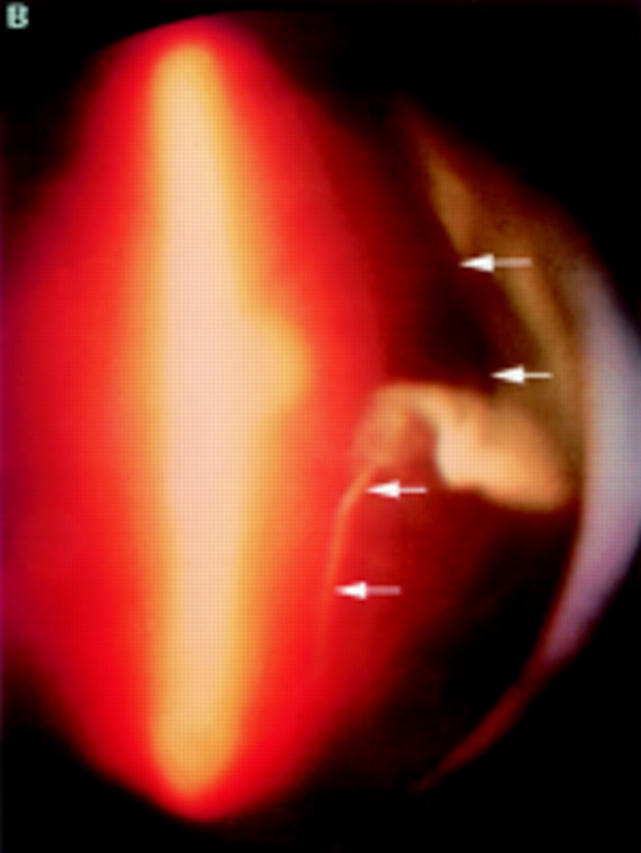
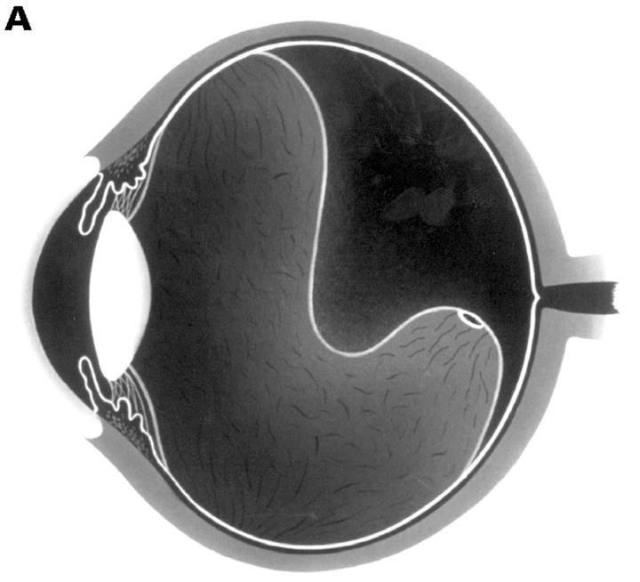
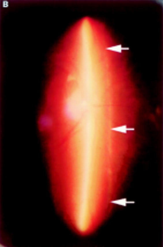
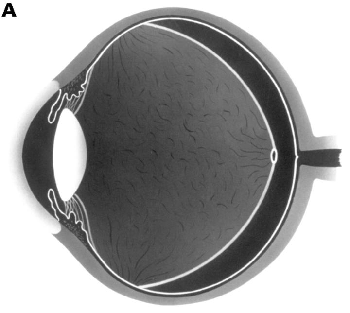
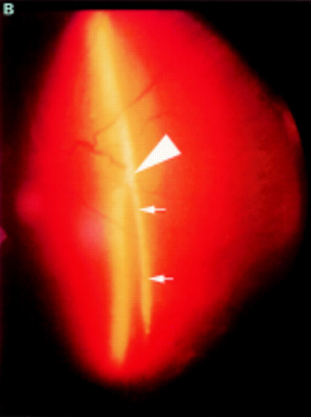
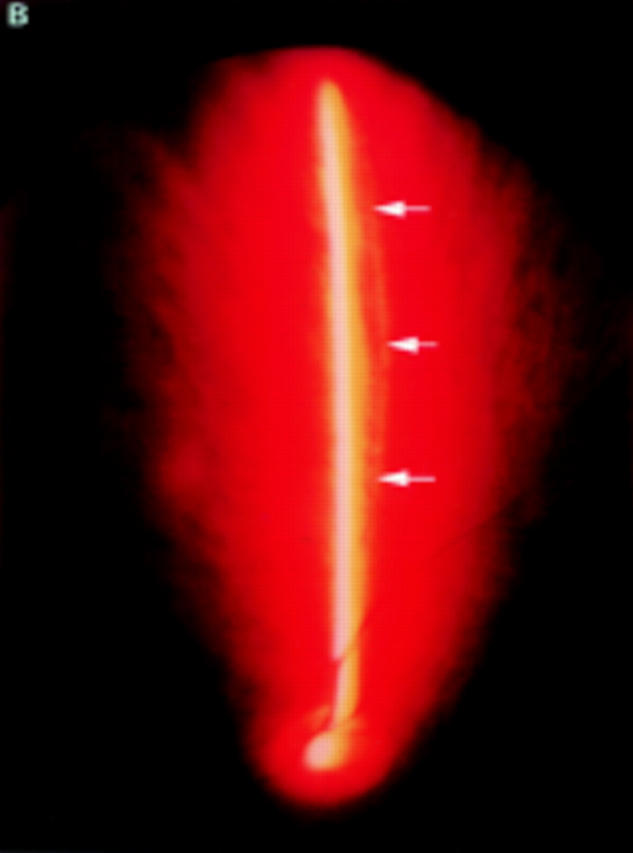
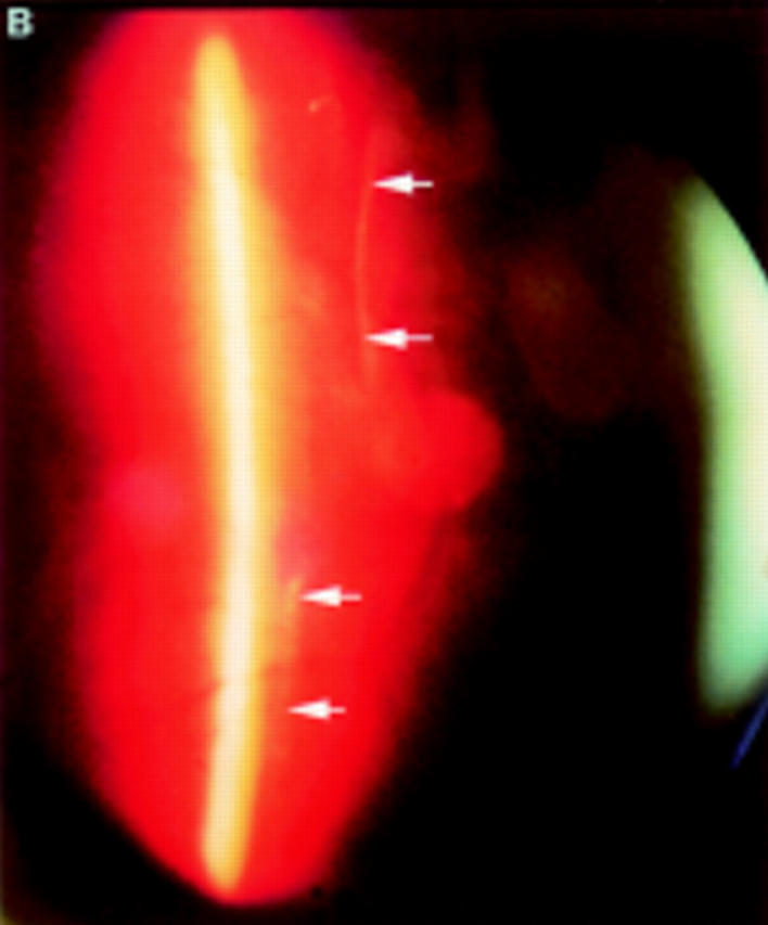
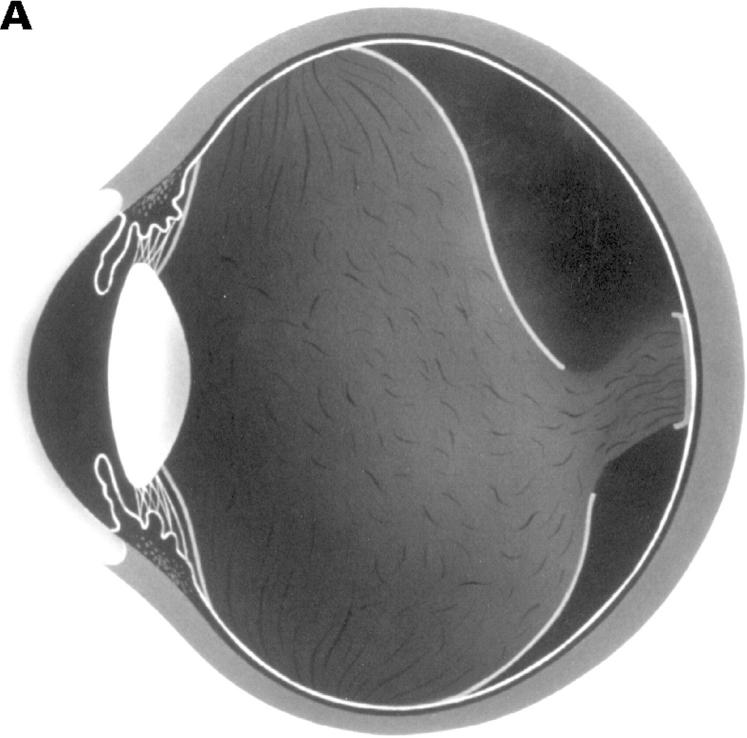
Methods: 400 consecutive eyes were examined using biomicroscopy and vitreous photography and classified the PVD variations-complete PVD with collapse, complete PVD without collapse, partial PVD with thickened posterior vitreous cortex (TPVC), or partial PVD without TPVC.

Results: In each PVD type, the most frequently seen ocular pathologies were as follows: in complete PVD with collapse (186 eyes), age related changes without vitreoretinal diseases (77 eyes, 41.4%) and high myopia (55 eyes, 29.6%); in complete PVD without collapse (39 eyes), uveitis (23 eyes, 59.0%) and central retinal vein occlusions (8 eyes, 20.5%); in partial PVD with TPVC (64 eyes), proliferative diabetic retinopathy (30 eyes, 46.9%); and inpartial PVD without TPVC (111 eyes), age related changes without vitreoretinal diseases (62 eyes, 55.9%). This PVD categorisation was significantly associated with the prevalence of each vitreoretinal disease (p < 0.0001, chi 2 test on contingency table).

Conclusions: PVD variations can be classified into four types, which is clinically useful because each type corresponds well to specific vitreoretinal changes.


Figures

Figure 1
Figure 1
(A) Schematic sketch of complete posterior vitreous detachment (PVD) with collapse. The posterior vitreous cortex has a characteristically sigmoidal shape. (B) In a patient who complained of floaters in his visual field, the biomicroscopic vitreous examination revealed complete PVD. The vitreous gel was mildly condensed and, with ocular movement, the posterior vitreous cortex (arrows) oscillated in a smooth, wavy motion. This case was classified as complete PVD with collapse.
Figure 1
Figure 1
(A) Schematic sketch of complete posterior vitreous detachment (PVD) with collapse. The posterior vitreous cortex has a characteristically sigmoidal shape. (B) In a patient who complained of floaters in his visual field, the biomicroscopic vitreous examination revealed complete PVD. The vitreous gel was mildly condensed and, with ocular movement, the posterior vitreous cortex (arrows) oscillated in a smooth, wavy motion. This case was classified as complete PVD with collapse.
Figure 2
Figure 2
(A) Schematic sketch of complete posterior vitreous detachment (PVD) without collapse. The posterior vitreous cortex is convex and shallowly detached. (B) In a patient with uveitis, the posterior vitreous cortex (arrows) is shallowly detached from the retina; vitreous opacities are observed in the vitreous gel and the posterior vitreous cortex is convex. The vitreous gel is condensed and has minimal mobility even with rapid ocular movement. This case was classified as complete PVD without collapse.
Figure 2
Figure 2
(A) Schematic sketch of complete posterior vitreous detachment (PVD) without collapse. The posterior vitreous cortex is convex and shallowly detached. (B) In a patient with uveitis, the posterior vitreous cortex (arrows) is shallowly detached from the retina; vitreous opacities are observed in the vitreous gel and the posterior vitreous cortex is convex. The vitreous gel is condensed and has minimal mobility even with rapid ocular movement. This case was classified as complete PVD without collapse.
Figure 3
Figure 3
(A) Schematic sketch of partial posterior vitreous detachment (PVD) with a thickened posterior vitreous cortex (TPVC), which exhibits minimal movement with ocular movement. (B) In a patient with proliferative diabetic retinopathy, the fundus examination by indirect ophthalmoscopy revealed tractional retinal detachment. Biomicroscopic vitreous examination revealed vitreous traction upon the detached retina (arrowhead) with a condensed posterior vitreous cortex (arrows). This case was classified as partial PVD with TPVC.
Figure 3
Figure 3
(A) Schematic sketch of partial posterior vitreous detachment (PVD) with a thickened posterior vitreous cortex (TPVC), which exhibits minimal movement with ocular movement. (B) In a patient with proliferative diabetic retinopathy, the fundus examination by indirect ophthalmoscopy revealed tractional retinal detachment. Biomicroscopic vitreous examination revealed vitreous traction upon the detached retina (arrowhead) with a condensed posterior vitreous cortex (arrows). This case was classified as partial PVD with TPVC.
Figure 4
Figure 4
(A) Schematic sketch of partial posterior vitreous detachment (PVD) without a thickened posterior vitreous cortex (TPVC), which exhibits some mobility with ocular movement. (B) In a patient who complained of flashes in his right eye, the fundus examination by indirect ophthalmoscopy did not reveal retinal disease. The biomicroscopic vitreous examination showed localised PVD in the upper quadrants of the right eye. The vitreous gel was moderately liquefied, and with ocular movement, the posterior vitreous cortex (arrows) was not condensed and had a smooth, wavy motion. This case was classified as partial PVD without TPVC.
Figure 4
Figure 4
(A) Schematic sketch of partial posterior vitreous detachment (PVD) without a thickened posterior vitreous cortex (TPVC), which exhibits some mobility with ocular movement. (B) In a patient who complained of flashes in his right eye, the fundus examination by indirect ophthalmoscopy did not reveal retinal disease. The biomicroscopic vitreous examination showed localised PVD in the upper quadrants of the right eye. The vitreous gel was moderately liquefied, and with ocular movement, the posterior vitreous cortex (arrows) was not condensed and had a smooth, wavy motion. This case was classified as partial PVD without TPVC.
Figure 5
Figure 5
(A) Schematic sketch of another type of partial posterior vitreous detachment (PVD) without a thickened posterior vitreous cortex (TPVC). The vitreous gel was attached to the macular area through a round defect in the posterior vitreous cortex. (B) In a patient who complained of floaters in his visual field, the fundus examination by indirect ophthalmoscopy did not reveal retinal disease. The biomicroscopic vitreous examination revealed complete PVD except at the macular area. The vitreous gel remained attached to the macular area through a round defect in the posterior vitreous cortex. Upon ocular movement, the posterior vitreous cortex (arrows) was not condensed and had a smooth, wavy motion. This case also was classified as partial PVD without TPVC.
Figure 5
Figure 5
(A) Schematic sketch of another type of partial posterior vitreous detachment (PVD) without a thickened posterior vitreous cortex (TPVC). The vitreous gel was attached to the macular area through a round defect in the posterior vitreous cortex. (B) In a patient who complained of floaters in his visual field, the fundus examination by indirect ophthalmoscopy did not reveal retinal disease. The biomicroscopic vitreous examination revealed complete PVD except at the macular area. The vitreous gel remained attached to the macular area through a round defect in the posterior vitreous cortex. Upon ocular movement, the posterior vitreous cortex (arrows) was not condensed and had a smooth, wavy motion. This case also was classified as partial PVD without TPVC.

Comment in

References

    1. Arch Ophthalmol. 1965 Dec;74(6):741-51 - PubMed
    1. Graefes Arch Clin Exp Ophthalmol. 1995 Jul;233(7):430-4 - PubMed
    1. Arch Ophthalmol. 1966 Feb;75(2):238-46 - PubMed
    1. Trans Am Acad Ophthalmol Otolaryngol. 1967 Jul-Aug;71(4):642-52 - PubMed
    1. Albrecht Von Graefes Arch Klin Exp Ophthalmol. 1975;193(1):33-56 - PubMed