

Human papilloma virus in neoplastic and non-neoplastic conditions of the external eye
- PMID: 9290377
- PMCID: PMC1722242
- DOI: 10.1136/bjo.81.7.595
Human papilloma virus in neoplastic and non-neoplastic conditions of the external eye
Abstract
Aim: Human papilloma virus (HPV) types 16 and 18 have been associated with neoplastic conditions of the conjuctiva. However, the presence of this virus has not been reported in non-neoplastic disorders of the external eye nor has it been studied in normal conjunctival tissues.
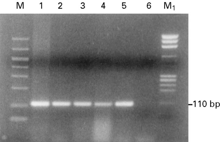
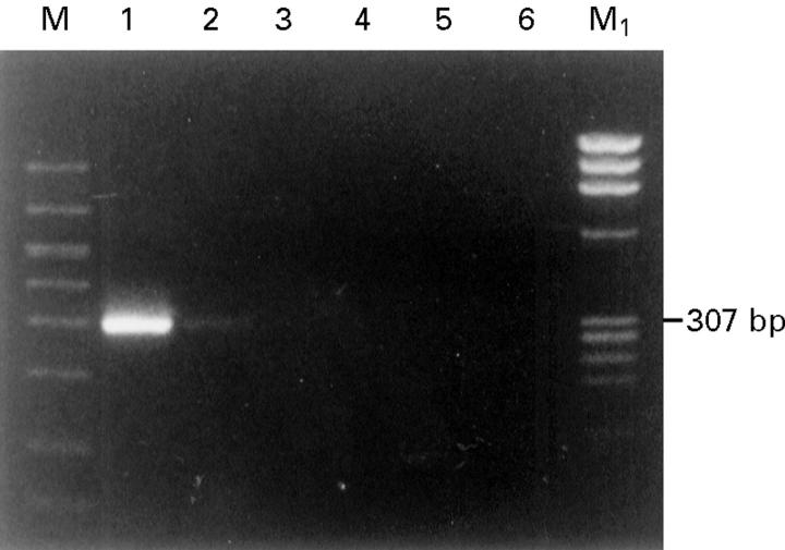
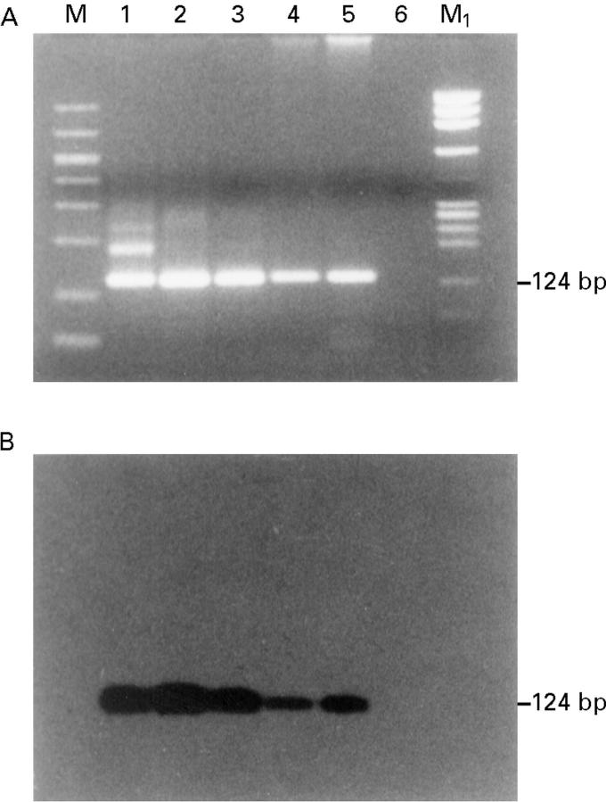
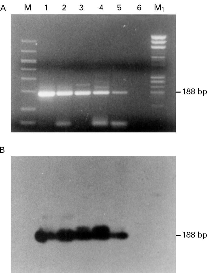
Methods: Ninety six paraffin embedded tissue specimens with neoplastic and non-neoplastic lesions and 19 conjunctiva samples free from overt disease were studied for HPV types 16 and 18 positivity with the polymerase chain reaction.
Results: HPV types 16 and 18 DNA were identified in 57% of in situ squamous cell carcinoma, in 55% of invasive squamous cell carcinoma, in 20% of climatic droplet keratopathy, in 35% of scarred corneas, and in 32% of normal conjunctival tissue obtained during routine cataract extractions.
Conclusion: These findings indicate that HPV types 16 and 18 are detectable with the polymerase chain reaction not only in epithelial neoplasms of the ocular mucous membrane but also in non-neoplastic lesions as well as in apparently healthy conjunctiva.
Figures
Similar articles
-
Failure to detect human papillomavirus DNA in malignant epithelial neoplasms of conjunctiva by polymerase chain reaction.Am J Clin Pathol. 2002 Mar;117(3):429-36. doi: 10.1309/RVUP-QMU3-5X6W-3CQ1. Am J Clin Pathol. 2002. PMID: 11888082
-
Bilateral HPV Positive Squamous Cell Carcinoma In Situ of Conjunctiva.Ophthalmic Plast Reconstr Surg. 2018 Jan/Feb;34(1):e1-e3. doi: 10.1097/IOP.0000000000000947. Ophthalmic Plast Reconstr Surg. 2018. PMID: 28622197
-
Human papillomavirus in corneal and conjunctival carcinoma.Aust N Z J Ophthalmol. 1997 Aug;25(3):211-5. doi: 10.1111/j.1442-9071.1997.tb01394.x. Aust N Z J Ophthalmol. 1997. PMID: 9296295
-
Human papillomavirus infection and ocular surface disease (Review).Int J Oncol. 2019 May;54(5):1503-1510. doi: 10.3892/ijo.2019.4755. Epub 2019 Mar 19. Int J Oncol. 2019. PMID: 30896784 Free PMC article. Review.
-
Human Papillomavirus Related Neoplasia of the Ocular Adnexa.Viruses. 2021 Aug 2;13(8):1522. doi: 10.3390/v13081522. Viruses. 2021. PMID: 34452388 Free PMC article. Review.
Cited by
-
Risk factors for conjunctival squamous cell neoplasia: a matched case-control study.Br J Ophthalmol. 2003 Apr;87(4):396-8. doi: 10.1136/bjo.87.4.396. Br J Ophthalmol. 2003. PMID: 12642297 Free PMC article.
-
Increased Ki-67 proliferative index and absence of P16INK4 in CIN-HPV related pathogenic pathways different from cervical squamous intraepithelial lesion.Br J Ophthalmol. 2006 Jul;90(7):894-9. doi: 10.1136/bjo.2005.086314. Epub 2006 Mar 15. Br J Ophthalmol. 2006. PMID: 16540490 Free PMC article.
-
HPV-Associated Squamous Cell Carcinoma of the Eyelid: Diagnostic Utility of p16 Immunohistochemistry and mRNA In Situ Hybridization.Head Neck Pathol. 2023 Dec;17(4):889-898. doi: 10.1007/s12105-023-01582-6. Epub 2023 Sep 21. Head Neck Pathol. 2023. PMID: 37735287 Free PMC article.
-
Does human papillomavirus cause pterygium?Br J Ophthalmol. 2003 Jul;87(7):806-8. doi: 10.1136/bjo.87.7.806. Br J Ophthalmol. 2003. PMID: 12812871 Free PMC article. No abstract available.
-
Novel low-cost approach to the treatment of ocular surface squamous neoplasia using pattern scanning laser photocoagulation.Arq Bras Oftalmol. 2020 Nov-Dec;83(6):505-510. doi: 10.5935/0004-2749.20200094. Arq Bras Oftalmol. 2020. PMID: 33470278 Free PMC article.
References
MeSH terms
Substances
LinkOut - more resources
Full Text Sources
Medical