. 1997 Sep 15;186(6):859-65.
doi: 10.1084/jem.186.6.859.

Rapid effector function in CD8+ memory T cells

Affiliations

Rapid effector function in CD8+ memory T cells

A Lalvani et al. J Exp Med. .

Abstract

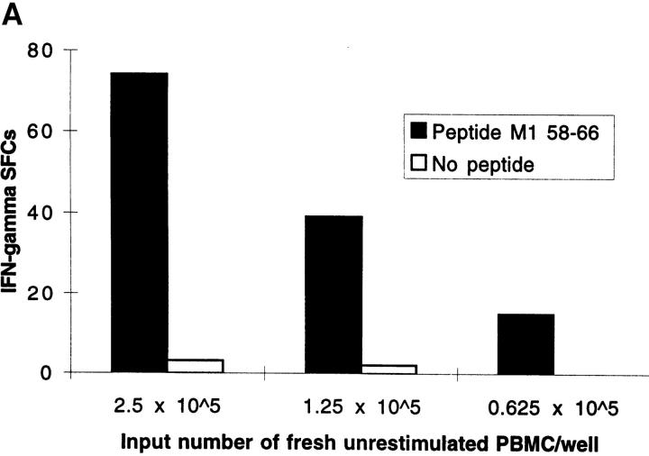
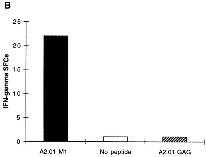
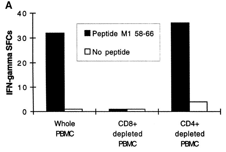
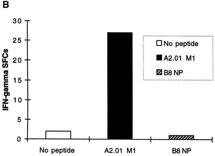
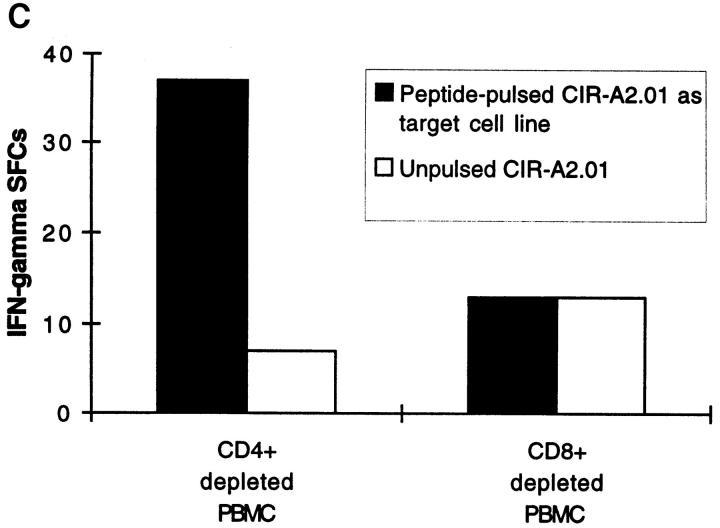
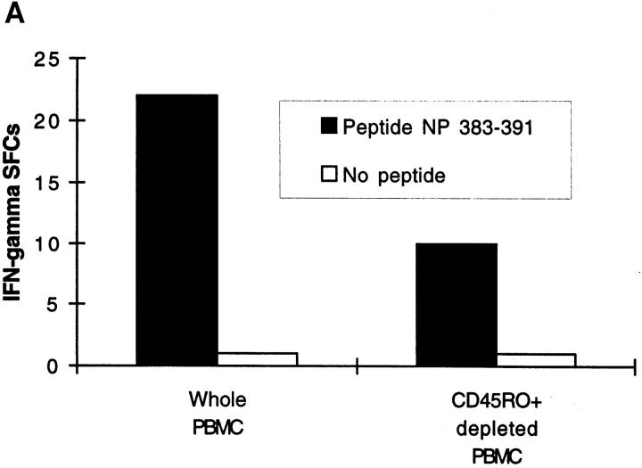
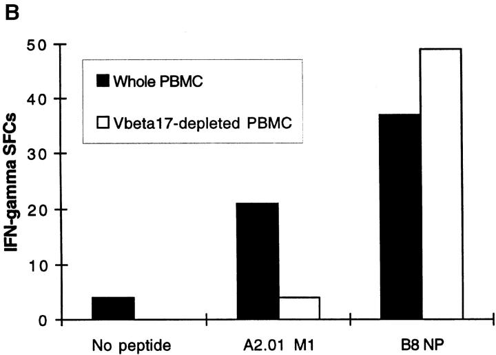
The nature of the CD8+ T cells that underlie antiviral protective immunological memory in vivo is unclear. We have characterized peptide-specific CD8+ T lymphocytes directly ex vivo from peripheral blood in humans with past exposure to influenza virus, using single cell interferon gamma (IFN-gamma) release as a measure of effector function. In individuals in the memory state with respect to influenza virus infection, unrestimulated antigen-specific CD8+ T cells displayed IFN-gamma release within 6 h of antigen contact, identifying a population of memory CD8+ T cells that exhibit effector function without needing to divide and differentiate over several days. We have quantified circulating CD8+ effector T cells specific for six different MHC class I-restricted influenza virus epitopes. Enumeration of these CD8+ T cells gives frequencies of peptide-specific T cells that correlate with, but are in general severalfold higher than, CTL precursor frequencies derived from limiting dilution analysis, indicating that this novel population of memory CD8+ T cells has hitherto been undetected by standard means. The phenotype of these cells, which persist at a low frequency long after recovery from an acute viral infection, suggests that they play a role in protective immunological memory.


Figures

Figure 1
Figure 1
(A) Freshly isolated, unrestimulated, peptide-specific, memory CD8+ T cells secrete IFN-γ within 6 h of antigen contact. Enumeration of IFN-γ SFCs in a 6 h ELISPOT assay performed with freshly isolated PBMC from donor SM (class I HLA haplotype: A2.01, A24; B44, B14). The HLA-A2.01–restricted peptide M1 58–66 was used at a final concentration of 2 μM. Mean values from duplicate wells are shown. (B) Irrelevant peptides do not induce IFN-γ SFCs. Using fresh PBMCs from HIV-uninfected donor SC (class I HLA haplotype: A2.01, A28; B7, B14) in a 6 h ELISPOT assay, quantitation of IFN-γ SFCs shows a response specific for the influenza HLA-A2.01–restricted peptide M1 58–66 compared with no response to the HLA-A2.01–restricted peptide from the HIV GAG protein, GAG 77-85 (SLYNTVATL).
Figure 1
Figure 1
(A) Freshly isolated, unrestimulated, peptide-specific, memory CD8+ T cells secrete IFN-γ within 6 h of antigen contact. Enumeration of IFN-γ SFCs in a 6 h ELISPOT assay performed with freshly isolated PBMC from donor SM (class I HLA haplotype: A2.01, A24; B44, B14). The HLA-A2.01–restricted peptide M1 58–66 was used at a final concentration of 2 μM. Mean values from duplicate wells are shown. (B) Irrelevant peptides do not induce IFN-γ SFCs. Using fresh PBMCs from HIV-uninfected donor SC (class I HLA haplotype: A2.01, A28; B7, B14) in a 6 h ELISPOT assay, quantitation of IFN-γ SFCs shows a response specific for the influenza HLA-A2.01–restricted peptide M1 58–66 compared with no response to the HLA-A2.01–restricted peptide from the HIV GAG protein, GAG 77-85 (SLYNTVATL).
Figure 2
Figure 2
(A) Influenza virus–specific CD8+ memory T cells with rapid effector function are CD8+. Freshly isolated PBMCs from donor SC were tested against the influenza HLA-A2.01–restricted peptide M1 58–66 at a concentration of 2 μM in a 14 h ELISPOT assay before or after depletion of CD4+ or CD8+ cells. Input cell numbers were 5 × 10⁵/ well predepletion, and mean values from duplicate wells are shown. (B and C) Unrestimulated influenza virus–specific memory CD8+ T cells are HLA class I restricted. (B) Peptide epitopes restricted by HLA class I alleles not present in the donor, do not elicit IFN-γ SFCs. Freshly isolated PBMCs from donor SC (class I HLA haplotype: A2.01, A28; B7, B14) were tested in an ELISPOT assay in the absence of peptide and in the presence of the influenza HLA-A2.01–restricted peptide M1 58–66 or the irrelevant influenza HLA-B8-restricted peptide NP 380–388, each at a concentration of 1 μM. (C) Freshly isolated CD8+ T cells recognize the HLA-A2.01–restricted peptide M1 58–66 presented by HLA-A2.01 on the surface of the cell line CIR-A2.01. Fresh PBMCs from donor WB (HLA class I haplotype: A1, A2; B7, B8) were depleted of CD4+ or CD8+ cells; after depletion, 3 × 10⁵ cells were plated out per well, along with 2 × 10⁴ CIR-A2.01 cells prepulsed with 2 μM of the influenza HLA-A2.01–restricted peptide M1 58–66 and washed three times. Control wells had equal numbers of peptide-unpulsed CIR-A2.01.
Figure 2
Figure 2
(A) Influenza virus–specific CD8+ memory T cells with rapid effector function are CD8+. Freshly isolated PBMCs from donor SC were tested against the influenza HLA-A2.01–restricted peptide M1 58–66 at a concentration of 2 μM in a 14 h ELISPOT assay before or after depletion of CD4+ or CD8+ cells. Input cell numbers were 5 × 10⁵/ well predepletion, and mean values from duplicate wells are shown. (B and C) Unrestimulated influenza virus–specific memory CD8+ T cells are HLA class I restricted. (B) Peptide epitopes restricted by HLA class I alleles not present in the donor, do not elicit IFN-γ SFCs. Freshly isolated PBMCs from donor SC (class I HLA haplotype: A2.01, A28; B7, B14) were tested in an ELISPOT assay in the absence of peptide and in the presence of the influenza HLA-A2.01–restricted peptide M1 58–66 or the irrelevant influenza HLA-B8-restricted peptide NP 380–388, each at a concentration of 1 μM. (C) Freshly isolated CD8+ T cells recognize the HLA-A2.01–restricted peptide M1 58–66 presented by HLA-A2.01 on the surface of the cell line CIR-A2.01. Fresh PBMCs from donor WB (HLA class I haplotype: A1, A2; B7, B8) were depleted of CD4+ or CD8+ cells; after depletion, 3 × 10⁵ cells were plated out per well, along with 2 × 10⁴ CIR-A2.01 cells prepulsed with 2 μM of the influenza HLA-A2.01–restricted peptide M1 58–66 and washed three times. Control wells had equal numbers of peptide-unpulsed CIR-A2.01.
Figure 3
Figure 3
(A) The majority of effectors detected in the ex vivo ELISPOT assay bear the memory-associated cell-surface marker CD45RO. Fresh PBMCs from donor SD were depleted of CD45RO+ cells and plated out at 5 × 10⁵/well with or without the HLA-B27.05–restricted peptide NP 383– 391. FACS® analysis confirmed 67% depletion of CD45RO+ CD8+ T cells from PBMCs. (B) All detectable CD8+ effectors specific for the A2.01-restricted peptide M1 58–66 bear TCRs with the Vβ17 gene segment. The ELISPOT assay was performed with freshly isolated whole PBMCs or PBMCs depleted of Vβ17+ T cells; FACS® analysis confirmed that the CD8+ Vβ17+ T cells initially present among whole PBMCs were depleted by 90%. Cells were seeded at 5 × 10⁵/well in the presence of the HLA-A2.01–restricted peptide M1 58–66 or the HLA-B8–restricted peptide NP 380–388. PBMCs were from donor WB (HLA class I haplotype: A1, A2.01; B7, B8).
Figure 3
Figure 3
(A) The majority of effectors detected in the ex vivo ELISPOT assay bear the memory-associated cell-surface marker CD45RO. Fresh PBMCs from donor SD were depleted of CD45RO+ cells and plated out at 5 × 10⁵/well with or without the HLA-B27.05–restricted peptide NP 383– 391. FACS® analysis confirmed 67% depletion of CD45RO+ CD8+ T cells from PBMCs. (B) All detectable CD8+ effectors specific for the A2.01-restricted peptide M1 58–66 bear TCRs with the Vβ17 gene segment. The ELISPOT assay was performed with freshly isolated whole PBMCs or PBMCs depleted of Vβ17+ T cells; FACS® analysis confirmed that the CD8+ Vβ17+ T cells initially present among whole PBMCs were depleted by 90%. Cells were seeded at 5 × 10⁵/well in the presence of the HLA-A2.01–restricted peptide M1 58–66 or the HLA-B8–restricted peptide NP 380–388. PBMCs were from donor WB (HLA class I haplotype: A1, A2.01; B7, B8).

References

    1. Ahmed R, Gray D. Immunological memory and protective immunity: understanding their relation. Science (Wash DC) 1996;272:54–60. - PubMed
    1. Sprent J. T and B memory cells. Cell. 1994;76:315–322. - PubMed
    1. Sprent J, Tough DF. Lymphocyte lifespan and memory. Science (Wash DC) 1994;265:1395–1400. - PubMed
    1. Kuendig TM, Bachmann MF, Oehen S, Hoffmann UW, Simard JJL, Kalberer CP, Pircher H, Ohashi PS, Hengartner H, Zinkernagel RM. On the role of antigen in maintaining cytotoxic T-cell memory. Proc Natl Acad Sci USA. 1996;93:9716–9723. - PMC - PubMed
    1. Oehen S, Waldner H, Kuendig TM, Hengartner H, Zinkernagel RM. Antivirally protective cytotoxic T cell memory to lymphocytic choriomeningitis virus is governed by persisting antigen. J Exp Med. 1992;176:1273–1281. - PMC - PubMed

Publication types

MeSH terms