. 1997 Sep 16;94(19):10034-9.
doi: 10.1073/pnas.94.19.10034.

ActA is a dimer

Affiliations

ActA is a dimer

P Mourrain et al. Proc Natl Acad Sci U S A. .

Abstract

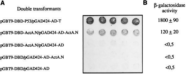
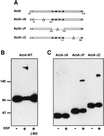
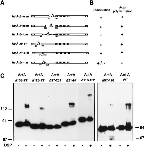
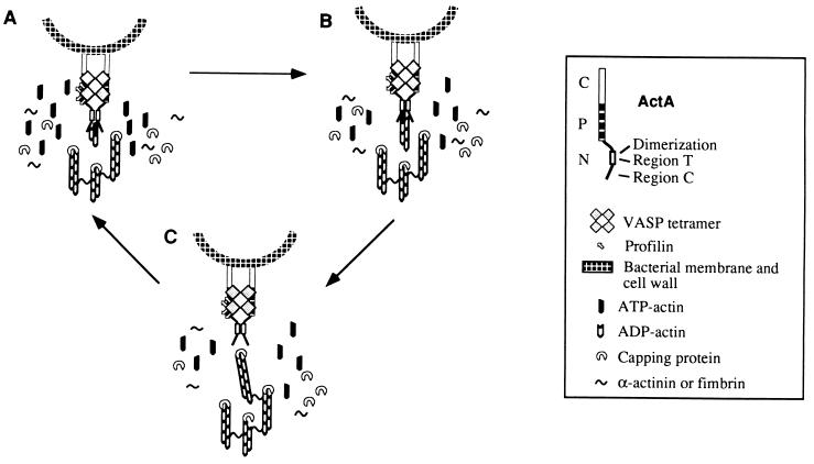
ActA, a surface protein of Listeria monocytogenes, is able to induce continuous actin polymerization at the rear of the bacterium, in the cytosol of the infected cells. Its N-terminal domain is sufficient to induce actin tail formation and movement. Here, we demonstrate, using the yeast two-hybrid system, that the N-terminal domain of ActA may form homodimers. By using chemical cross-linking to explore the possibility that ActA could be a multimer on the surface of the bacteria, we show that ActA is a dimer. Cross-linking experiments on various L. monocytogenes strains expressing different ActA variants demonstrated that the region spanning amino acids 97-126, and previously identified as critical for actin tail formation, is also critical for dimer formation. A model of actin polymerization by L. monocytogenes, involving the ActA dimer, is presented.


Figures

Figure 1
Figure 1
Qualitative and quantitative analysis of β-gal activity in the yeast two-hybrid system. (A) Filter assay: five independent colonies of cotransformants were patched onto filters layered over synthetic medium lacking Trp and Leu, and incubated overnight at 30°C. Filters were incubated in the X-Gal solution during 30 min for positive control (pGBT9-DBD-p53/pGAD424-AD-T) and 3–4 h for (pGBT9-DBD-ActA.N/pGAD424-AD-ActA.N). (B) β-Gal activity of yeast cotransformed with indicated plasmids was measured using 4-methyl-umbelliferyl β-d-galactopiranoside (MUG) as the substrate. The results represent the average ± SD of triplicate samples from three independent transformants. Activities were computed as [(fluorescence units/hour/yeast) × 100,000].
Figure 2
Figure 2
Detection of cross-linked ActA by SDS/PAGE and immunoblotting. (A) Schematic representation of the ActA protein and the ActA variants. (B) L. monocytogenes LO28 wild-type strain grown in broth was treated with DSP (100 μM) as described. Protein extracts were separated by SDS/PAGE (7%) and analyzed by immunoblotting using anti-ActA affinity purified antibodies (A18K). Treatment of cross-linked proteins with β-mercaptoethanol (5%) before loading reverse the presence of the 190-kDa protein. (C) L. monocytogenes ΔactA producing different ActA variants were grown in broth and treated with DSP (10 μM). Total protein extracts of bacteria expressing ActA-ΔN, ActA-ΔP and ActA-ΔC were separated by SDS/PAGE (7%) and analyzed by immunoblotting using anti-ActA affinity purified antibodies (A18K).
Figure 3
Figure 3
Identification of the minimal region of ActA necessary for DSP cross-linking. (A) Schematic representation of the ActA variants used for the cross-linking experiments. (B) Properties of the variants. (C) Cross-linking experiments: L. monocytogenes producing different ActA variants were grown in broth and treated with DSP. Total protein extracts of bacteria producing ActA-Δ158–231, ActA-Δ126–231 ActA-Δ97–231, ActA-Δ21–97, ActA-Δ116–122, and ActA-Δ97–126 were separated by SDS/PAGE (9%) and analyzed by immunoblotting using anti-ActA affinity purified antibodies (A18K). Bacteria were treated with 40 μM (ActA-Δ158–231), 10 μM (ActA-Δ126–231, ActA-Δ97–126, and ActA-Δ116–122), and 20 μM (ActA-Δ97–231 and ActA-Δ21–97) of DSP.
Figure 4
Figure 4
Hypothetical model of actin assembly by ActA dimers. The N-terminal domain of ActA contains two regions critical for actin based motility. Both are required for actin tail formation but each has in addition a specific role to maintain the dynamics of the process. Region T (116–122) is necessary for actin tail formation, and region C (21–97) is necessary for continuous actin filament elongation. The region of ActA spanning amino acids 97–126 is necessary for both dimerization of ActA and actin polymerization. (A) Generation of free barbed ends either by nucleation or uncapping or severing. Dimerization of ActA.N could allow ActA either to nucleate actin, or to interact with a host nucleator or to displace a capping protein, or to sever actin filaments. (B) Elongation. Once nucleation has occurred, two ActA monomers bind the actin filament through region 21–97, which keeps the barbed ends uncapped. ActA dimerization favors recruitment of VASP–profilin–actin complexes on the central proline rich region of ActA, bringing polymerization competent actin monomers in the vicinity of the bacterium, resulting in filament elongation. (C) Release. Actin filament elongation continues until capping, release, and cross-linking to the performed tail take place. When the filament is released, the ActA dimer is immediately available for a new cycle of nucleation, elongation, and release.

References

    1. Cossart P. Curr Opin Cell Biol. 1995;7:94–101. - PubMed
    1. Theriot J. Annu Rev Cell Dev Biol. 1995;11:213–239. - PubMed
    1. Lasa I, Cossart P. Trends Cell Biol. 1996;6:109–114. - PubMed
    1. Domann E, Wehland J, Rohde M, Pistor S, Hartl M, Goebel W, Leimeister-Wächter M, Wuenscher M, Chakraborty T. EMBO J. 1992;11:1981–1990. - PMC - PubMed
    1. Kocks C, Gouin E, Tabouret M, Berche P, Ohayon H, Cossart P. Cell. 1992;68:521–531. - PubMed

Publication types

MeSH terms

LinkOut - more resources