

Structural mimicry of a native protein by a minimized binding domain
- PMID: 9294166
- PMCID: PMC23311
- DOI: 10.1073/pnas.94.19.10080
Structural mimicry of a native protein by a minimized binding domain
Abstract
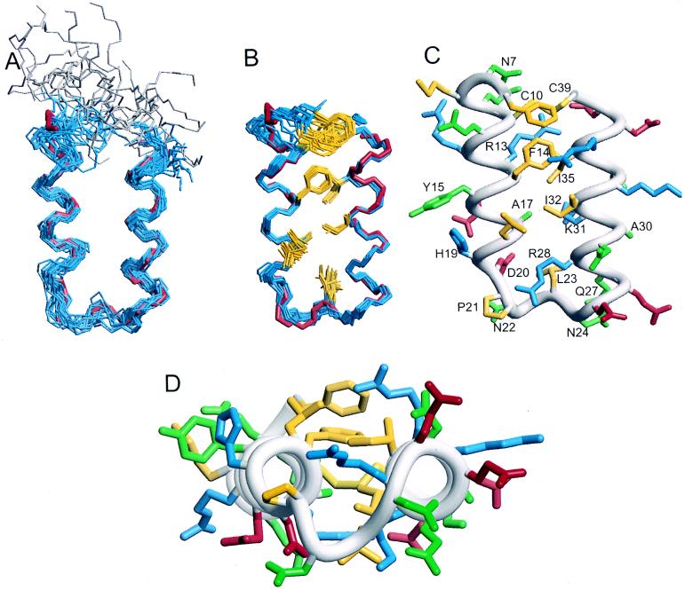
The affinity between molecules depends both on the nature and presentation of the contacts. Here, we observe coupling of functional and structural elements when a protein binding domain is evolved to a smaller functional mimic. Previously, a 38-residue form of the 59-residue B-domain of protein A, termed Z38, was selected by phage display. Z38 contains 13 mutations and binds IgG only 10-fold weaker than the native B-domain. We present the solution structure of Z38 and show that it adopts a tertiary structure remarkably similar to that observed for the first two helices of B-domain in the B-domain/Fc complex [Deisenhofer, J. (1981) Biochemistry 20, 2361-2370], although it is significantly less stable. Based on this structure, we have improved on Z38 by designing a 34-residue disulfide-bonded variant (Z34C) that has dramatically enhanced stability and binds IgG with 9-fold higher affinity. The improved stability of Z34C led to NMR spectra with much greater chemical shift dispersion, resulting in a more precisely determined structure. Z34C, like Z38, has a structure virtually identical to the equivalent region from native protein A domains. The well-defined hydrophobic core of Z34C reveals key structural features that have evolved in this small, functional domain. Thus, the stabilized two-helix peptide, about half the size and having one-third of the remaining residues altered, accurately mimics both the structure and function of the native domain.
Figures

References
-
- McKnight C J, Doering D S, Matsudaira P T, Kim P S. J Mol Biol. 1996;260:126–134. - PubMed
-
- Kalia Y N, Brocklehurst S M, Hipps D S, Appella E, Sakaguchi K, Perham R N. J Mol Biol. 1993;230:323–341. - PubMed
-
- Gouda H, Torigoe H, Saito A, Sato M, Arata Y, Shimada I. Biochemistry. 1992;31:9665–9672. - PubMed
-
- Starovasnik M A, Skelton N J, O’Connell M P, Kelley R F, Reilly D, Fairbrother W J. Biochemistry. 1996;35:15558–15569. - PubMed
-
- Jendeberg L, Tashiro M, Tejero R, Lyons B A, Uhlen M, Montelione G T, Nilsson B. Biochemistry. 1996;35:22–31. - PubMed
MeSH terms
Substances
Associated data
- Actions
- Actions
- Actions
- Actions
LinkOut - more resources
Full Text Sources
Other Literature Sources
