. 1997 Sep 16;94(19):10116-21.
doi: 10.1073/pnas.94.19.10116.

The C-type lectin domains of lecticans, a family of aggregating chondroitin sulfate proteoglycans, bind tenascin-R by protein-protein interactions independent of carbohydrate moiety

Affiliations

The C-type lectin domains of lecticans, a family of aggregating chondroitin sulfate proteoglycans, bind tenascin-R by protein-protein interactions independent of carbohydrate moiety

A Aspberg et al. Proc Natl Acad Sci U S A. .

Abstract

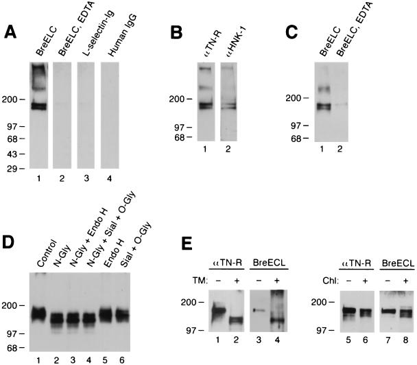
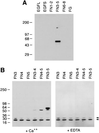
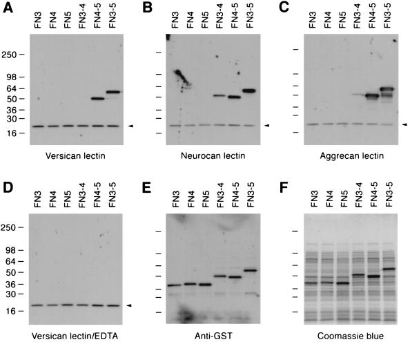
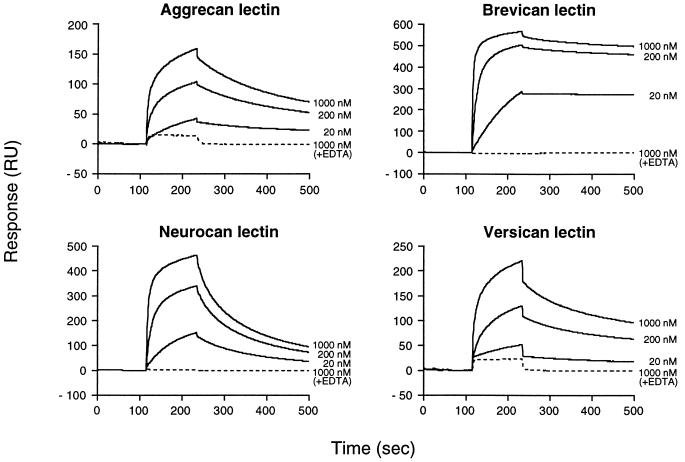
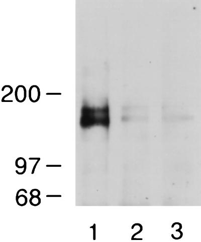
The lecticans are a family of chondroitin sulfate proteoglycans including aggrecan, versican, neurocan, and brevican. The C-terminal globular domains of lecticans are structurally related to selectins, consisting of a C-type lectin domain flanked by epidermal growth factor and complement regulatory protein domains. The C-type lectin domain of versican has been shown to bind tenascin-R, an extracellular matrix protein specifically expressed in the nervous system, and the interaction was presumed to be mediated by a carbohydrate-protein interaction. In this paper, we show that the C-type lectin domain of brevican, another lectican that is specifically expressed in the nervous system, also binds tenascin-R. Surprisingly, this interaction is mediated by a protein-protein interaction through the fibronectin type III domains 3-5 of tenascin-R, independent of any carbohydrates or sulfated amino acids. The lectin domains of versican and other lecticans also bind the same domain of tenascin-R by protein-protein interactions. Surface plasmon resonance analysis revealed that brevican lectin has at least a 10-fold higher affinity than the other lectican lectins. Tenascin-R is coprecipitated with brevican from adult rat brain extracts, suggesting that tenascin-R and brevican form complexes in vivo. These results demonstrate that the C-type lectin domain can interact with fibronectin type III domains through protein-protein interactions, and suggest that brevican is a physiological tenascin-R ligand in the adult brain.


Figures

Figure 1
Figure 1
Brevican ELC chimera binds tenascin-R. (A) Brevican ELC chimera binds to 160/180 kDa bands in ligand blotting. Soluble extracts of rat brain were resolved in an 8–16% gradient SDS/PAGE under nonreducing conditions and transferred to nitrocellulose filters. Filters were probed in the absence (lanes 1, 3, and 4) or presence (lane 2) of 20 mM EDTA with brevican ELC chimera (lanes 1 and 2), l-selectin-Ig chimera (21) (lane 3), or human IgG (lane 4). (B) Bands at 160 and 180 kDa are recognized by anti-tenascin-R and anti-HNK-1 monoclonal antibodies. Soluble extracts from adult rat brain were probed in immunoblotting with anti-rat tenascin-R monoclonal antibody 596 (11) (lane 1) or anti-HNK-1 monoclonal antibody (lane 2). (C) Tenascin-R is precipitated with brevican ELC chimera. Soluble brain extracts were precipitated with brevican ELC chimera and protein A-agarose in the absence (lane 1) or presence (lane 2) of 20 mM EDTA, and the precipitated materials were probed by immunoblotting with anti-tenascin-R monoclonal antibody 596. (D) Enzymatic deglycosylation does not affect the binding of brevican ELC chimera to tenascin-R. Soluble brain extracts were treated without any enzyme (lane 1); with N-glycosidase F (lane 2); with N-glycosidase F and endo-β-acetylglucosaminidase H (lane 3); with N-glycosidase F, sialidase, and O-glycanase (lane 4); with endo-β-acetylglucosaminidase H (lane 5); or with sialidase and O-glycanase (lane 6). Treated samples were probed with brevican ELC chimera in ligand blotting. (E) Metabolic deglycosylation and desulfation do not affect the binding of brevican ELC chimera to tenascin-R. B9 cells, which secrete the 180 kDa form of tenascin-R, were treated with tunicamycin (lanes 1–4) or chlorate (lanes 5–8). Culture supernatants from the treated B9 cells were immunoblotted with anti-tenascin-R monoclonal antibody 596 (lanes 1, 2, 5, and 6) or brevican ELC chimera (lanes 3, 4, 7, and 8).
Figure 2
Figure 2
Brevican lectin binds tenascin-R fragments produced as bacterial fusion proteins. (A) Fusion proteins representing various tenascin-R domains were probed with brevican ELC chimera by ligand blotting. EGFL, EGF repeats with cysteine-rich sequences; EGFS, EGF repeats without cysteine-rich sequences; FN1–2, FN3–5, and FN6–8, FNIII domains 1–2, 3–5, and 6–8, respectively; FG, fibrinogen-like domain (see ref. for details). (B) Ligand blotting with brevican rCLD. Lysates of bacteria expressing GST fusion proteins representing the following FNIII domains were probed with biotinylated brevican rCLD. Ligand blotting was performed in the presence of 5 mM CaCl2 (Left) or 5 mM EDTA (Right). FN3, domain 3; FN4, domain 4; FN5, domain 5; FN3–4, domain 3–4; FN4–5, domain 4–5; FN3–5, domains 3–5. Arrowheads indicate nonspecific bands reactive to peroxidase-conjugated avidin used in these assays.
Figure 3
Figure 3
The lectin domains of versican, neurocan, and aggrecan bind bacterial fusion proteins containing FNIII domain 4 of tenascin-R. Lysates of bacteria expressing GST fusion proteins representing different domains of tenascin-R were probed with the biotinylated rCLDs of versican (A and D), neurocan (B), and aggrecan (C) in the presence of 5 mM CaCl2 (AC) or 5 mM EDTA (D) in ligand blotting. (E) The same blot was probed with anti-GST antiserum. (F) Coomassie brilliant blue staining of the gel loaded with identical amounts of lysates as in AE. The fusion proteins used in this experiment are the same as described in the legend of Fig. 2. Arrowheads indicate nonspecific bands reactive to peroxidase-conjugated avidin.
Figure 4
Figure 4
Surface plasmon resonance measurements of the interactions between lectican lectins and tenascin-R. Mono Q-purified tenascin-R FN3–5 fragments at different concentrations were injected over flow cells coupled with individual lectican rCLDs. Solid lines indicate response traces in the presence of 1 mM CaCl2 and broken lines in the presence of 5 mM EDTA. Injection of the tenascin-R fragment started at 115 sec and ended at 235 sec. The response trace average between 75–100 sec was normalized to 0.
Figure 5
Figure 5
Tenascin-R coimmunoprecipitates with brevican from adult rat brain extracts. Soluble brain extracts were incubated with anti-brevican monoclonal antibody RB18 (lanes 1 and 2) in the presence of 5 mM CaCl2 (lane 1) or 20 mM EDTA (lane 2). In a control experiment, the incubation was performed without the antibody in the presence of 5 mM CaCl2 (lane 3). The samples were precipitated with protein G-agarose and then probed in ligand blotting assay with brevican ELC chimera.

References

    1. Ruoslahti E. Glycobiology. 1996;6:489–492. - PubMed
    1. Doege K, Sasaki M, Horigan E, Hassell J R, Yamada Y. J Biol Chem. 1987;262:17757–17767. - PubMed
    1. Zimmermann D R, Ruoslahti E. EMBO J. 1989;8:2975–2981. - PMC - PubMed
    1. Rauch U, Karthikeyan L, Maurel P, Margolis R U, Margolis R K. J Biol Chem. 1992;267:19536–19547. - PubMed
    1. Yamada H, Watanabe K, Shimonaka M, Yamaguchi Y. J Biol Chem. 1994;269:10119–10126. - PubMed

Publication types

LinkOut - more resources