. 1997 Sep 16;94(19):10161-6.
doi: 10.1073/pnas.94.19.10161.

Exploring the folding free energy surface of a three-helix bundle protein

Affiliations

Exploring the folding free energy surface of a three-helix bundle protein

Z Guo et al. Proc Natl Acad Sci U S A. .

Abstract

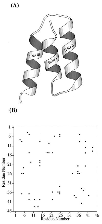
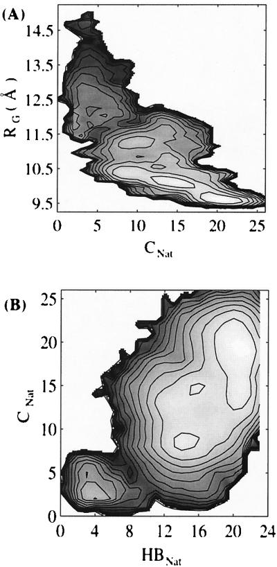
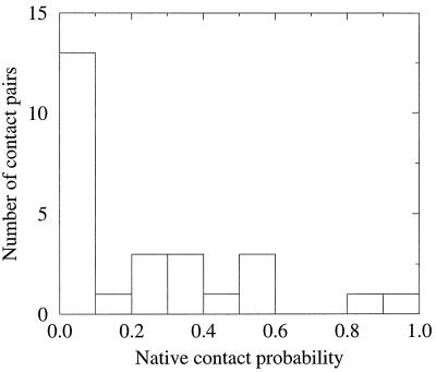
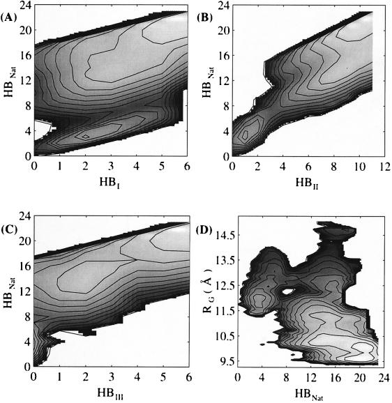
The multidimensional free energy surface for a small fast folding helical protein is explored based on first-principle calculations. The model represents the 46-residue segment from fragment B of staphylococcal protein A. The relationship between collapse and tertiary structure formation, and the order of collapse and secondary structure formation, are investigated. We find that the initial collapse process gives rise to a transition state with about 30% of the native tertiary structure and 50-70% of the native helix content. We also observe two distinct distributions of native helix in this collapsed state (Rg approximately 12 A), one with about 20% of the native helical hydrogen bonds, the other with near 70%. The former corresponds to a local minimum. The barrier from this metastable state to the native state is about 2 kBT. In the latter case, folding is essentially a downhill process involving topological assembly. In addition, the order of formation of secondary structure among the three helices is examined. We observe cooperative formation of the secondary structure in helix I and helix II. Secondary structure in helix III starts to form following the formation of certain secondary structure in both helix I and helix II. Comparisons of our results with those from theory and experiment are made.


Figures

Figure 1
Figure 1
(A) Ribbon model of the native structure of the 46-residue segment of fragment B of staphylococcal protein A. (B) The center of geometry based native contact map from the simulation of two 1.1-ns native trajectories. Only contacts between residues i and j (ji + 4) are considered.
Figure 2
Figure 2
Diagram illustrating the sampling strategy for free energy calculations of protein folding. The positions of the ellipses represent initial conditions used in generating sampling about conformational basins along the folding reaction coordinate, denoted by Rg and increasing from top to bottom in the figure. The lines illustrate connectivity between sampling of different initial conditions. Both sufficient initial conditions to span the reaction coordinate and a path of connectivity between all sampling basins are requirements for accurate calculation of free energy surfaces for protein folding.
Figure 3
Figure 3
(A) Contour plot of the free energy as a function of total number of native contacts (CNat) and radius of gyration (Rg). (B) Contour plot of the free energy as a function of total number of native hydrogen bonds (HBNat) and total number of native contacts (CNat). Contour levels are drawn at 0.5-kcal/mol intervals.
Figure 4
Figure 4
Distribution of native contacts in the transition state.
Figure 5
Figure 5
Contour plot of the free energy as a function of (A) total number of native hydrogen bonds (HBNat) and total number of native hydrogen bonds in helix I (HBI); (B) total number of native hydrogen bonds (HBNat) and total number of native hydrogen bonds in helix II (HBII); (C) total number of native hydrogen bonds (HBNat) and total number of native hydrogen bonds in helix III (HBIII); (D) total number of native hydrogen bonds (HBNat) and radius of gyration (Rg). Contour levels are drawn at 0.5-kcal/mol intervals.

References

    1. Kim P S, Baldwin R L. Annu Rev Biochem. 1990;59:631–660. - PubMed
    1. Karplus M, Weaver D L. Biopolymers. 1979;18:1421–1437.
    1. Ptitsyn O B. J Protein Chem. 1987;6:273–293.
    1. Onuchic J N, Wolynes P G, Luthey-Schulten Z, Socci N D. Proc Natl Acad Sci USA. 1995;92:3626–3630. - PMC - PubMed
    1. Ballew R M, Sabelko J, Gruebele M. Proc Natl Acad Sci USA. 1996;93:5759–5764. - PMC - PubMed

Publication types

LinkOut - more resources