. 1997 Sep 16;94(19):10356-61.
doi: 10.1073/pnas.94.19.10356.

The epitopes for some antiphospholipid antibodies are adducts of oxidized phospholipid and beta2 glycoprotein 1 (and other proteins)

Affiliations

The epitopes for some antiphospholipid antibodies are adducts of oxidized phospholipid and beta2 glycoprotein 1 (and other proteins)

S Hörkkö et al. Proc Natl Acad Sci U S A. .

Abstract

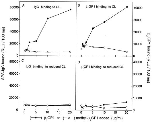
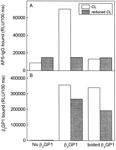
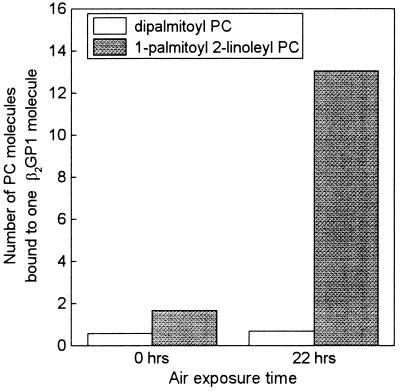
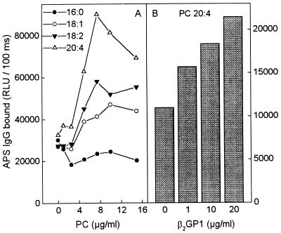
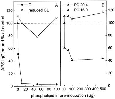
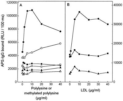
Circulating autoantibodies to phospholipids (aPLs), such as cardiolipin (CL), are found in patients with antiphospholipid antibody syndrome (APS). We recently demonstrated that many aPLs bound to CL only after it had been oxidized (OxCL), but not to a reduced CL analogue that could not undergo oxidation. We now show that the neoepitopes recognized by some aPLs consist of adducts formed between breakdown products of oxidized phospholipid and associated proteins, such as beta2 glycoprotein 1 (beta2GP1). Addition of human beta2GP1, polylysine, native low-density lipoprotein, or apolipoprotein AI to OxCL-coated wells increased the anticardiolipin antibody (aCL) binding from APS sera that first had been diluted so that no aCL binding to OxCL could be detected. No increase in aCL binding was observed when these proteins were added to wells coated with reduced CL. The ability of beta2GP1, polylysine, or low-density lipoprotein to be a "cofactor" for aCL binding to OxCL was greatly reduced when the proteins were methylated. Incubation of beta2GP1 with oxidized 1-palmitoyl-2-linoleyl-[1-14C]-phosphatidylcholine (PC), but not with dipalmitoyl-[1-14C]-PC, led to formation of covalent adducts with beta2GP1 recognized by APS sera. These data suggest that the reactive groups of OxCL, such as aldehydes generated during the decomposition of oxidized polyunsaturated fatty acids, form covalent adducts with beta2GP1 (and other proteins) and that these are epitopes for aCLs. Knowledge that the epitopes recognized by many aPLs are adducts of oxidized phospholipid and associated proteins, including beta2GP1, may give new insights into the pathogenic events underlying the clinical manifestations of APS.


Figures

Figure 1
Figure 1
(A and C) Chemiluminescent immunoassay for IgG binding from a serum pool of 10 APS patients to OxCL and reduced CL in the absence or presence of increasing amounts of native (•) or methylated (○) human β2GP1. The serum pool was first diluted (1:500) so that no antibody binding to OxCL was detected (0 point). Increasing amounts of native or methylated human β2GP1 were added to the sera before addition to the PL-coated wells. Binding of IgG to OxCL and reduced CL was measured with alkaline phosphatase-labeled goat anti-human-IgG antibody. (B and D) In parallel wells the binding of β2GP1 and methylated β2GP1 was detected with alkaline phosphatase-labeled goat anti-human-β2GP1 antibody. Preliminary data indicated that this antibody bound equally well to native and methylated β2GP1. RLU, relative light units. Each point is the mean of triplicate determinations.
Figure 2
Figure 2
(A) Chemiluminescent immunoassay for IgG binding from a serum pool of five APS patients to OxCL and reduced CL. Native β2GP1 or β2GP1 boiled for 30 min (5 μg/ml in 3% BSA-TBS) was incubated in the PL-coated wells for 1 hr. The serum pool was first diluted 1:750 so that minimal antibody binding to OxCL was detected. The IgG bound was measured using alkaline phosphatase-labeled goat anti-human-IgG antibody. (B) In parallel wells the binding of β2GP1 to OxCL and reduced CL was detected with goat anti-human-β2GP1 antibody, which then was detected with alkaline phosphatase-labeled rabbit anti-goat-IgG secondary antibody. Each point is the mean of triplicate determinations.
Figure 3
Figure 3
Binding of dipalmitoyl-[1-14C]-PC and 1-palmitoyl-2-linoleyl-[1-14C]-PC to β2GP1. PCs were dried under nitrogen, exposed to air for 0 or 22 hr and then incubated with β2GP1 for 6 hr in the presence of 10 mM NaCNBH3. The amount of PC bound to β2GP1 was determined after solubilizing the mixture in n-octyl glucoside and precipitating protein with 10% trichloroacetic acid.
Figure 4
Figure 4
(A) Chemiluminescent immunoassay for IgG binding from a serum pool of five APS patients (1:100 dilution) to different PC: 1,2-dipalmitoyl-PC (16:0), 1,2-dioleoyl-PC (18:1), 1,2-dilinoleyl-PC (18:2), and 1,2-diarachidonyl-PC (20:4). (B) IgG binding from the same serum pool to 1,2-diarachidonyl-PC (20:4) in the presence of β2GP1. The serum pool was first diluted 1:750 so that minimal antibody binding to OxCL was detected. The IgG bound was measured using alkaline phosphatase-labeled goat anti-human-IgG antibody. Each point is the mean of triplicate determinations.
Figure 5
Figure 5
Competition immunoassay of the binding of IgG from APS sera to OxCL. A serum pool from five APS patients (1:100 dilution) was preincubated with OxCL (•), reduced CL (○), 1,2-diarachidonyl-[20:4]-PC (▴), or 1,2-dipalmitoyl-[16:0]-PC (▵). After preincubation, the immune complexes were pelleted by centrifugation and the supernatants tested for aCL IgG binding to OxCL. Results are expressed as percentage of control (control = APS sera preincubated without any PL). Each point is the mean of four replicate determinations.
Figure 6
Figure 6
Chemiluminescent immunoassay for IgG binding from a serum pool of five APS patients to OxCL (circles), reduced CL (squares), and ethanol-only control wells (triangles). (A) Binding in the presence of polylysine (solid figures) or methylated polylysine (open figures). (B) Binding in the presence of LDL. Each compound (in TBS) was incubated for 10 min in wells coated with OxCL, reduced CL, or ethanol. The serum pool was first diluted 1:500 so that minimal antibody binding to OxCL was detected. The IgG binding was measured with alkaline phosphatase-labeled goat anti-human-IgG antibody. Each point is the mean of four replicate determinations.

References

    1. Alarcon-Segovia D, Deleze M, Oria C, Sanchez-Guerrero J, Gomez-Pancheco L, Cabiedes J, Fernandez L, Ponce de Leon S. Medicine. 1989;58:353–363. - PubMed
    1. Asherson R A, Khamashta M A, Ordi-Ros J, Derksen R H W M, Machin S J, Barquinero J, Out H H, Harris E N, Vilardell-Torres M, Hughes G R V. Medicine. 1989;68:366–374. - PubMed
    1. Harris E N, Pierangeli S S. Springer Semin Immunopathol. 1994;16:223–245. - PubMed
    1. Silver R M, Draper M L, Scott J R, Lyon J L, Reading J, Branch D W. Obstet Gynecol. 1994;83:372–377. - PubMed
    1. Lockshin M D. New Engl J Med. 1995;333:667. - PubMed

Publication types

LinkOut - more resources