. 1997 Sep 16;94(19):10379-84.
doi: 10.1073/pnas.94.19.10379.

The product of ORF O located within the domain of herpes simplex virus 1 genome transcribed during latent infection binds to and inhibits in vitro binding of infected cell protein 4 to its cognate DNA site

Affiliations

The product of ORF O located within the domain of herpes simplex virus 1 genome transcribed during latent infection binds to and inhibits in vitro binding of infected cell protein 4 to its cognate DNA site

G Randall et al. Proc Natl Acad Sci U S A. .

Abstract

The partially overlapping ORF P and ORF O are located within the domains of the herpes simplex virus 1 genome transcribed during latency. Earlier studies have shown that ORF P is repressed by infected cell protein 4 (ICP4), the major viral regulatory protein, binding to its cognate site at the transcription initiation site of ORF P. The ORF P protein binds to p32, a component of the ASF/SF2 alternate splicing factors; in cells infected with a recombinant virus in which ORF P was derepressed there was a significant decrease in the expression of products of key regulatory genes containing introns. We report that (i) the expression of ORF O is repressed during productive infection by the same mechanism as that determining the expression of ORF P; (ii) in cells infected at the nonpermissive temperature for ICP4, ORF O protein is made in significantly lower amounts than the ORF P protein; (iii) the results of insertion of a sequence encoding 20 amino acids between the putative initiator methionine codons of ORF O and ORF P suggest that ORF O initiates at the methionine codon of ORF P and that the synthesis of ORF O results from frameshift or editing of its RNA; and (iv) glutathione S-transferase-ORF O fusion protein bound specifically ICP4 and precluded its binding to its cognate site on DNA in vitro. These and earlier results indicate that ORF P and ORF O together have the capacity to reduce the synthesis or block the expression of regulatory proteins essential for viral replication in productive infection.


Figures
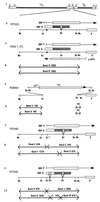
Figure 1
Figure 1
Schematic representations of sequence arrangements of recombinant virus genomes. Lines 1 and 3, representation of the HSV-1 (F) genome. The lines represent unique long (UL) and short (US) sequences that are flanked by inverted repeats ab and b′a′ and a′c′ and ca, respectively, shown as rectangles. Line 2, representation of recombinant virus R7520 (15), which contains the cytomegalovirus (CMV) epitope inserted in the DraIII site, in-frame with ORF O. The rectangular boxes and arrows represent gene domains and direction of transcription of the ORF O, ORF P, and γ134.5 genes in the inverted repeat sequence b′a′. The identical sequences in the inverted orientation map in the ab repeat. The closed circle denotes a wild-type ICP4 binding site. Line 5, the corresponding domain of R3659 (16). The StuI–BstEII sequences encoding the ORF O, ORF P, and γ134.5 genes were replaced in both repeats by the chimeric α27-tk gene (21). Line 7, the corresponding domain of the recombinant virus R7540. The α27-tk gene of the recombinant R3659 was replaced with sequences containing a mutated ICP4 binding site with a diagnostic EcoRI endonuclease site, and a CMV glycoprotein B (gB) epitope containing a diagnostic EcoRI site in the DraIII (Dr) site in-frame with ORF O. Line 9, the corresponding domain of the recombinant virus R7548. Here the α27-tk of R3659 was replaced with the CMV epitope in the DraIII (Dr) site in frame with ORF O, a mutated ICP4 binding site, and an additional CMV gB epitope inserted in the SacI site, also in-frame with ORF O. The second insertion was within the predicted ORF O gene, upstream of the sequences subsequently shown to be the ORF P coding sequences. Lines 4, 6, 8, and 10, the expected sizes of fragments detected by hybridization of the 1,800-bp NcoI fragment with electrophoretically separated digests of viral DNAs with NcoI–EcoRI (the first band set per virus), diagnostic of the ICP4 binding site mutation; or NcoI–XbaI (the second band set per virus), diagnostic of the CMV epitope tag insertion. The arrows in these lines denote restriction cleavage sites present in the respective viruses and therefore the DNA fragment boundaries. HSV-1(F) would be expected to yield bands A and B, respectively; R3659 would be expected to yield bands C and D; R7540 would be expected to yield bands E, F, and G; R7548 would be expected to yield bands H, J, K, L, and M, respectively. Dr, DraIII; St, StuI; Nc, NcoI; Bs, BstEII; Sc, SacI; Ec1, the introduced EcoRI site.
Figure 2
Figure 2
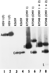
Autoradiographic image of electrophoretically separated viral DNA fragments containing sequences in the domain of the ORF O/ORF P/γ134.5 genes. Viral DNAs were digested with either NcoI and EcoRI (lanes 1, 3, 5, and 7) or NcoI and XbaI (lanes 2, 4, 6, and 8). They were then subjected to electrophoresis on a 28-cm, 0.85% agarose gel and transferred to a Zeta probe (Bio-Rad) by capillary action in 0.5 M NaOH. The membrane was rinsed in 2× SSC (0.3 M NaCl/0.015 M Na citrate), prehybridized in 30% formamide, 6× SSC, 1% milk, 1% SDS, and 100 μg single-stranded calf thymus DNA per ml for 30 min at 68°C. A total of 106 cpm of denatured, 32P-labeled pRB4794 was then added overnight and the blot was rinsed as recommended by the manufacturer. Autoradiographic images on Kodak XAR-5 film were overexposed to detect smaller fragments. The expected sizes of the fragments generated by cleavages (bands A through M) are shown in Fig. 1.
Figure 3
Figure 3
Photograph of infected cell proteins electrophoretically separated on a denaturing polyacrylamide gel and reacted with the CH28-2 mouse mAb to the CMV epitope. (A) Replicate Vero cell cultures grown in 25-cm2 flasks were infected with 10 pfu of HSV-1(F), R7519 (CMV–ORF P), or R7520 (CMV–ORF O) per cell, maintained at 37°C for 4 or 18 h (lanes 1–5) or maintained at 39.5°C for 24 h (lanes 6–8), harvested, solubilized, electrophoretically separated on 15% polyacrylamide denaturing gels, transferred to a nitrocellulose sheet, and reacted first with mouse mAb CH28-2 (20) and second with goat anti-mouse IgG conjugated to horseradish peroxidase, and horseradish peroxidase electrochemiluminescent substrate. Blots were exposed to Kodak XAR-5 film for 1 min. (B) Replicate Vero cell cultures grown in 25-cm2 flasks were infected with 10 pfu of HSV-1(F), R7520 (CMV–ORF O), or R7540 (P++/O++, CMV 1) per cell, maintained at 37°C for 2, 12, or 22 h (lanes 1–7) or maintained at 39.5°C for 22 h (lanes 8–10), harvested, solubilized, electrophoretically separated on 12.5% polyacrylamide denaturing gels, transferred to a nitrocellulose sheet, and reacted with mouse monoclonal antiserum to CMV gB, then goat anti-mouse IgG conjugated to alkaline phosphatase, and alkaline phosphatase substrate.
Figure 4
Figure 4
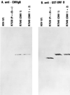
Photograph of infected cell proteins electrophoretically separated on a denaturing polyacrylamide gel and reacted with (A) rabbit polyclonal antiserum specific for GST–ORF O or with (B) mAb CH28-2. Replicate Vero cell cultures grown in 25-cm2 flasks were infected with 10 pfu of HSV-1(F), R7530 (P++/O++), R7540 (CMV 1), or R7548 (CMV 1+2) per cell, maintained at 37°C for 22 h, harvested, solubilized, electrophoretically separated on 12.5% polyacrylamide denaturing gels, transferred to a nitrocellulose sheet, and reacted with (A) rabbit polyclonal antiserum to ORF O or (B) mouse monoclonal antiserum to CMV gB followed by secondary antibodies conjugated to alkaline phosphatase as described in the legend to Fig. 3. R7540 (CMV 1) and R7548 (CMV 1+2) both have mutated ICP4 binding sites, designated (P++/O++).
Figure 5
Figure 5
(A) Autoradiographic image of [35S]methionine-labeled infected cell proteins from total cell lysates or of proteins bound to GST fusion proteins electrophoretically separated on denaturing gels. (B) Photograph of the same gel reacted with monoclonal antiserum to ICP4. Replicate HeLa cells grown in 150-cm2 flasks were mock-infected (lanes 1–4) or infected with HSV-1(F) (lanes 5–8) and maintained at 37°C. At 4 h after infection the medium was replaced with mixture 199 lacking methionine but supplemented with 1% calf serum and 50 μCi (1 Ci = 37 GBq) of [35S]methionine. After additional incubation for 4 h, the cells were harvested, solubilized by vigorous pipetting in lysis buffer (10 mM Hepes, pH 7.6/250 mM NaCl/10 mM MgCl2/1% Triton X-100/0.5 mM phenylmethylsulfonyl flouride/2 mM benzamidine). The lysates were precleared with 20 μg of GST and divided into three identical samples to which were added 10 μg of GST–ORF PC (lanes 2 and 6), GST–ORF PN (lanes 3 and 7), or GST–ORF O (lanes 4 and 8) fusion proteins, respectively, and reacted at 4°C for 3 h. Glutathione-conjugated agarose beads were then added to the mixtures and allowed to react for 30 min at 4°C. The beads were collected by low speed centrifugation and washed four times with 50 volumes of lysis buffer. The proteins adhering to the beads were then solubilized, electrophoretically separated on a denaturing 15% polyacrylamide gel, transferred to a nitrocellulose sheet, and exposed to Kodak XAR-5 film for 2 days. (B) Photograph of the blot shown in A after reaction first with mouse mAb specific for ICP4, H1114 (30) (Goodwin Cancer Research Institute), followed by goat-anti-mouse antibody conjugated to alkaline phosphatase.
Figure 6
Figure 6
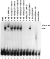
Autoradiographic image of a gel retardation assay showing the interaction of ICP4 with a high affinity DNA site in the presence or absence of GST-ORF O. Nuclear extracts (1.5 μg) were reacted with 2 × 10⁴ cpm of a 32P-labeled probe DNA in 25 μl of a solution containing 20 mM Tris (pH 7.6), 50 mM KCl, 0.05% Nonidet P-40, 5% glycerol, 1 mM EDTA, 1 mg BSA per ml, 10 mM 2-mercaptoethanol, and 3 μg poly(dI⋅dC). The DNA probes were as follows: the 112-bp MscI–SacI fragment from pRB4794 (13), which is nucleotides −87 to +25 relative to the ORF P transcription initiation site (lanes 1–11), and probeΔICP4bs (lanes 12 and 13), which is the same fragment as above from pRB4855 (14), which contains a mutated ICP4 binding site. The probes were dephosphorylated by shrimp alkaline phosphatase and 5′ labeled with [γ-32P]dATP using T4 polynucleotide kinase. The ICP4–DNA complex was supershifted by the addition of the mAb H943 to the reaction mixture as originally described by Kristie and Roizman (26). The contents of each reaction mixture is described above the corresponding lane. NE, nuclear extract; GST-ORF P, GST-ORF PC (Fig. 5). The electrophoretically separated samples in a 4% nondenaturing polyacrylamide gel were dried and exposed to Kodak XAR-5 film for 4 h.

Similar articles

Cited by

References

    1. Roizman B. Proc Natl Acad Sci USA. 1996;93:11307–11312. - PMC - PubMed
    1. Roizman B, Sears A E. In: Fields’ Virology. 3rd Ed. Fields B N, Knipe D M, Howley P, Chanock R M, Hirsch M S, Melnick J L, Monath T P, Roizman B, editors. New York: Lipcincott–Raven; 1996. pp. 2231–2295.
    1. Mitchell W J, Lirette R P, Fraser N. J Gen Virol. 1990;71:125–132. - PubMed
    1. Spivack J G, Fraser N W. J Virol. 1987;61:3841–3847. - PMC - PubMed
    1. Stevens J G, Wagner E K, Devi-Rao G B, Cook M L, Feldman L T. Science. 1987;235:1056–1059. - PubMed

Publication types