. 1997 Sep 16;94(19):10385-90.
doi: 10.1073/pnas.94.19.10385.

Yersinia signals macrophages to undergo apoptosis and YopJ is necessary for this cell death

Affiliations

Yersinia signals macrophages to undergo apoptosis and YopJ is necessary for this cell death

D M Monack et al. Proc Natl Acad Sci U S A. .

Abstract

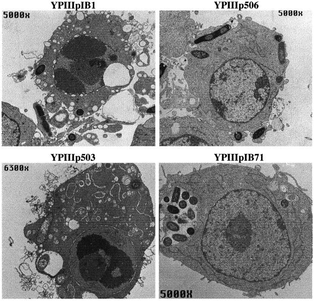
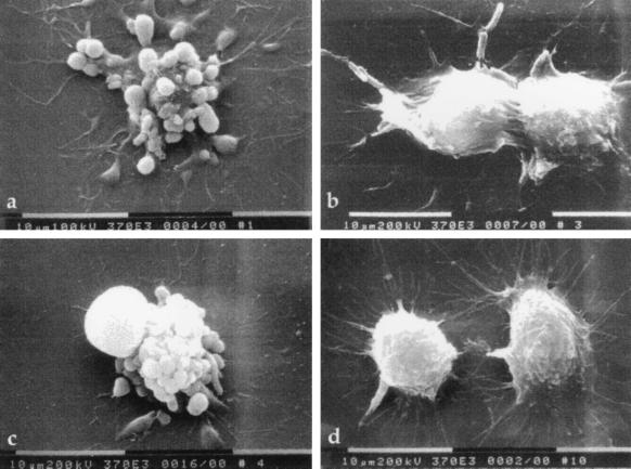
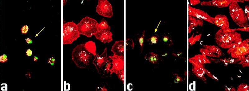
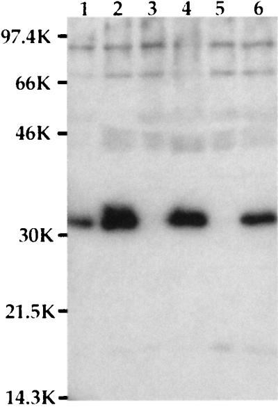
Pathogenic Yersinia spp. carry a large common plasmid that encodes a number of essential virulence determinants. Included in these factors are the Yersinia-secreted proteins called Yops. We analyzed the consequences of wild-type and mutant strains of Yersinia pseudotuberculosis interactions with the macrophage cell line RAW264. 7 and murine bone marrow-derived macrophages. Wild-type Y. pseudotuberculosis kills approximately 70% of infected RAW264.7 macrophages and marrow-derived macrophages after an 8-h infection. We show that the cell death mediated by Y. pseudotuberculosis is apoptosis. Mutant Y. pseudotuberculosis that do not make any Yop proteins no longer cause host cell death. Attachment to host cells via invasin or YadA is necessary for the cell death phenotype. Several Yop mutant strains that fail to express one or more Yop proteins were engineered and then characterized for their ability to cause host cell death. A mutant with a polar insertion in YpkA Ser/Thr kinase that does not express YpkA or YopJ is no longer able to cause apoptosis. In contrast, a mutant no longer making YopE or YopH (a tyrosine phosphatase) induces apoptosis in macrophages similar to wild type. When yopJ is added in trans to the ypkAyopJ mutant, the ability of this strain to signal programmed cell death in macrophages is restored. Thus, YopJ is necessary for inducing apoptosis. The ability of Y. pseudotuberculosis to promote apoptosis of macrophages in cell culture suggests that this process is important for the establishment of infection in the host and for evasion of the host immune response.


Figures

Figure 1
Figure 1
Transmission electron micrographs of apoptotic RAW264.7 macrophages infected with Y. pseudotuberculosis. Macrophages were infected with YPIIIpIB1 wild-type Y. pseudotuberculosis, YPIIIp506 (ypkAyopJ mutant), YPIIIp503 (yopEyopH mutant), or YPIIIpIB71 Yop mutant for 6 h. Note the chromatin condensation, cytoplasmic vacuolization, and swollen endoplasmic reticulum in macrophages infected with YPIIIpIB1 and YPIIIp503. In contrast, the macrophages infected with YPIIIp506 or YPIIIpIB71 appear normal despite the presence of bacteria.
Figure 2
Figure 2
Scanning electron micrographs of infected RAW264.7 macrophages. Macrophages were infected for 6 h with (a) wild-type Y. pseudotuberculosis YPIIIpIB1, (b) YPIIIp506, (c) YPIIIp503, or (d) uninfected control. Note membrane blebbing on the surface of cells infected with YPIIIpIB1 and YPIIIp503. Macrophages infected with YPIIIp506 appear slightly rounded compared to uninfected cells, but the membrane blebbing is absent.
Figure 3
Figure 3
Infections with (a) YPIIIpIB1, (b) YPIIIp506, (c) YPIIIp503, or (d) YPIIIpIB71 for 8 h. TUNEL reaction was used to label 3′-OH termini with fluorescein. Anti-Y. pseudotuberculosis primary antibody and anti-rabbit 7-amino-4-methylcoumarin-3-acetic acid (AMCA) secondary antibody were used to label bacteria and rhodamine phalloidin was used to label actin filaments. Images of epifluorescence were scanned into Adobe photoshop and aligned to make a composite. The arrow indicates an infected macrophage positive for TUNEL reaction.
Figure 4
Figure 4
YopJ expression. Supernatants from bacteria grown as described were precipitated with trichloroacetic acid and run on SDS/PAGE. The proteins were transferred to nitrocellulose, and the 32.5-kDa YopJ protein was detected with anti-YopJ antibody. Lanes: 1, YPIIpIB1; 2, YPIIIp503; 3, YPIIIp506; 4, YPIIIp506(pyopJ); 5, YPIIIp506(pACYC184); 6, YPIIIp507(pyopJ).

Similar articles

Cited by

References

    1. Cornelis G R, Wolf-Watz H. Mol Microbiol. 1997;23:861–867. - PubMed
    1. Isberg R R, Falkow S. Nature (London) 1985;317:262–264. - PubMed
    1. Miller V L, Falkow S. Infect Immun. 1988;56:1242–1248. - PMC - PubMed
    1. Yang Y, Merriam J J, Mueller J P, Isberg R R. Infect Immun. 1996;64:2483–2489. - PMC - PubMed
    1. Bolin I, Wolf-Watz H. Infect Immun. 1984;43:72–78. - PMC - PubMed

Publication types

Substances

LinkOut - more resources