. 1997 Sep 16;94(19):10461-6.
doi: 10.1073/pnas.94.19.10461.

RGS proteins reconstitute the rapid gating kinetics of gbetagamma-activated inwardly rectifying K+ channels

Affiliations

RGS proteins reconstitute the rapid gating kinetics of gbetagamma-activated inwardly rectifying K+ channels

C A Doupnik et al. Proc Natl Acad Sci U S A. .

Abstract

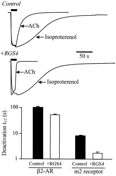
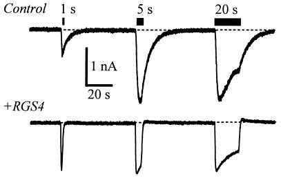
G protein-gated inward rectifier K+ (GIRK) channels mediate hyperpolarizing postsynaptic potentials in the nervous system and in the heart during activation of Galpha(i/o)-coupled receptors. In neurons and cardiac atrial cells the time course for receptor-mediated GIRK current deactivation is 20-40 times faster than that observed in heterologous systems expressing cloned receptors and GIRK channels, suggesting that an additional component(s) is required to confer the rapid kinetic properties of the native transduction pathway. We report here that heterologous expression of "regulators of G protein signaling" (RGS proteins), along with cloned G protein-coupled receptors and GIRK channels, reconstitutes the temporal properties of the native receptor --> GIRK signal transduction pathway. GIRK current waveforms evoked by agonist activation of muscarinic m2 receptors or serotonin 1A receptors were dramatically accelerated by coexpression of either RGS1, RGS3, or RGS4, but not RGS2. For the brain-expressed RGS4 isoform, neither the current amplitude nor the steady-state agonist dose-response relationship was significantly affected by RGS expression, although the agonist-independent "basal" GIRK current was suppressed by approximately 40%. Because GIRK activation and deactivation kinetics are the limiting rates for the onset and termination of "slow" postsynaptic inhibitory currents in neurons and atrial cells, RGS proteins may play crucial roles in the timing of information transfer within the brain and to peripheral tissues.


Figures

Figure 1
Figure 1
Effects of RGS1–4 on the temporal and steady-state properties of m2 receptor-evoked GIRK currents recorded from Xenopus oocytes. (a) Representative traces of ACh-evoked GIRK currents (Kir3.1/Kir3.2) recorded from oocytes not expressing (Control) or expressing RGS4 (+RGS4). (b Top) Deactivation time constants (τdeact) derived from exponential fits to the GIRK current deactivation phase from “Control” oocytes (i.e., not injected with RGS cRNA) or oocytes injected with cRNA (10 ng) for RGS1, RGS2, RGS3, or RGS4. (Middle) Activation time constants (τact) derived from exponential fits of the GIRK activation phase in oocytes expressing the various RGS proteins. (Lower) Effects of RGS proteins on “basal” GIRK currents (IK,basal, open bars) measured as the inward current induced by changing the external [K+] from 0 to 20 mM. Solid bars are the amplitudes of inward GIRK currents induced by 1 μM ACh (IK,ACh) in 20 mM external [K+]. All bars are the mean ± SEM from 12–15 oocytes from the same three batches.
Figure 2
Figure 2
ACh dose-effect relations for kinetic and steady-state GIRK gating properties. (Top) Deactivation time constants derived from exponential fits to the GIRK currents after washout of various ACh concentrations. Solid circles are values without RGS4 expression; open circles are with RGS4 expression (mean ± SEM from four oocytes of the same batch). (Middle) Activation time constant of ACh-evoked GIRK currents as a function of ACh concentration with (open circle) and without (solid circle) coexpression of RGS4. The time course for GIRK activation was fit with an exponential function plus a sloping base line. (Lower) Steady-state ACh dose-response curve for GIRK responses with (open circles) and without (solid circles) RGS4 expression. Current amplitudes were normalized to the currents evoked by 10 μM ACh for each cell (1–2 μA) and are from the same records used to derive the kinetic data shown in Top and Middle. The data (solid circles) were fitted with a Hill function (solid line) having an EC50 value of 48 nM.
Figure 3
Figure 3
RGS4 selectively accelerates the gating of Gα(i/o) receptor-coupled GIRK currents. Xenopus oocytes coexpressing the m2 receptor, β2-AR, Gαs, Kir3.1, and Kir3.2 elicit GIRK currents in response to either ACh (1 μM) or isoproterenol (1 μM). (Upper) Superimposed ACh- and isoproterenol-evoked GIRK current records are from the same oocyte either expressing (+RGS4) or not expressing RGS4 (Control), and have been normalized for comparison. (Lower) Summary of the deactivation half times (t1/2) for ACh-evoked and β2-AR-evoked GIRK currents with (open bar) and without (solid bar) RGS4 coexpression. (Bars are the mean ± SEM from 6–10 oocytes from two batches of oocytes.)
Figure 4
Figure 4
RGS4 expression in GIRK-transfected CHO cells elicits currents that mimic native atrial GIRK currents in their temporal gating properties. (a Upper) Current traces from a rat atrial myocyte and from CHO cells transfected with the m2 receptor, Kir3.1, Kir3.2, and without (control) or with (+RGS4) RGS4 cDNA. ACh (1 μM) was applied for 10 s. The time constant for solution changes was <100 ms. (Scale bars = 200 pA and 5 s.) Exponential fits to the current deactivation were used to derive τdeact. (Lower) Deactivation time constants (τdeact) for atrial (n = 6) and CHO cells transfected with (n = 22) or without (n = 23) RGS4. (Bars = mean ± SEM.) (b Upper) Activation phase of current traces for cells exposed to increasing concentrations of ACh. Current amplitudes were normalized to the currents evoked at 10 μM ACh. (Lower) Summary of the GIRK activation time constant (τact) as a function of ACh concentration obtained from at least four cells without (control) and with RGS4 coexpression (mean ± SEM). The holding potential was −80 mV.
Figure 5
Figure 5
Effects of RGS4 on GIRK current waveforms produced by varying durations of m2 receptor activation. Current traces from transfected CHO cells are as described in Fig. 4, exposed to 1 μM ACh for 1, 5, and 20 s. Holding potential, −80 mV.

References

    1. De Vries L, Mousli M, Wurmser A, Farquhar M G. Proc Natl Acad Sci USA. 1995;92:11916–11920. - PMC - PubMed
    1. Druey K M, Blumer K J, Kang V H, Kehrl J H. Nature (London) 1996;379:742–746. - PubMed
    1. Koelle M R, Horvitz H R. Cell. 1996;84:115–125. - PubMed
    1. Berman D M, Wilkie T M, Gilman A G. Cell. 1996;86:445–452. - PubMed
    1. Watson N, Linder M E, Druey K M, Kehrl J H, Blumer K J. Nature (London) 1996;383:172–177. - PubMed

Publication types

LinkOut - more resources