

Distinct contributions of high- and low-voltage-activated calcium currents to afterhyperpolarizations in cholinergic nucleus basalis neurons of the guinea pig
- PMID: 9295377
- PMCID: PMC6573441
- DOI: 10.1523/JNEUROSCI.17-19-07307.1997
Distinct contributions of high- and low-voltage-activated calcium currents to afterhyperpolarizations in cholinergic nucleus basalis neurons of the guinea pig
Abstract
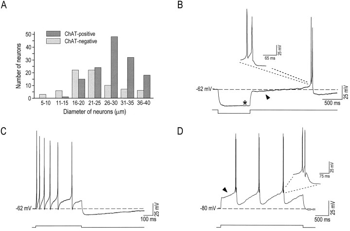
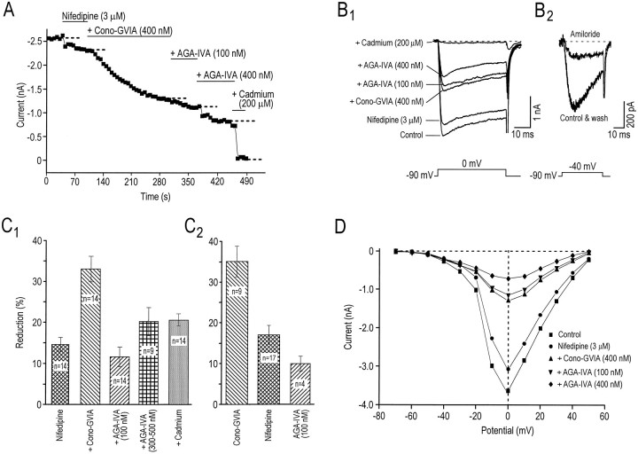
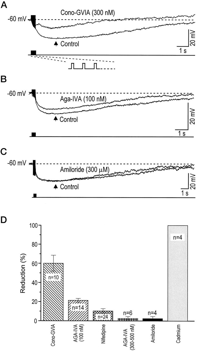
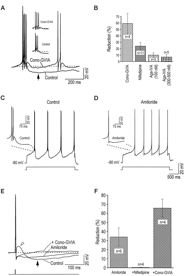
The contributions made by low- (LVA) and high-voltage-activated (HVA) calcium currents to afterhyperpolarizations (AHPs) of nucleus basalis (NB) cholinergic neurons were investigated in dissociated cells. Neurons with somata >25 microM were studied because 80% of them stained positively for choline acetyltransferase and had electrophysiological characteristics identical to those of cholinergic NB neurons previously recorded in basal forebrain slices. Calcium currents of cholinergic NB neurons first were dissected pharmacologically into an amiloride-sensitive LVA and at least five subtypes of HVA currents. Approximately 17% of the total HVA current was sensitive to nifedipine (3 microM), 35% to omega-conotoxin-GVIA (200-400 nM), 10% to omega-Agatoxin-IVA (100 nM), and 20% to omega-Agatoxin-IVA (300-500 nM), suggesting the presence of L-, N-, P-, and Q-type channels, respectively. A remaining current (R-type) resistant to these antagonists was blocked by cadmium (100-200 microM). We then assessed pharmacologically the role that LVA and HVA currents had in activating the apamin-insensitive AHP elicited by a long train of action potentials (sAHP) and the AHP evoked either by a short burst of action potentials or by a single action potential (mAHP) that is known to be apamin-sensitive. During sAHPs, approximately 60% of the hyperpolarization was activated by calcium flowing through N-type channels and approximately 20% through P-type channels, whereas T-, L-, and Q-type channels were not involved significantly. In contrast, during mAHPs, N- and T-type channels played key roles (approximately 60 and 30%, respectively), whereas L-, P-, and Q-type channels were not implicated significantly. It is concluded that in cholinergic NB neurons various subtypes of calcium channels can differentially activate the apamin-sensitive mAHP and the apamin-insensitive sAHP.
Figures
Similar articles
-
Specificity in the interaction of high-voltage-activated Ca2+ channel types with Ca2+-dependent afterhyperpolarizations in magnocellular supraoptic neurons.J Neurophysiol. 2018 Oct 1;120(4):1728-1739. doi: 10.1152/jn.00285.2018. Epub 2018 Jul 18. J Neurophysiol. 2018. PMID: 30020842 Free PMC article.
-
Biophysical and pharmacological diversity of high-voltage-activated calcium currents in layer II neurones of guinea-pig piriform cortex.J Physiol. 1999 Aug 1;518 ( Pt 3)(Pt 3):705-20. doi: 10.1111/j.1469-7793.1999.0705p.x. J Physiol. 1999. PMID: 10420008 Free PMC article.
-
Control of spontaneous firing patterns by the selective coupling of calcium currents to calcium-activated potassium currents in striatal cholinergic interneurons.J Neurosci. 2005 Nov 2;25(44):10230-8. doi: 10.1523/JNEUROSCI.2734-05.2005. J Neurosci. 2005. PMID: 16267230 Free PMC article.
-
Calcium channel currents in acutely dissociated intracardiac neurons from adult rats.J Neurophysiol. 1997 Apr;77(4):1769-78. doi: 10.1152/jn.1997.77.4.1769. J Neurophysiol. 1997. PMID: 9114235
-
Whole-cell and single-channel calcium currents in guinea pig basal forebrain neurons.J Neurophysiol. 1994 Jun;71(6):2359-76. doi: 10.1152/jn.1994.71.6.2359. J Neurophysiol. 1994. PMID: 7931521
Cited by
-
Similar nicotinic excitability responses across the developing hippocampal formation are regulated by small-conductance calcium-activated potassium channels.J Neurophysiol. 2018 May 1;119(5):1707-1722. doi: 10.1152/jn.00426.2017. Epub 2018 Jan 31. J Neurophysiol. 2018. PMID: 29384449 Free PMC article.
-
Recruitment of apical dendritic T-type Ca2+ channels by backpropagating spikes underlies de novo intrinsic bursting in hippocampal epileptogenesis.J Physiol. 2007 Apr 15;580(Pt. 2):435-50. doi: 10.1113/jphysiol.2007.127670. Epub 2007 Feb 1. J Physiol. 2007. PMID: 17272342 Free PMC article.
-
Ca(2+) signaling by T-type Ca(2+) channels in neurons.Pflugers Arch. 2009 Mar;457(5):1161-72. doi: 10.1007/s00424-008-0582-6. Epub 2008 Sep 11. Pflugers Arch. 2009. PMID: 18784939 Review.
-
Enhanced calcium buffering in F344 rat cholinergic basal forebrain neurons is associated with age-related cognitive impairment.J Neurophysiol. 2009 Oct;102(4):2194-207. doi: 10.1152/jn.00301.2009. Epub 2009 Aug 12. J Neurophysiol. 2009. PMID: 19675291 Free PMC article.
-
Chronic Intermittent Ethanol Exposure Dysregulates Nucleus Basalis Magnocellularis Afferents in the Basolateral Amygdala.eNeuro. 2022 Nov 15;9(6):ENEURO.0164-22.2022. doi: 10.1523/ENEURO.0164-22.2022. Print 2022 Nov-Dec. eNeuro. 2022. PMID: 36280288 Free PMC article.
References
-
- Akaike N. T-type calcium channel in mammalian CNS neurones. Comp Biochem Physiol [C] 1991;98:31–40. - PubMed
-
- Alonso A, Khateb A, Fort P, Jones BE, Mühlethaler M. Differential oscillatory properties of cholinergic and non-cholinergic nucleus basalis neurons in guinea pig brain slice. Eur J Neurosci. 1996;8:169–182. - PubMed
-
- Bertrand D, Buisson B, Krause RM, Bertrand S. Electrophysiology: a method to investigate the functional properties of ligand-gated channels. J Recept Signal Transduct Res. 1997;17:227–242. - PubMed
Publication types
MeSH terms
Substances
LinkOut - more resources
Full Text Sources