Androgen mitigates axotomy-induced decreases in calbindin expression in motor neurons
- PMID: 9295385
- PMCID: PMC6573455
- DOI: 10.1523/JNEUROSCI.17-19-07396.1997
Androgen mitigates axotomy-induced decreases in calbindin expression in motor neurons
Abstract
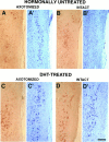
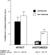
Androgens can rescue axotomized motor neurons from cell death. Here we examine a possible mechanism for this trophic action in juvenile Xenopus laevis: regulation of a calcium-binding protein, calbindin, after axotomy. Western analysis revealed that a monoclonal antibody to calbindin D specifically recognizes a single approximately 28 kDa band in X. laevis CNS and rat cerebellum. Retrograde transport of peroxidase combined with immunohistochemistry demonstrated that somata, axons, and synaptic terminals of laryngeal motor neurons in nucleus (N.) IX-X of X. laevis are calbindin-positive. The number of calbindin-positive cells was compared in the intact and axotomized sides of N.IX-X of gonadectomized males that were either hormonally untreated or DHT-treated for 1 month. Although axotomy decreased the number of calbindin-positive cells by 86% in hormonally untreated males, the decrease was only 56% in DHT-treated animals. Compared with hormonally untreated animals, the number of calbindin-labeled cells in N.IX-X of DHT-treated males was increased in both the intact (14%) and axotomized sides (75%). We conclude that axotomy decreases and that DHT enhances calbindin immunoreactivity in N.IX-X. Axotomy-induced decrease in calbindin immunoreactivity precedes cell loss in N.IX-X and may impair the capacity of motor neurons to regulate cytoplasmic calcium. Androgen-mediated maintenance of calbindin expression is thus a candidate cellular mechanism for trophic maintenance of hormone target neurons.
Figures


Similar articles
-
Trophic effects of androgen: receptor expression and the survival of laryngeal motor neurons after axotomy.J Neurosci. 1996 Nov 1;16(21):6625-33. doi: 10.1523/JNEUROSCI.16-21-06625.1996. J Neurosci. 1996. PMID: 8824303 Free PMC article.
-
Axotomy induces transient calbindin D28K immunoreactivity in hypoglossal motoneurons in vivo.Cell Calcium. 1997 Nov;22(5):367-72. doi: 10.1016/s0143-4160(97)90021-x. Cell Calcium. 1997. PMID: 9448943
-
Trophic effects of androgen: development and hormonal regulation of neuron number in a sexually dimorphic vocal motor nucleus.J Neurobiol. 1999 Sep 5;40(3):375-85. J Neurobiol. 1999. PMID: 10440737
-
Differential expression of calbindin and calmodulin in motoneurons after hypoglossal axotomy.Brain Res. 1998 Mar 9;786(1-2):181-8. doi: 10.1016/s0006-8993(97)01458-3. Brain Res. 1998. PMID: 9555004
-
Androgen receptor mRNA expression in Xenopus laevis CNS: sexual dimorphism and regulation in laryngeal motor nucleus.J Neurobiol. 1996 Aug;30(4):556-68. doi: 10.1002/(SICI)1097-4695(199608)30:4<556::AID-NEU10>3.0.CO;2-D. J Neurobiol. 1996. PMID: 8844518
Cited by
-
Increased T-type Ca2+ channel activity as a determinant of cellular toxicity in neuronal cell lines expressing polyglutamine-expanded human androgen receptors.Mol Cell Biochem. 2000 Jan;203(1-2):23-31. doi: 10.1023/a:1007010020228. Mol Cell Biochem. 2000. PMID: 10724329
-
Afferent input is necessary for seasonal growth and maintenance of adult avian song control circuits.J Neurosci. 2001 Apr 1;21(7):2320-9. doi: 10.1523/JNEUROSCI.21-07-02320.2001. J Neurosci. 2001. PMID: 11264307 Free PMC article.
-
Neuroprotective actions of androgens on motoneurons.Front Neuroendocrinol. 2009 Jul;30(2):130-41. doi: 10.1016/j.yfrne.2009.04.005. Epub 2009 Apr 23. Front Neuroendocrinol. 2009. PMID: 19393684 Free PMC article. Review.
-
Gonadal steroid attenuation of developing hamster facial motoneuron loss by axotomy: equal efficacy of testosterone, dihydrotestosterone, and 17-beta estradiol.J Neurosci. 2005 Apr 20;25(16):4004-13. doi: 10.1523/JNEUROSCI.5279-04.2005. J Neurosci. 2005. PMID: 15843602 Free PMC article.
-
A neuroendocrine basis for the hierarchical control of frog courtship vocalizations.Front Neuroendocrinol. 2011 Aug;32(3):353-66. doi: 10.1016/j.yfrne.2010.12.006. Epub 2010 Dec 28. Front Neuroendocrinol. 2011. PMID: 21192966 Free PMC article. Review.
References
-
- Abercrombie M. Estimation of nuclear population from microtome sections. Anat Rec. 1946;94:239–247. - PubMed
-
- Baimbridge KG, Celio MR, Rogers JH. Calcium-binding proteins in the nervous system. Trends Neurosci. 1992;15:303–308. - PubMed
-
- Bao-Kuan H, Alexianu ME, Colom LV, Mohamed AH, Serrano F, Appel SH. Expression of calbindin-D28k in motoneuron hybrid cells after retroviral infection with calbindin-D28k cDNA prevents amyotrophic lateral sclerosis Ig-G-mediated cytotoxicity. Proc Natl Acad Sci USA. 1996;93:6796–6801. - PMC - PubMed
-
- Barr ML, Hamilton JD. A quantitative study of certain morphological changes in spinal motoneurons during axon reaction. J Comp Neurol. 1948;89:93–121. - PubMed
-
- Beato M, Herrlich P, Schütz G. Steroid hormone receptors: many actors in search of a plot. Cell. 1995;83:851–857. - PubMed
Publication types
MeSH terms
Substances
Grants and funding
LinkOut - more resources
Full Text Sources
