. 1997 Oct 1;17(19):7425-32.
doi: 10.1523/JNEUROSCI.17-19-07425.1997.

The swiss cheese mutant causes glial hyperwrapping and brain degeneration in Drosophila

Affiliations

The swiss cheese mutant causes glial hyperwrapping and brain degeneration in Drosophila

D Kretzschmar et al. J Neurosci. .

Abstract

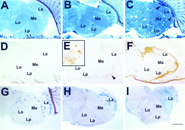
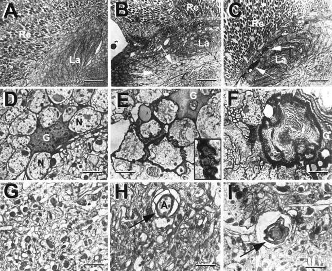
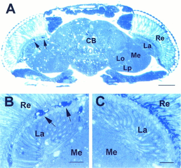
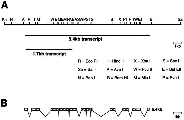
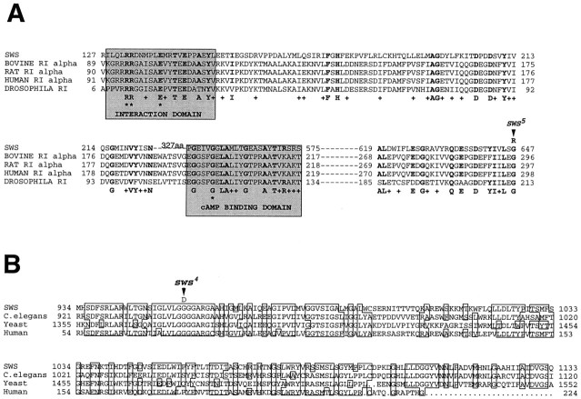
Swiss cheese (sws) mutant flies develop normally during larval life but show age-dependent neurodegeneration in the pupa and adult and have reduced life span. In late pupae, glial processes form abnormal, multilayered wrappings around neurons and axons. Degeneration first becomes evident in young flies as apoptosis in single scattered cells in the CNS, but later it becomes severe and widespread. In the adult, the number of glial wrappings increases with age. The sws gene is expressed in neurons in the brain cortex. The conceptual 1425 amino acid protein shows two domains with homology to the regulatory subunits of protein kinase A and to conceptual proteins of yet unknown function in yeast, worm, and human. Sequencing of two sws alleles shows amino acid substitutions in these two conserved domains. It is suggested that the novel SWS protein plays a role in a signaling mechanism between neurons and glia that regulates glial wrapping during development of the adult brain.


Figures

Fig. 1.
Fig. 1.
Progression of brain degeneration insws flies. A–C, One micrometer horizontal plastic head sections stained with toluidine blue. Vacuolization and darkly stained structures (arrowheadsin C) increase with advancing age. These are accompanied by a thinning of the cortex (long arrow inC). A, Wild type, age 20 d (arrows indicate white areas in the lobula plate that are not vacuoles but cross-sections of giant fibers). B, sws1 5 d old. C, sws1 20 d old. D–F,Apoptosis staining on 8 μm horizontal cryosections. D,Wild type 20 d old. E, In a 5-d-oldsws1 fly, single cells are intensely stained (arrowhead and inset). Single dying cells are already indicated by the dark toluidine blue staining in B (arrows). F, In a 20-d-old sws1 fly, there is widespread staining, suggesting apoptosis in most cortical cell bodies.G–I, Effect of degeneration on LacZ-expressing cells of the glial-specific enhancer–trap line rC56: 1 μm horizontal plastic brain sections stained with X-galactosidase (X-Gal).G, Heterozygous rC56/wild-type fly 10 d old. H, 8-d-oldsws4/y;rC56/wild-type fly. The number of cells showing LacZ staining is decreased. I, Fourteen-day-oldsws4/y;rC56/wild-type brain. The LacZ staining is almost completely absent, indicating loss of expression or degeneration of many glial cells. Brains were dissected and stained with X-Gal and then embedded in plastic, sectioned, and counterstained with pyronin Y. All flies were raised at 25°C. La, Lamina; Me, medulla;Lo, lobula; Lp, lobula plate. Anterior is at the top. Scale bar: A–I, 50 μm;inset in E, 15 μm.
Fig. 2.
Fig. 2.
Multilayered glial membranes in swsmutant brains as seen in EM horizontal sections. A, D, G, Wild type. A, Retina and lamina.D, Cell bodies of the lamina cortex, showing a glial cell recognizable by its dark cytoplasm. Extensions of the glial cell surround various neurons with single-layered wrappings.G, Medulla neuropil, a tangle of synapses. B, E, H, In newly eclosed sws1flies, glial processes form multiple sheaths around neurons in the cortex (arrows in B, inset inE) and around axons in the neuropil (arrow inH). C–I, In 7-d-oldsws1 flies, large membrane whorls are visible (F, I). These are especially prominent in the lamina cortex (arrows in C). Flies were kept at 25°C. Re, Retina, La, lamina; G, glial cell; N, neuron;A, axon. Scale bars: A–C, 3 μm;D–I, 0.5 μm.
Fig. 3.
Fig. 3.
Survival curves for the five swsalleles at 29°C. At least 200 flies were tested per allele.
Fig. 4.
Fig. 4.
Head section ofsws4 mosaic fly, 8 d old.A, Left side, B, Darkly stained bodies characteristic of the sws mutation (arrows). These are missing toward the wild-type side at the right (A, C). The few vacuoles on the wild-type side may be caused by mutant neurons projecting from the mutant side to the wild-type side.Re, Retina; La, lamina;Me, medulla; CB, central brain;Lo, lobula; Lp, lobula plate. Scale bars:A, 50 μm; B, C, 10 μm.
Fig. 5.
Fig. 5.
Genomic map of the sws/olfE region and structure of the cDNA. A, Top line, Restriction map of the genomic fragment used for the rescue experiment. The two transcripts are indicated below. B, Exon–intron structure of the 5.4 kb transcript. The open reading frame isstriped.
Fig. 6.
Fig. 6.
In situ hybridization ofsws RNA on wild-type cryosections. A, The antisense probe derived from pBS-E75 detects sws mRNA in the entire brain cortex, suggesting widespread expression in cortical cell bodies. B, Control, using a sense RNA probe.Re, Retina; La, lamina;Me, medulla; Lo, lobula;Lp, lobula plate. Scale bar, 50 μm.
Fig. 7.
Fig. 7.
Conceptual sequence of the SWS protein. Possible transmembrane domains are underlined. The locations of the sws1, sws4, andsws5 mutations are identified byarrows.
Fig. 8.
Fig. 8.
Comparison of the SWS conceptual protein with other, known sequences. A, Similarity of SWS to regulatory subunit type Iα of cAMP-dependent protein kinase from various species. The domains interacting with the catalytic subunit and the cAMP-binding site are shaded. The two arginines and the glutamic acid in the interaction domain (asterisks) are the only amino acids shared by all the types of regulatory subunits (RIα, RIβ, RIIα, and RIIβ). One of the important amino acids in the cAMP-binding domain is the conserved glycine (asterisk), which, when mutated in mouse lymphoma cells, inactivates the binding site (Taylor et al., 1990). Amino acids conserved between SWS and at least one of the regulatory subunits are shown in boldface; similar ones are indicated by aplus. The amino acid substitution insws5 is indicated (arrow). B, Similarity of a second segment of SWS to conceptual proteins from sequencing projects onSaccharomyces cerevisiae, C. elegans, and human. Amino acids conserved in at least three of the sequences areboxed. The amino acid substitution insws4 is indicated (arrow).

Similar articles

Cited by

References

    1. Altschul S, Gish W, Miller W, Myers EW, Lipman DJ. Basic local alignment search tool. J Mol Biol. 1990;215:403–410. - PubMed
    1. Ashburner M. Drosophila: a laboratory manual. Cold Spring Harbor Laboratory; Cold Spring Harbor, NY: 1989.
    1. Barres BA, Raff MC. Control of oligodendrocyte number in the developing rat optic nerve. Neuron. 1994;12:935–942. - PubMed
    1. Bergoffen J, Scherer SS, Wang S, Scott MO, Bone LJ, Paul DL, Chen K, Lensch MW, Chance PF, Fischbeck KH. Connexin mutations in X-linked Charcot-Marie-Tooth disease. Science. 1993;262:2039–2042. - PubMed
    1. Brendel V, Bucher P, Nourbakhsh I, Blaisdell BE, Karlin S. Methods and algorithms for statistical analysis of protein sequences. Proc Natl Acad Sci USA. 1992;89:2002–2006. - PMC - PubMed

Publication types

Associated data

LinkOut - more resources