. 1997 Oct 1;17(19):7532-40.
doi: 10.1523/JNEUROSCI.17-19-07532.1997.

Rapid seizure-induced reduction of benzodiazepine and Zn2+ sensitivity of hippocampal dentate granule cell GABAA receptors

Affiliations

Rapid seizure-induced reduction of benzodiazepine and Zn2+ sensitivity of hippocampal dentate granule cell GABAA receptors

J Kapur et al. J Neurosci. .

Abstract

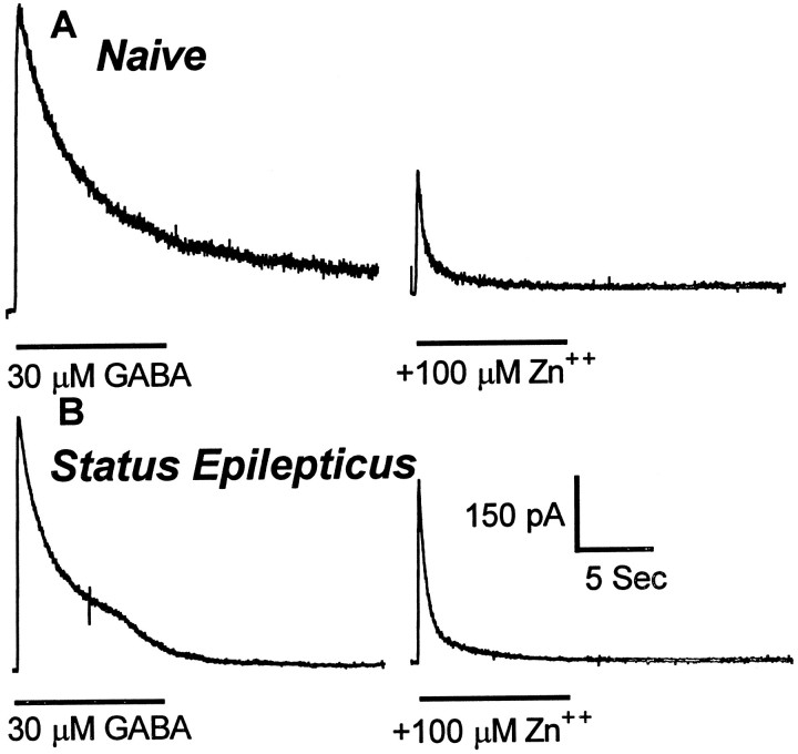
Fast synaptic inhibition in the forebrain is mediated primarily by GABA acting on GABAA receptors (GABARs). GABARs are regulated by numerous positive (barbiturates, benzodiazepines, and neurosteroids) and negative (picrotoxin, bicuculline, and Zn2+) allosteric modulators. The sensitivity of GABARs to GABA and to allosteric modulators changes gradually during normal development, during development of chronic epilepsy, and after prolonged exposure to GABAR agonists. Here we report the development of rapid functional plasticity of GABARs occurring over 45 min of continuous seizures (status epilepticus) in rats. Seizures induced in rats by administration of lithium followed by pilocarpine were readily terminated by the benzodiazepine diazepam when administered early during the seizures (after 10 min of seizures). However, during status epilepticus, there was a substantial reduction of diazepam potency for termination of the seizures. To determine whether the loss of sensitivity of the animals to diazepam was caused by an alteration of GABAR functional properties, we obtained whole-cell GABAR currents from hippocampal dentate granule cells isolated acutely from control rats and from rats undergoing status epilepticus. GABAR properties were characterized by determining GABA sensitivity and the sensitivity of GABARs to regulation by benzodiazepines, barbiturates, and Zn2+. When compared with those from naive controls, GABAR currents from rats undergoing status epilepticus were less sensitive to diazepam and Zn2+ but retained their sensitivity to GABA and pentobarbital. We conclude that the prolonged seizures of status epilepticus rapidly altered the functional properties of hippocampal dentate granule cell GABARs.


Figures

Fig. 1.
Fig. 1.
Diazepam was effective in controlling brief (10 min) seizures but lost efficacy after prolonged (45 min) seizures. Seizures were induced in 70–150 gm rats by intraperitoneal injection of LiCl at 3 mEq/kg followed 16–24 hr later by intraperitoneal injection of pilocarpine at 50 mg/kg. Behavioral seizures started within 1–5 min in all rats. Diazepam was administered 10 min (filled boxes, solid line;n = 14) or 45 min (filled circles, dashed line; n = 12) after pilocarpine injection. The percent of rats that stopped having seizures within 5 min of diazepam injection was plotted against the log of the diazepam dose. The data were fitted to a sigmoidal dose–response curve with the maximum fixed to 100% and the minimum to 0%. The ED50 values were derived from the equation that best fit the data.
Fig. 2.
Fig. 2.
Stabilization of GABAR currents after access. GABAR currents elicited immediately on access from dentate granule cells. Traces were from two neurons, thetop from a cell isolated from a control animal and thebottom from an animal undergoing status epilepticus. The durations of GABA application were indicated by horizontal bars. Two minutes elapsed between each GABA application.A, GABAR currents elicited from hippocampal dentate granule cells isolated from control animals rapidly increased to a relatively stable amplitude. B, GABAR currents elicited from hippocampal dentate granule cells isolated from animals undergoing status epilepticus took longer to stabilize and showed a greater increase in amplitude.
Fig. 3.
Fig. 3.
Run-up of GABAR currents after access. Granule cell GABAR peak currents were normalized to the initial current evoked by 10 μm GABA after access. Means ± SEMs of peak normalized GABAR currents from five neurons from animals undergoing status epilepticus and four neurons from control animals were plotted.
Fig. 4.
Fig. 4.
GABA concentration dependency. GABA concentration and normalized GABAR peak current relationships were plotted for 17 neurons isolated from control animals and for 9 neurons isolated from animals undergoing status epilepticus. Concentration–response data were obtained after stabilization of currents. Eachpoint represented the mean of normalized peak currents, and the error bars showed SEMs. The line was the best fit of data to a sigmoidal function. The EC50 and Imax were derived from the equation for the sigmoidal function that best fitted the data.
Fig. 5.
Fig. 5.
Diazepam enhancement of GABAR currents in dentate granule cells from control animals and in cells isolated from rats after 45 min of seizures. Diazepam at 300 nm enhanced GABAR current in dentate granule cells from control animals but did not enhance current in cells isolated from rats after 45 min of seizures. The traces are from two different neurons.Horizontal bars showed the duration of application of the drug. A, Diazepam (300 nm) was applied with 10 μm GABA to a dentate granule cell from a control animal. B, Diazepam (300 nm) was applied with 6 μm GABA to a granule cell isolated from a rat after status epilepticus. A lower concentration of GABA was used to compensate for a small leftward shift of the GABA concentration–response curve in cells from animals undergoing status epilepticus (equipotent GABA concentration).
Fig. 6.
Fig. 6.
Diazepam concentration–dentate granule cell GABAR current enhancement relationships. Diazepam concentration–response curves were obtained for neurons isolated from control animals (filled boxes, solid line;n = 9) and for neurons isolated from animals undergoing status epilepticus (filled circles,dashed line; n = 12). Higher concentrations of diazepam inhibited GABAR current as reported previously (De Deyn and Macdonald, 1988).
Fig. 7.
Fig. 7.
Zn2+ inhibition of GABAR currents in dentate granule cells from control animals and from animals undergoing status epilepticus. Zn2+ (100 μm) inhibited GABAR currents in dentate granule cells from control animals more than it did in granule cells from animals undergoing status epilepticus. The traces are from two different neurons. Zn2+ (100 μm) was coapplied with 30 μm GABA. Horizontal barsshow the duration of application of the drug. A,Traces from a dentate granule cell isolated from a control animal. B, Traces from a granule cell isolated from an animal undergoing status epilepticus.
Fig. 8.
Fig. 8.
Zn2+ concentration–dentate granule cell GABAR current reduction relationships. Zn2+ concentration–dentate granule cell GABAR current inhibition relationships were obtained from neurons isolated from control animals (filled boxes, solid line; n = 12) and from neurons isolated from animals undergoing status epilepticus (filled circles, dashed line; n = 12). The lines were the best fit of the data to a sigmoidal function. The IC50 and Hill slope (nH) were derived from the equation for the sigmoidal function that best fitted the data.
Fig. 9.
Fig. 9.
Pentobarbital enhancement of GABAR currents from dentate granule cells from control animals and from cells isolated from animals undergoing status epilepticus. Pentobarbital (30 μm) equally enhanced GABAR currents in dentate granule cells from control animals and in granule cells from animals undergoing status epilepticus. The traces are from two different neurons. Pentobarbital (30 μm) was coapplied with 10 μm GABA. Horizontal bars show the duration of application of the drug. A, Tracesfrom a dentate granule cell isolated from a control animal.B, Traces from a granule cell isolated from an animal undergoing status epilepticus.
Fig. 10.
Fig. 10.
Pentobarbital concentration–dentate granule cell GABAR current enhancement relationships. Pentobarbital concentration–dentate granule cell GABAR current enhancement relationships were obtained for neurons isolated from control animals (filled boxes, solid line;n = 7) and for neurons isolated from animals undergoing status epilepticus (filled circles,dashed line; n = 6). The lines were the best fit of the data to a sigmoidal function. The EC50and Hill slope were derived from the equation for the sigmoidal function that best fitted the data.

References

    1. Akaike N, Hattori K, Inomata N, Oomura Y. Gamma-Aminobutyric-acid- and pentobarbitone-gated chloride currents in internally perfused frog sensory neurones. J Physiol (Lond) 1985;360:367–386. - PMC - PubMed
    1. Bertram EH, Lothman EW. Morphometric effects of intermittent kindled seizures and limbic status epilepticus in the dentate gyrus of the rat. Brain Res. 1993;603:25–31. - PubMed
    1. Buhl EH, Otis TS, Mody I. Zinc-induced collapse of augmented inhibition by GABA in a temporal lobe epilepsy model. Science. 1996;271:369–373. - PubMed
    1. Cavazos JE, Das I, Sutula TP. Neuronal loss induced in limbic pathways by kindling: evidence for induction of hippocampal sclerosis by repeated brief seizures. J Neurosci. 1994;14:3106–3121. - PMC - PubMed
    1. Chen QX, Wong RK. Intracellular Ca2+ suppressed a transient potassium current in hippocampal neurons. J Neurosci. 1991;11:337–343. - PMC - PubMed

Publication types

MeSH terms