. 1997 Sep 22;138(6):1181-92.
doi: 10.1083/jcb.138.6.1181.

Transportin-mediated nuclear import of heterogeneous nuclear RNP proteins

Affiliations

Transportin-mediated nuclear import of heterogeneous nuclear RNP proteins

M C Siomi et al. J Cell Biol. .

Abstract

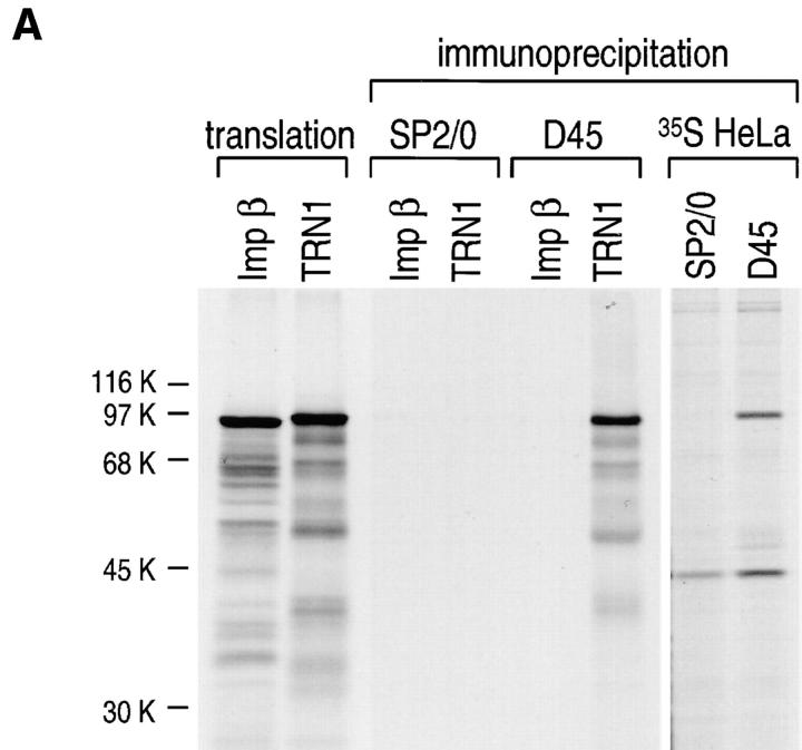
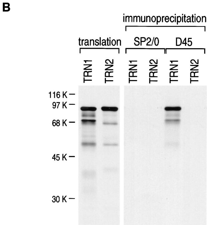
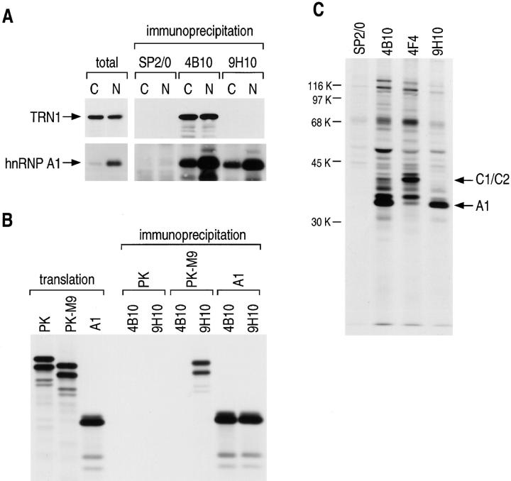
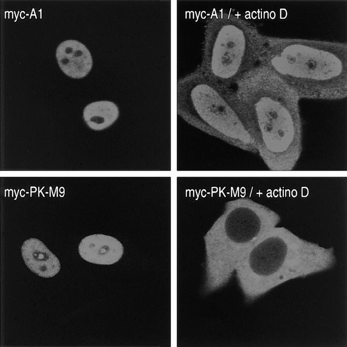
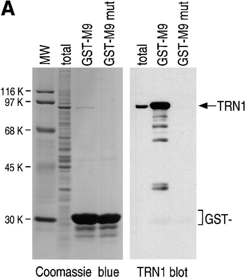
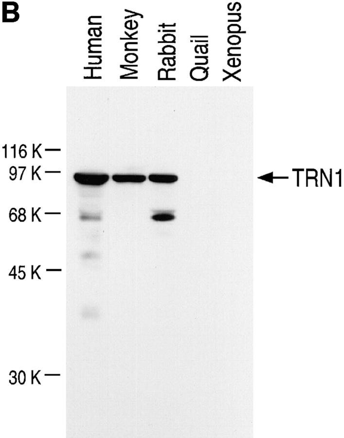
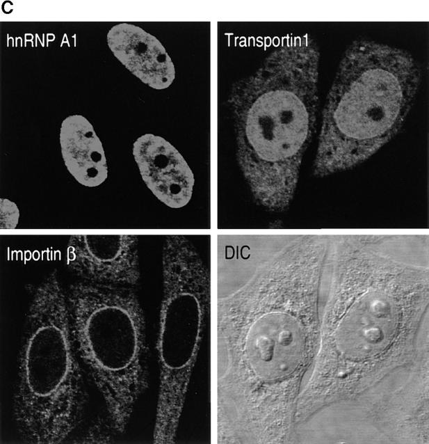
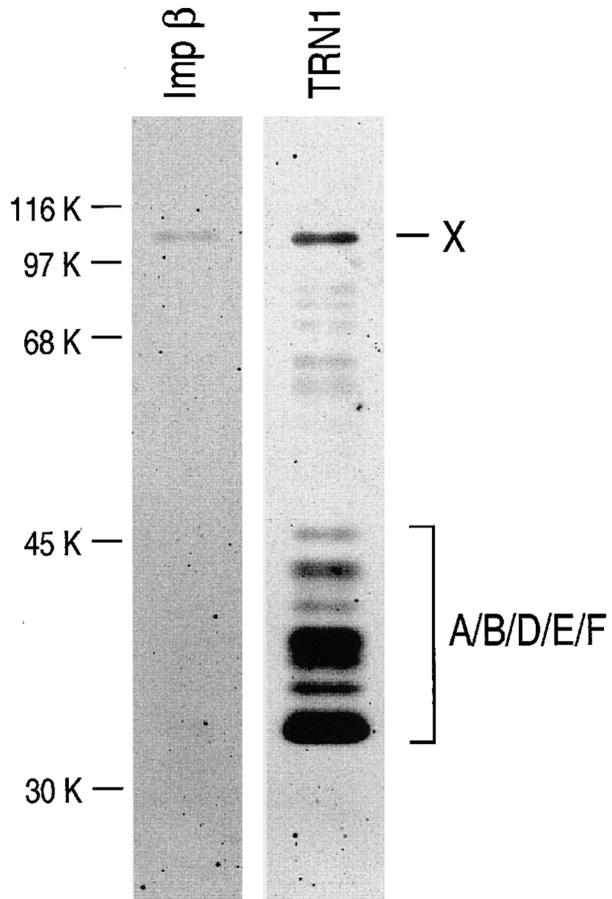
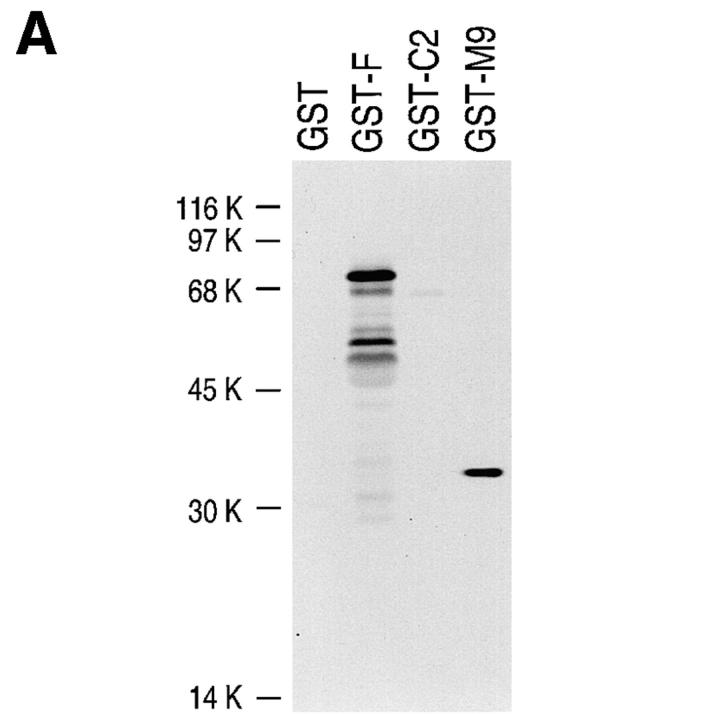
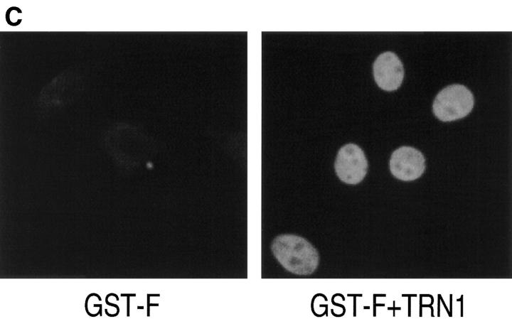
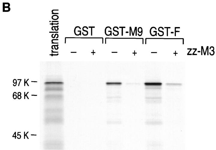
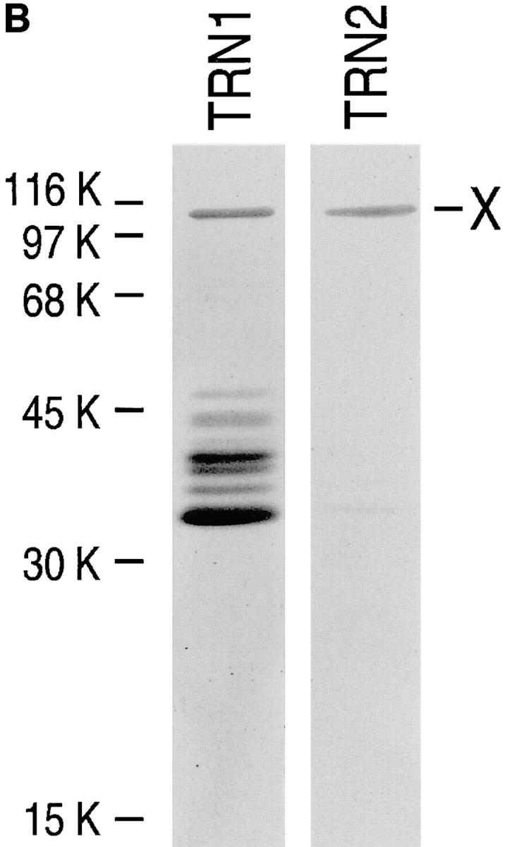
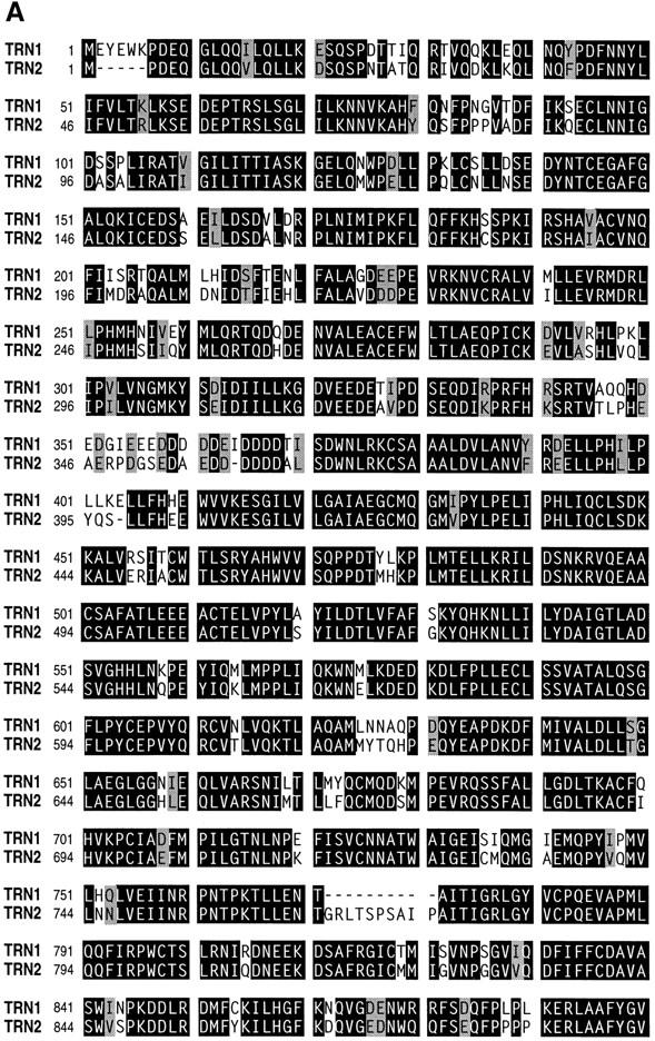
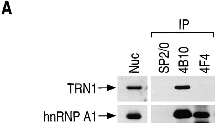
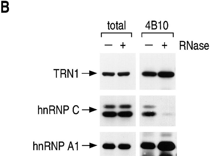
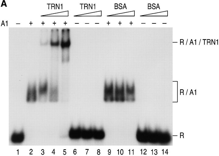
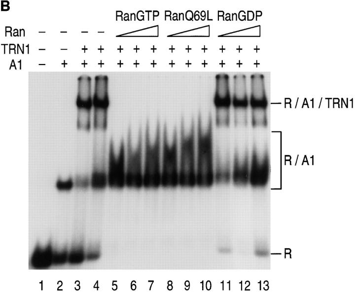
Heterogeneous nuclear ribonucleoprotein (hnRNP) A1 is an abundant nuclear protein that plays an important role in pre-mRNA processing and mRNA export from the nucleus. A1 shuttles rapidly between the nucleus and the cytoplasm, and a 38-amino acid domain, M9, serves as the bidirectional transport signal of A1. Recently, a 90-kD protein, transportin, was identified as the mediator of A1 nuclear import. In this study, we show that transportin mediates the nuclear import of additional hnRNP proteins, including hnRNP F. We have also isolated and sequenced a novel transportin homolog, transportin2, which may differ from transportin1 in its substrate specificity. Immunostaining shows that transportin1 is localized both in the cytoplasm and the nucleoplasm, and nuclear rim staining is also observed. The nuclear localization of A1 is dependent on ongoing RNA polymerase II transcription. Interestingly, a pyruvate kinase-M9 fusion, which normally localizes in the nucleus, also accumulates in the cytoplasm when RNA polymerase II is inhibited. Thus, M9 itself is a specific sensor for transcription-dependent nuclear transport. Transportin1-A1 complexes can be isolated from the cytoplasm and the nucleoplasm, but transportin1 is not detectable in hnRNP complexes. RanGTP causes dissociation of A1-transportin1 complexes in vitro. Thus, it is likely that after nuclear import, A1 dissociates from transportin1 by RanGTP and becomes incorporated into hnRNP complexes, where A1 functions in pre-mRNA processing.


Figures

Figure 4
Figure 4
Specificity of the mono-clonal antibody for transportin1, D45. (A) Transportin1 (TRN1) and importin β (Impβ) were transcribed–translated in vitro in the presence of [35S]methionine (translation). Immunoprecipitations were carried out with D45 and SP2/0 (as a control) in the presence of the ionic detergent EmpigenBB, and the bound fraction of the translated products was analyzed by SDS-PAGE and visualized by fluorography (immunoprecipitation). Products of transcription–translation reaction are shown as translation. Additional immunoprecipitation was carried out from total HeLa extract labeled with [35S]methionine under the same conditions. Note that D45 reacts specifically with transportin1 and does not cross-react to importin β. The positions of molecular mass markers are indicated on the left. (B) Immunoprecipitation of transportins 1 and 2 with D45. Immunoprecipitation was carried out with D45 using transportins 1 (TRN1) and 2 (TRN2) transcribed–translated in vitro in the presence of [35S]methionine (translation) as described above. D45 does not cross-react to transportin2.
Figure 4
Figure 4
Specificity of the mono-clonal antibody for transportin1, D45. (A) Transportin1 (TRN1) and importin β (Impβ) were transcribed–translated in vitro in the presence of [35S]methionine (translation). Immunoprecipitations were carried out with D45 and SP2/0 (as a control) in the presence of the ionic detergent EmpigenBB, and the bound fraction of the translated products was analyzed by SDS-PAGE and visualized by fluorography (immunoprecipitation). Products of transcription–translation reaction are shown as translation. Additional immunoprecipitation was carried out from total HeLa extract labeled with [35S]methionine under the same conditions. Note that D45 reacts specifically with transportin1 and does not cross-react to importin β. The positions of molecular mass markers are indicated on the left. (B) Immunoprecipitation of transportins 1 and 2 with D45. Immunoprecipitation was carried out with D45 using transportins 1 (TRN1) and 2 (TRN2) transcribed–translated in vitro in the presence of [35S]methionine (translation) as described above. D45 does not cross-react to transportin2.
Figure 8
Figure 8
M9 is not accessible in hnRNP complexes. (A) Cytoplasmic (C) and nucleoplasmic (N) fractions were prepared from HeLa cells, and immunoprecipitations were carried out with 4B10 and 9H10 (anti-hnRNP A1 antibodies). Note that 9H10 can immunoprecipitate A1 (hnRNP A1); however, transportin1 (TRN1) is not detectable in the 9H10 immunoprecipitates from either compartment. (B) Epitope mapping of 4B10 and 9H10. The in vitro transcription– translation was carried out for PK, PK-M9, and full length hnRNP A1 (A1) in the presence of [35S]methionine (translation), and immunoprecipitation was performed using 4B10 and 9H10 in the presence of EmpigenBB. The bound fraction was analyzed by SDS-PAGE and visualized by fluorography. Both antibodies are capable of immunoprecipitating full length A1, but only 9H10 can immunoprecipitate PK-M9, indicating that the epitope of 9H10 is within the M9 region of A1. (C) 9H10 does not immunoprecipitate hnRNP complexes. Immunoprecipitations were carried out from the nucleoplasmic fraction of HeLa cells labeled with [35S]methionine using 4B10, 4F4, and 9H10. After immunoprecipitation, all proteins were analyzed by SDS-PAGE and visualized by fluorography. Proteins corresponding to hnRNP C1/C2 proteins are not observed in the 9H10 immunoprecipitate, indicating that 9H10 can not immunoprecipitate hnRNP complexes.
Figure 6
Figure 6
M9 confers the transcription sensitivity to nuclear localization of A1. Transfection of HeLa cells was carried out with either myc-full length A1 (myc-A1) or myc–PK-M9 (Siomi and Dreyfuss, 1995), and the transfected cells were then incubated in the presence (+ actino D) or absence of actinomycin D (5 μg/ml) for 4 h. Afterwards, immunofluorescence microscopy was carried out using an anti-myc antibody as described in Fig. 5 C.
Figure 5
Figure 5
(A) M9-containing protein specifically interacts with transportin1 among all the cytoplasmic proteins from HeLa cells. GST-M9 or the import-defective GST-M9 mutant (G274 to A; Michael et al., 1995b ) on glutathione-Sepharose (both indicated by GST-) was incubated with the cytoplasmic fraction from HeLa cells in the presence of 400 mM NaCl. The total HeLa cytoplasmic fraction and the bound fraction to the GST-fusion proteins were analyzed by SDS-PAGE and either visualized by Coomassie staining (Coomassie blue) or by immunoblotting with D45 (TRN1 blot). Transportin1 specifically interacting with GST-M9 but not with the mutant (GST-M9 mut) is indicated by TRN1 with an arrow. GST- indicates the GST-fusion proteins bound on glutathione-Sepharose beads. The positions of molecular mass markers (MW) are indicated on the left. (B) Zoo blot analysis with D45. Approximately equal amounts of total proteins from HeLa (Human), COS (Monkey), QT-6 (Quail), and XL177 (Xenopus) cells and rabbit reticulocyte lysate (Rabbit) were separated by SDS-PAGE, transferred to a nitrocellulose membrane, and probed with D45. The immunoblot signals were visualized with the ECL kit (Amersham). D45 cross-reacts to protein bands of similar mobility to human transportin1 in monkey and rabbit (indicated by TRN1 with an arrow), but not in quail and frog. (C) Subcellular localization of transportin1 in HeLa cells. HeLa cells grown on glass coverslips were fixed with 2% formaldehyde, permeabilized with 0.1% Triton X-100, and incubated with either anti-hnRNP A1 protein, 4B10 (Choi and Dreyfuss, 1984; Piñol-Roma et al., 1988), anti-importin β, 3E9 (Chi et al., 1995), or D45. The primary antibodies were recognized with FITC-conjugated goat anti–mouse antibodies, and the confocal images of the protein staining were analyzed on a Leica confocal microscope. Transportin1 is localized both in the cytoplasm and the nucleoplasm and is also accumulated in the nuclear rim as seen for importin β (Chi et al., 1995).
Figure 5
Figure 5
(A) M9-containing protein specifically interacts with transportin1 among all the cytoplasmic proteins from HeLa cells. GST-M9 or the import-defective GST-M9 mutant (G274 to A; Michael et al., 1995b ) on glutathione-Sepharose (both indicated by GST-) was incubated with the cytoplasmic fraction from HeLa cells in the presence of 400 mM NaCl. The total HeLa cytoplasmic fraction and the bound fraction to the GST-fusion proteins were analyzed by SDS-PAGE and either visualized by Coomassie staining (Coomassie blue) or by immunoblotting with D45 (TRN1 blot). Transportin1 specifically interacting with GST-M9 but not with the mutant (GST-M9 mut) is indicated by TRN1 with an arrow. GST- indicates the GST-fusion proteins bound on glutathione-Sepharose beads. The positions of molecular mass markers (MW) are indicated on the left. (B) Zoo blot analysis with D45. Approximately equal amounts of total proteins from HeLa (Human), COS (Monkey), QT-6 (Quail), and XL177 (Xenopus) cells and rabbit reticulocyte lysate (Rabbit) were separated by SDS-PAGE, transferred to a nitrocellulose membrane, and probed with D45. The immunoblot signals were visualized with the ECL kit (Amersham). D45 cross-reacts to protein bands of similar mobility to human transportin1 in monkey and rabbit (indicated by TRN1 with an arrow), but not in quail and frog. (C) Subcellular localization of transportin1 in HeLa cells. HeLa cells grown on glass coverslips were fixed with 2% formaldehyde, permeabilized with 0.1% Triton X-100, and incubated with either anti-hnRNP A1 protein, 4B10 (Choi and Dreyfuss, 1984; Piñol-Roma et al., 1988), anti-importin β, 3E9 (Chi et al., 1995), or D45. The primary antibodies were recognized with FITC-conjugated goat anti–mouse antibodies, and the confocal images of the protein staining were analyzed on a Leica confocal microscope. Transportin1 is localized both in the cytoplasm and the nucleoplasm and is also accumulated in the nuclear rim as seen for importin β (Chi et al., 1995).
Figure 1
Figure 1
Transportin1 interaction with HeLa single-stranded DNA-binding proteins. Nucleoplasmic ssDNA-binding proteins (3 μg total protein each) were separated by 10% SDS-PAGE, transferred to nitrocellulose, and probed with either 35S- labeled importin β (Imp β) or transportin1 (TRN1), which were synthesized in a coupled transcription–translation reticulocyte lysate system. A/B/D/E/F indicates the protein bands (likely hnRNP A, B, D, E, and F by size) specifically interacting with transportin1. X indicates an artifact band that appears even after probing with an unprogrammed control reticulocyte lysate (data not shown). The molecular weight standards are shown to the left.
Figure 2
Figure 2
Transportin1 interaction with hnRNP F. (A) GST-hnRNP fusion proteins (GST, GST-F, GST-C2, and GST-M9; 2 μg in each lane) bound to nitrocellulose blots were probed with 35S-labeled transportin1, prepared as described in Fig. 1. (B) Transportin1 binding to hnRNP F can be competed by the transportin1-binding domain of hnRNP A1. GST-fusion proteins (GST, GST-M9, and GST-F; 2 μg each) bound to glutathione-Sepharose beads were incubated with 35S-transportin1 (10 μl from a 50 μl reaction) in the absence (−) or presence (+) of a 10-fold molar excess (over fusion protein) of zz-M3 peptide (Pollard et al., 1996). Transportin1 bound to the indicated fusion protein was eluted with 2× electrophoresis sample buffer and detected by SDS-PAGE and fluorography. Product of transcription–translation reaction was shown as translation (1 μl from a 50 μl reaction). (C) Transportin1 mediates the nuclear import of hnRNP F. Digitonin-permeabilized HeLa cells were incubated with GST-F (100 μg/ml) in the presence or absence of transportin1 (50 μg/ml). Import was detected with mouse monoclonal anti–GST-antibody, followed by indirect immunofluorescence with FITC-conjugated goat anti–mouse IgG.
Figure 2
Figure 2
Transportin1 interaction with hnRNP F. (A) GST-hnRNP fusion proteins (GST, GST-F, GST-C2, and GST-M9; 2 μg in each lane) bound to nitrocellulose blots were probed with 35S-labeled transportin1, prepared as described in Fig. 1. (B) Transportin1 binding to hnRNP F can be competed by the transportin1-binding domain of hnRNP A1. GST-fusion proteins (GST, GST-M9, and GST-F; 2 μg each) bound to glutathione-Sepharose beads were incubated with 35S-transportin1 (10 μl from a 50 μl reaction) in the absence (−) or presence (+) of a 10-fold molar excess (over fusion protein) of zz-M3 peptide (Pollard et al., 1996). Transportin1 bound to the indicated fusion protein was eluted with 2× electrophoresis sample buffer and detected by SDS-PAGE and fluorography. Product of transcription–translation reaction was shown as translation (1 μl from a 50 μl reaction). (C) Transportin1 mediates the nuclear import of hnRNP F. Digitonin-permeabilized HeLa cells were incubated with GST-F (100 μg/ml) in the presence or absence of transportin1 (50 μg/ml). Import was detected with mouse monoclonal anti–GST-antibody, followed by indirect immunofluorescence with FITC-conjugated goat anti–mouse IgG.
Figure 3
Figure 3
(A) Amino acid sequence alignment of transportin1 with transportin2. Identical amino acids between transportins 1 (TRN1; Pollard et al., 1996) and 2 (TRN2) are indicated by black boxes, and similar amino acids are boxed in gray. Dashed lines specify gaps in the sequences. (B) Far Western blotting on ssDNA-binding proteins with transportin2. The blots with ssDNA-binding proteins were prepared as described in Fig. 1 and probed with the indicated 35S-labeled protein, which was synthesized in a coupled transcription–translation reticulocyte lysate system. TRN1 and TRN2 indicate transportins 1 and 2, respectively. X indicates the same artifact band observed and mentioned in Fig. 1.
Figure 3
Figure 3
(A) Amino acid sequence alignment of transportin1 with transportin2. Identical amino acids between transportins 1 (TRN1; Pollard et al., 1996) and 2 (TRN2) are indicated by black boxes, and similar amino acids are boxed in gray. Dashed lines specify gaps in the sequences. (B) Far Western blotting on ssDNA-binding proteins with transportin2. The blots with ssDNA-binding proteins were prepared as described in Fig. 1 and probed with the indicated 35S-labeled protein, which was synthesized in a coupled transcription–translation reticulocyte lysate system. TRN1 and TRN2 indicate transportins 1 and 2, respectively. X indicates the same artifact band observed and mentioned in Fig. 1.
Figure 7
Figure 7
Transportin1 is not associated with hnRNP complexes. (A) Immunoprecipitations (IP) were carried out using anti-hnRNP A1 (4B10) and anti-C (4F4) antibodies from the nucleoplasmic fraction (Nuc) of HeLa cells. As a control, SP2/0 was employed in this experiment. Afterwards, immunoblotting was performed with 4B10 and D45 to show the existence of hnRNP A1 and transportin1, respectively. Transportin1 (TRN1) is observed in the 4B10 immunoprecipitate, but it is not detectable in the 4F4 immunoprecipitate. (B) Transportin1-A1 interaction is not abolished by RNase treatment. Immunoprecipitation using 4B10 was carried out from the nucleoplasmic fraction of HeLa cells pre-incubated either with (+) or without (−) RNaseA (10 μg/ml). After RNase treatment, hnRNP complexes are no longer immunoprecipitated with 4B10, since they are dissociated by RNase treatment (note that there is no detectable hnRNP C proteins in the 4B10 immunoprecipitate after RNase digestion). In contrast, transportin1 is still in the 4B10 immunoprecipitate after RNase treatment.
Figure 7
Figure 7
Transportin1 is not associated with hnRNP complexes. (A) Immunoprecipitations (IP) were carried out using anti-hnRNP A1 (4B10) and anti-C (4F4) antibodies from the nucleoplasmic fraction (Nuc) of HeLa cells. As a control, SP2/0 was employed in this experiment. Afterwards, immunoblotting was performed with 4B10 and D45 to show the existence of hnRNP A1 and transportin1, respectively. Transportin1 (TRN1) is observed in the 4B10 immunoprecipitate, but it is not detectable in the 4F4 immunoprecipitate. (B) Transportin1-A1 interaction is not abolished by RNase treatment. Immunoprecipitation using 4B10 was carried out from the nucleoplasmic fraction of HeLa cells pre-incubated either with (+) or without (−) RNaseA (10 μg/ml). After RNase treatment, hnRNP complexes are no longer immunoprecipitated with 4B10, since they are dissociated by RNase treatment (note that there is no detectable hnRNP C proteins in the 4B10 immunoprecipitate after RNase digestion). In contrast, transportin1 is still in the 4B10 immunoprecipitate after RNase treatment.
Figure 9
Figure 9
RanGTP dissociates transportin1–A1-RNA complexes. (A) Transportin1 is capable of interacting with A1-RNA complexes. A1 and 32P-labeled RNA (A1 winner, Burd and Dreyfuss, 1994) were incubated in the presence of either GST-transportin1 (shown as GST-TRN1: lanes 3–5; 0.25, 0.5, and 1 μg, respectively) or BSA (lanes 9–11; 0.25, 0.5, and 1 μg), and the resultant complexes were subjected to 5% native polyacrylamide gel electrophoresis. Lanes 6–8 (with GST-TRN1) and 12–14 (with BSA) are showing the complexes when 32P-labeled RNA was incubated in the absence of hnRNP A1. Lanes 1 and 2 show where RNA itself (R) and A1-RNA complex (R/A1) migrate on the gel, respectively. The formation of a new complex of lower mobility is observed when A1 and RNA are incubated with GST-transportin1 (lanes 3–5; R/A1/TRN1). All incubations were carried out at 20°C for 10 min. (B) Addition of RanGTP or RanQ69L disrupts the transportin1–A1-RNA complex. After A1, 32P-labeled RNA (A1 winner) and transportin1 were pre-incubated on ice for 15 min to form a complex (lane 3), either binding buffer alone (lane 4), RanGTP (lanes 5–7; 0.4, 0.8, and 1.2 μg), RanQ69L (lanes 8–10; 0.4, 0.8, and 1.2 μg), or RanGDP (lanes 11–13; 0.4, 0.8, and 1.2 μg) was added and incubated at 20°C for another 10 min. The resultant complexes were analyzed as in Fig. 9 A. Lanes 1 and 2 show 32P-labeled RNA itself and A1-RNA complex, respectively.
Figure 9
Figure 9
RanGTP dissociates transportin1–A1-RNA complexes. (A) Transportin1 is capable of interacting with A1-RNA complexes. A1 and 32P-labeled RNA (A1 winner, Burd and Dreyfuss, 1994) were incubated in the presence of either GST-transportin1 (shown as GST-TRN1: lanes 3–5; 0.25, 0.5, and 1 μg, respectively) or BSA (lanes 9–11; 0.25, 0.5, and 1 μg), and the resultant complexes were subjected to 5% native polyacrylamide gel electrophoresis. Lanes 6–8 (with GST-TRN1) and 12–14 (with BSA) are showing the complexes when 32P-labeled RNA was incubated in the absence of hnRNP A1. Lanes 1 and 2 show where RNA itself (R) and A1-RNA complex (R/A1) migrate on the gel, respectively. The formation of a new complex of lower mobility is observed when A1 and RNA are incubated with GST-transportin1 (lanes 3–5; R/A1/TRN1). All incubations were carried out at 20°C for 10 min. (B) Addition of RanGTP or RanQ69L disrupts the transportin1–A1-RNA complex. After A1, 32P-labeled RNA (A1 winner) and transportin1 were pre-incubated on ice for 15 min to form a complex (lane 3), either binding buffer alone (lane 4), RanGTP (lanes 5–7; 0.4, 0.8, and 1.2 μg), RanQ69L (lanes 8–10; 0.4, 0.8, and 1.2 μg), or RanGDP (lanes 11–13; 0.4, 0.8, and 1.2 μg) was added and incubated at 20°C for another 10 min. The resultant complexes were analyzed as in Fig. 9 A. Lanes 1 and 2 show 32P-labeled RNA itself and A1-RNA complex, respectively.

Similar articles

Cited by

References

    1. Adam EA, Adam SA. Identification of cytosolic factors required for the nuclear localization sequence-mediated binding to the nuclear envelope. J Cell Biol. 1994;125:547–555. - PMC - PubMed
    1. Adam SA, Gerace L. Cytosolic proteins that specifically bind nuclear localization signals are receptors for nuclear import. Cell. 1991;66:837–847. - PubMed
    1. Adam SA, Marr RS, Gerace L. Nuclear protein import in permeabilized mammalian cells requires soluble cytoplasmic factors. J Cell Biol. 1990;111:807–816. - PMC - PubMed
    1. Aitchison JD, Blobel G, Rout MP. Kap104p: a karyopherin involved in the nuclear transport of messenger RNA binding proteins. Science (Wash DC) 1996;274:624–627. - PubMed
    1. Becker J, Melchior F, Gerke V, Bischoff FR, Ponstingl H, Wittinghofer A. RNA1 encodes a GTPase-activating protein specific for Gsp1p, the Ran/TC4 homologue of Saccharomyces cerevisiae. . J Biol Chem. 1995;270:11860–11865. - PubMed

Publication types

MeSH terms

Associated data