. 1997 Sep 22;138(6):1207-17.
doi: 10.1083/jcb.138.6.1207.

Leukemia inhibitory factor-dependent transcriptional activation in embryonic stem cells

Affiliations

Leukemia inhibitory factor-dependent transcriptional activation in embryonic stem cells

H Boeuf et al. J Cell Biol. .

Abstract

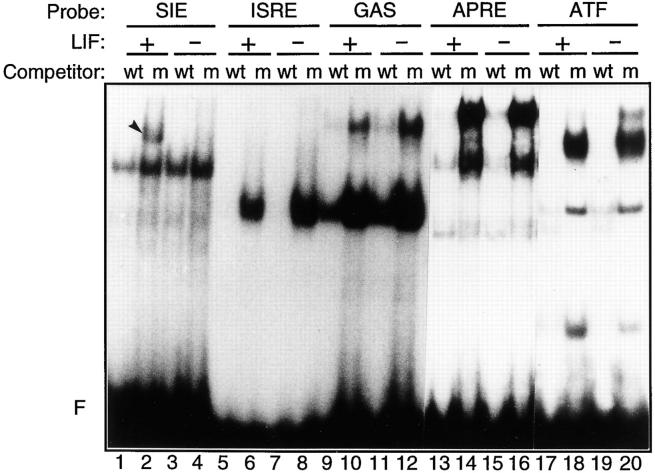
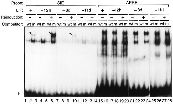
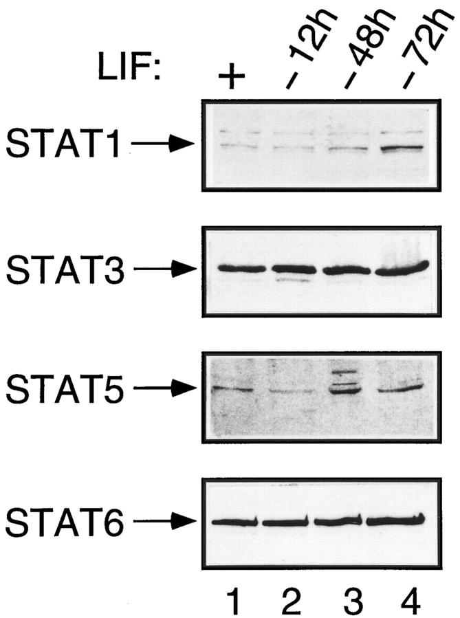
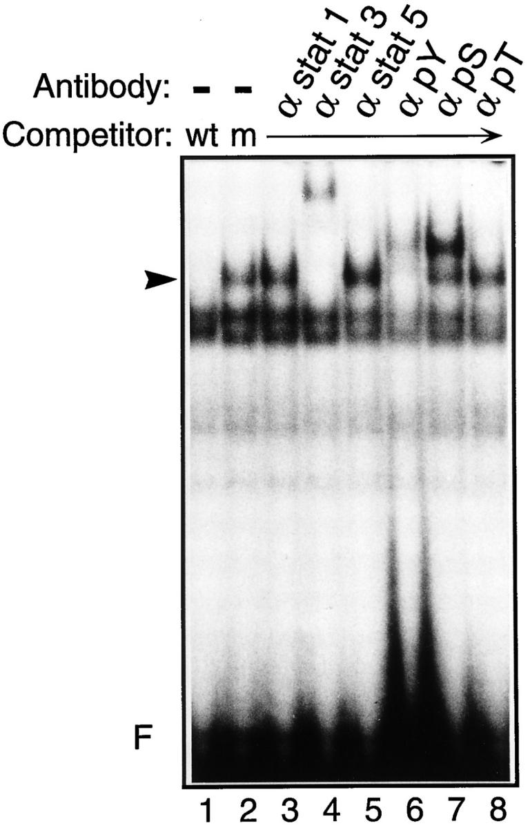
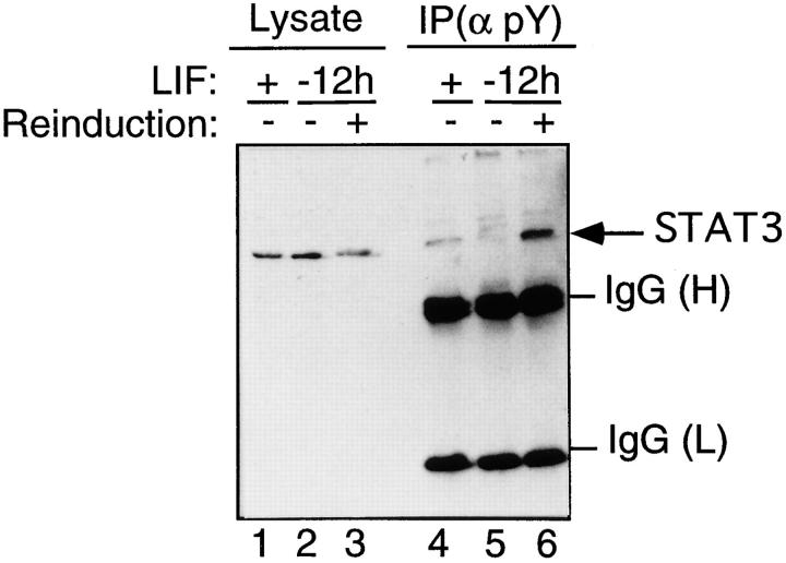
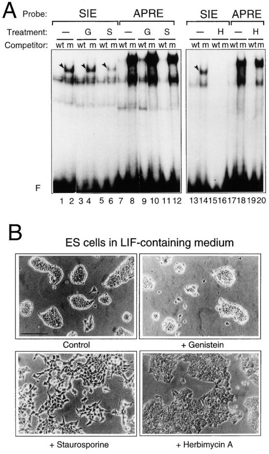
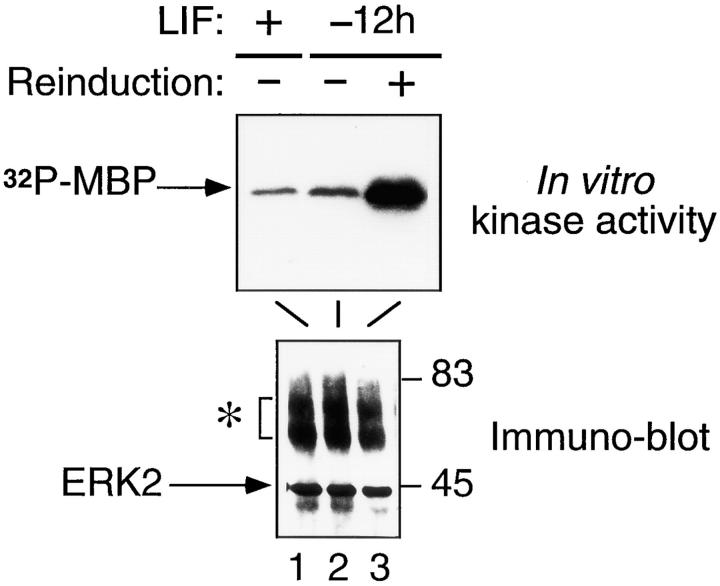
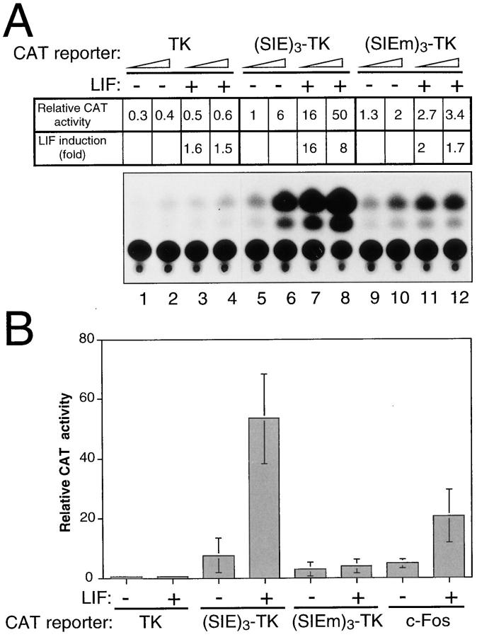
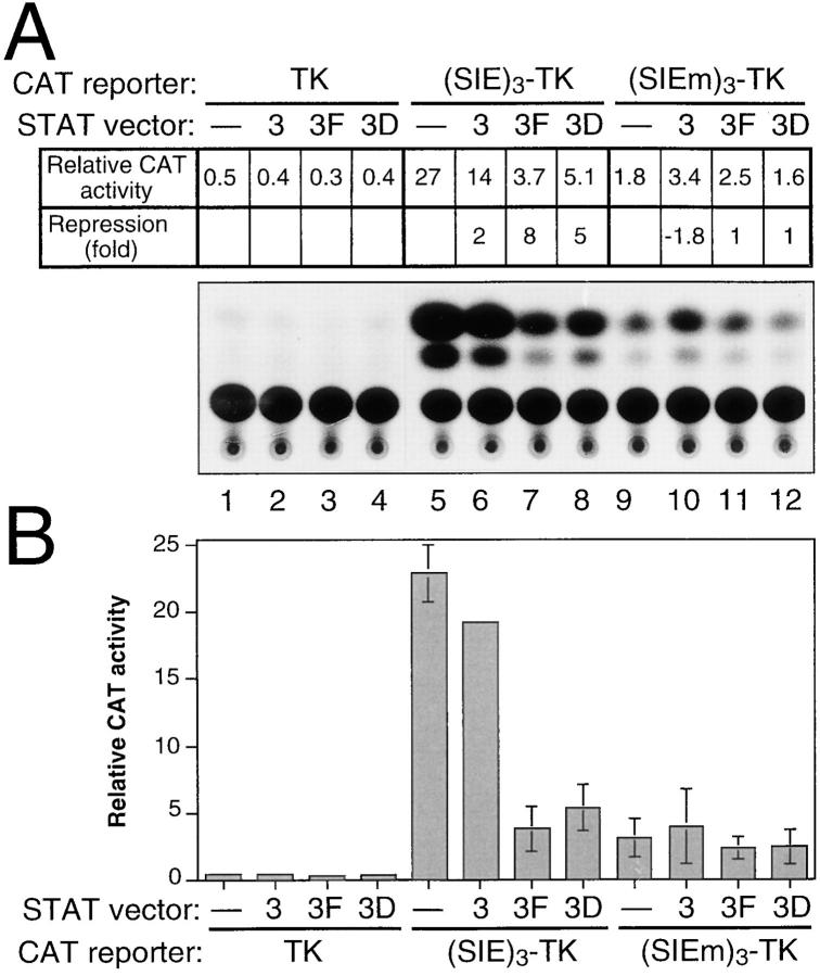
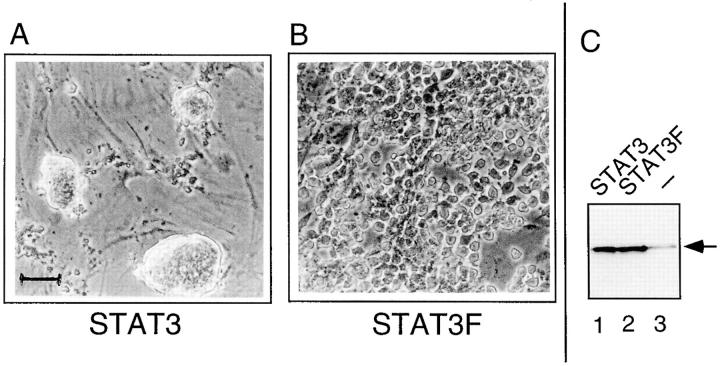
STAT transcription factors are induced by a number of growth factors and cytokines. Within minutes of induction, the STAT proteins are phosphorylated on tyrosine and serine residues and translocated to the nucleus, where they bind to their DNA targets. The leukemia inhibitory factor (LIF) mediates pleiotropic and sometimes opposite effects both in vivo and in cultured cells. It is known, for example, to prevent differentiation of embryonic stem (ES) cells in vitro. To get insights into LIF-regulated signaling in ES cells, we have analyzed protein-binding and transcriptional properties of STAT recognition sites in ES cells cultivated in the presence and in the absence of LIF. We have detected a specific LIF-regulated DNA-binding activity implicating the STAT3 protein. We show that STAT3 phosphorylation is essential for this LIF-dependent DNA-binding activity. The possibility that ERK2 or a closely related protein kinase, whose activity is modulated in a LIF-dependent manner, contributes to this phosphorylation is discussed. Finally, we show that the multimerized STAT3-binding DNA element confers LIF responsiveness to a minimal thymidine kinase promoter. This, together with our observation that overexpression of STAT3 dominant-negative mutants abrogates this LIF responsiveness, clearly indicates that STAT3 is involved in LIF-regulated transcriptional events in ES cells. Finally, stable expression of such a dominant negative mutant of STAT3 induces morphological differentiation of ES cells despite continuous LIF supply. Our results suggest that STAT3 is a critical target of the LIF signaling pathway, which maintains pluripotent cell proliferation.


Figures

Figure 1
Figure 1
A LIF-dependent DNA-binding activity is detected in ES cells. Nuclear extracts from ES cells constantly maintained in the presence of LIF (+) or grown in the absence of LIF for 12 h (−) were used in standard band-shift assays with the 5′ end- labeled SIE, ISRE, GAS, APRE, or ATF probes as indicated (see Materials and Methods). A 100-fold molar excess of each corresponding unlabeled wild-type oligonucleotide (wt) or of the mutated SIE oligonucleotide (m) was added as competitor to the binding reactions. F, the unbound probe. The arrowhead in lane 2 points to the LIF-regulated complex.
Figure 2
Figure 2
The LIF-dependent DNA-binding complex is reinducible by LIF up to 8 d without LIF. Nuclear extracts were prepared from ES cells that were maintained with LIF or without LIF for 12 h and 8 or 11 d (−12h, −8d, or −11d, respectively) and reinduced with LIF for 10 min where indicated. Standard band-shift assays were run with the 5′ end-labeled SIE or APRE probes in the presence of wild-type (wt) or mutant (m) SIE competitors, as indicated. The arrowheads refer to the LIF-regulated complexes.
Figure 3
Figure 3
STAT protein expression remains constant in the presence and absence of LIF. Nuclear extracts were prepared from ES cell cultures that were maintained in the presence of LIF (+) or those from which LIF was removed for 12, 48, or 72 h, as indicated. Immunoblot analyses were performed with specific anti-STAT antibodies, as described in Materials and Methods.
Figure 4
Figure 4
The LIF-dependent complex contains a STAT3-related protein and is phosphorylated both on tyrosine and serine residues. Nuclear extracts from ES cells maintained in the presence of LIF were preincubated for 30 min with 1 μg of anti-STAT1, anti-STAT3, or anti-STAT5 (α stat 1, α stat 3, or α stat 5, respectively), or with 0.5 μg of the antiphosphotyrosine, antiphosphoserine, or antiphosphothreonine (α pY, α pS, or α pT, respectively), as indicated. Standard band-shift assays were then performed with the 5′ end-labeled SIE probe and wild-type (wt) or mutant (m) SIE competitors, as indicated. The arrowhead points to the position of the LIF-dependent complex.
Figure 5
Figure 5
STAT3 is present in tyrosine-phosphorylated complexes only upon LIF treatment. Whole-cell extracts were prepared from ES cells that were maintained with LIF (+) or without LIF for 12 h (−12h) and reinduced or not with LIF for 10 min, as indicated. The extracts (20 μg) were loaded, either directly (Lysate) or after immunoprecipitation with the antiphosphotyrosine antibody [IP(α pY)], on SDS-polyacrylamide gels, and were analyzed with the anti-STAT3 antibody. Arrows point to bands corresponding to STAT3. The heavy (H) and light (L) chains of the anti-pY Igs are also revealed.
Figure 6
Figure 6
LIF-dependent complex is sensitive to Staurosporine and Herbimycin A. (A) The 5′ end-labeled SIE or APRE probes were used in the presence of wild-type (wt) or mutant (m) SIE competitors in standard band-shift assays with nuclear extracts derived from ES cells maintained with LIF and treated for 20 h with 27 μg/ml of Genistein (G), 100 nM Staurosporine (S), or 1 μg/ml Herbimycin A (H) when indicated. Arrowheads point to the LIF-regulated complexes. (B) Phase contrast micrographs of ES cells treated or not (control) with the different kinase inhibitors. Bar, 40 μm.
Figure 7
Figure 7
An ERK2-related MAP kinase is activated in ES cells reinduced by LIF. Nuclear extracts from ES cells maintained with LIF or without LIF for 12 h (−12h) and reinduced by LIF for 10 min, as indicated, were immunoprecipitated with the anti-ERK2 antibody, as described in Materials and Methods. Two thirds of the reaction were assayed in an in vitro kinase assay using MBP as an exogenous substrate (top). The remaining third of the reaction was analyzed by immunoblot with the anti-ERK2 antibody (bottom). The asterisk refers to the heavy chains of the Igs. Numbers on the right-hand side indicate the position of protein size markers (kD).
Figure 8
Figure 8
The SIE element confers LIF responsiveness to a heterologous promoter in ES cells. (A) ES cells maintained with (+) or without (−) LIF were transfected with 2 (odd lanes) or 5 μg (even lanes) of the CAT reporters: pBLCAT5 (referred to as TK), (SIE)3-TK, and (SIEm)3-TK, as indicated. The results of a typical CAT assay with corresponding quantification are presented. (B) Transfection experiments were performed as described above, but using 5 μg of each of the indicated CAT reporters and including the c-Fos CAT reporter. The mean (±SD) of four independent experiments performed with different plasmid preparations is plotted.
Figure 9
Figure 9
Dominant negative mutants of STAT3 negatively interfere with LIF-dependent promoter activity. (A) ES cells maintained with LIF were cotransfected with 5 μg of the CAT reporters [pBLCAT5 (referred to as TK), (SIE)3-TK, and (SIEm)3-TK], together with 3 μg of the empty pEF-BOS vector (lanes 1, 5, and 9), the pEF-BOS HA-STAT3 (lanes 2, 6, and 10), the pEF-BOS HA-STAT3F (lanes 3, 7, and 11), or the pEF-BOS HA-STAT3D (lanes 4, 8, and 12), as indicated. The results of a typical CAT assay with corresponding quantification are presented. (B) The mean (±SD) of three independent experiments performed as described above, with different plasmid preparations, is plotted. Depending on the experiment, cotransfection of the (SIE)3-TK CAT reporter with the pEF-BOS HA-STAT3 vector resulted either in a slight stimulation or slight repression of CAT activity compared to cotransfection with the empty pEF-BOS vector. This variation, probably reflecting differences in recipient cell conditions, generated artificially elevated SD values: the misleading error bar was therefore omitted on the corresponding column.
Figure 10
Figure 10
Stable expression of STAT3F dominant negative mutant induces ES cell differentiation. Phase contrast micrographs of Neomycin-resistant ES cell clones stably expressing the wild-type STAT3 (A) or the dominant negative STAT3F mutant (B). The selected clones were grown on feeder layers of mouse embryo fibroblasts (visible in A between the clumps of undifferentiated ES cells) and continuously maintained in the presence of LIF. (C) Western blot analysis of equivalent amounts of whole-cell extracts of STAT3- (lane 1) or STAT3F-transformed cells (lane 2), or untransformed ES cells (lane 3). The arrow points to the specific signal obtained with the monoclonal anti-STAT3 antibody. Bar, 100 μm.

Similar articles

Cited by

References

    1. Akira S, Nishio Y, Inoue M, Wang XJ, Wei S, Matsusaka T, Yoshida K, Sudo T, Naruto M, Kishimoto T. Molecular cloning of APRF, a novel IFN-stimulated gene factor 3 p91-related transcription factor involved in the gp130-mediated signaling pathway. Cell. 1994;77:63–71. - PubMed
    1. Akiyama T, Ishida J, Nakagawa S, Ogawara H, Watanabe S, Itoh N, Shibuya M, Fukami Y. Genistein, a specific inhibitor of tyrosine-specific protein kinases. J Biol Chem. 1987;262:5592–5595. - PubMed
    1. Boschart M, Kluppel M, Schmidt A, Schutz G, Luckow B. Reporter constructs with low background activity utilizing the CATgene. Gene (Amst) 1992;110:129–130. - PubMed
    1. Boulter CA, Agguzi A, Williams RL, Wagner EF, Evans MJ, Beddington R. Expression of v-src induces aberrant development and twinning in chimaeric mice. Development (Camb) 1991;111:357–366. - PubMed
    1. Boulton TG, Zhong Z, Wen Z, Darnell JE, Jr, Stahl N. STAT3 activation by cytokines utilizing gp130 and related transducers involves a secondary modification requiring an H7-sensitive kinase. Proc Natl Acad Sci USA. 1995;92:6915–6919. - PMC - PubMed

Publication types

MeSH terms