. 1997 Sep 22;138(6):1239-54.
doi: 10.1083/jcb.138.6.1239.

The role of ADP-ribosylation factor and phospholipase D in adaptor recruitment

Affiliations

The role of ADP-ribosylation factor and phospholipase D in adaptor recruitment

M A West et al. J Cell Biol. .

Abstract

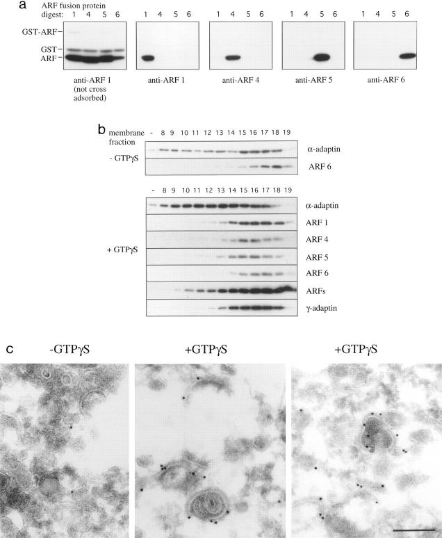
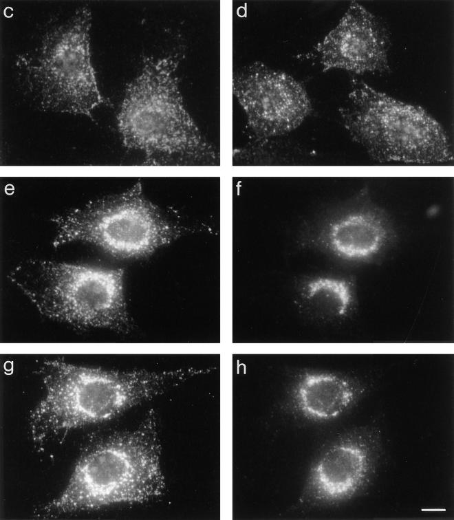
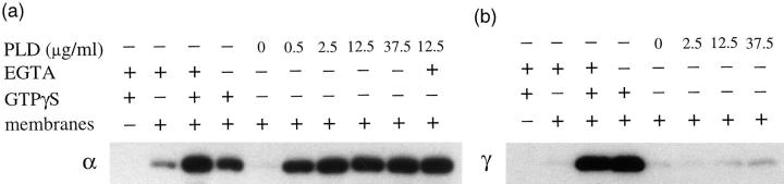
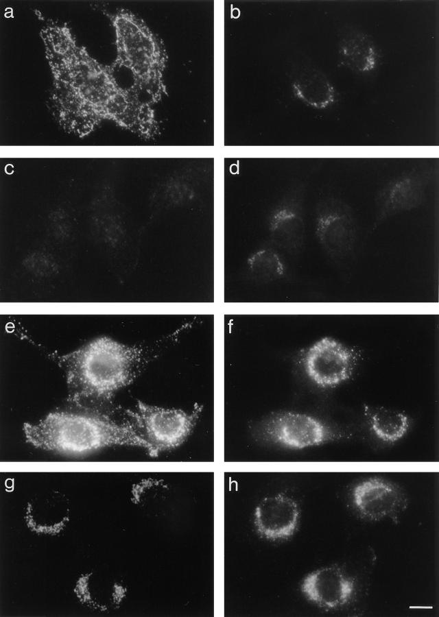
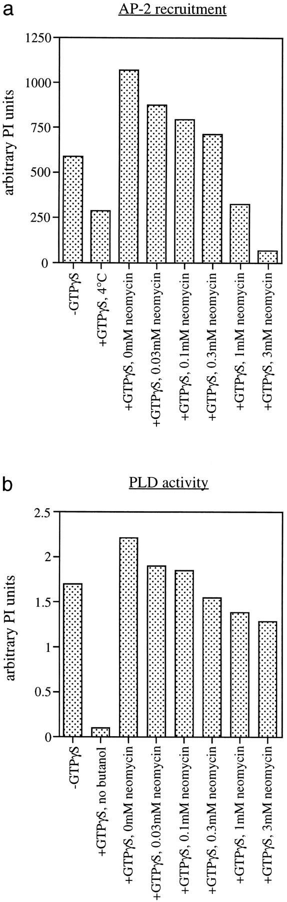
AP-1 and AP-2 adaptors are recruited onto the TGN and plasma membrane, respectively. GTPgammaS stimulates the recruitment of AP-1 onto the TGN but causes AP-2 to bind to an endosomal compartment (Seaman, M.N.J., C.L. Ball, and M.S. Robinson. 1993. J. Cell Biol. 123:1093-1105). We have used subcellular fractionation followed by Western blotting, as well as immunofluorescence and immunogold electron microscopy, to investigate both the recruitment of AP-2 adaptors onto the plasma membrane and their targeting to endosomes, and we have also examined the recruitment of AP-1 under the same conditions. Two lines of evidence indicate that the GTPgammaS-induced targeting of AP-2 to endosomes is mediated by ADP-ribosylation factor-1 (ARF1). First, GTPgammaS loses its effect when added to ARF-depleted cytosol, but this effect is restored by the addition of recombinant myristoylated ARF1. Second, adding constitutively active Q71L ARF1 to the cytosol has the same effect as adding GTPgammaS. The endosomal membranes that recruit AP-2 adaptors have little ARF1 or any of the other ARFs associated with them, suggesting that ARF may be acting catalytically. The ARFs have been shown to activate phospholipase D (PLD), and we find that addition of exogenous PLD has the same effect as GTPgammaS or Q71L ARF1. Neomycin, which inhibits endogenous PLD by binding to its cofactor phosphatidylinositol 4,5-bisphosphate, prevents the recruitment of AP-2 not only onto endosomes but also onto the plasma membrane, suggesting that both events are mediated by PLD. Surprisingly, however, neither PLD nor neomycin has any effect on the recruitment of AP-1 adaptors onto the TGN, even though AP-1 recruitment is ARF mediated. These results indicate that different mechanisms are used for the recruitment of AP-1 and AP-2.


Figures

Figure 1
Figure 1
Recruitment of cytosolic proteins onto rat liver membranes. (a) To determine the specificity of the anti-ARF antibodies raised in this study, samples of ARF–GST fusion proteins were digested with Factor X and the digests subjected to SDS-PAGE on 15% gels and Western blotted. Identical panels were probed with affinity-purified and cross-adsorbed anti-ARF antisera as indicated. Non–cross-adsorbed anti-ARF1 labels all four ARF isoforms, while after cross-adsorption each antibody is specific for its own isoform. ARF3, which is 97% identical to ARF1, was not included in this study. (b) Membranes from a Nycodenz gradient were incubated with pig brain cytosol in the presence or absence of GTPγS. The incubation mixture in this experiment and in all subsequent experiments also contained ATP and an ATP-regenerating system. After the incubation, membranes were pelleted and assayed by Western blotting for the various ARF isoforms, or for newly recruited AP-2 or AP-1 adaptor complexes using brain-specific anti–α- or species-specific anti–γ-adaptin antibodies, respectively. Significant amounts of both α-adaptin and ARF6 are associated with membranes even in the absence of GTPγS, although they show only partial overlap. In the presence of GTPγS, the binding of AP-2 is increased and the fractionation profile shifted to less dense membranes. The other ARF isoforms and AP-1 are also recruited onto membranes and show similar fractionation profiles to each other but not to AP-2. (c) Frozen thin sections were prepared from samples equivalent to 8–19 in b. The sections were labeled with brain-specific anti–α-adaptin, followed by 10-nm protein A gold. In the absence of GTPγS, AP-2 is recruited onto small vesicles, presumably derived from the plasma membrane, while in the presence of GTPγS, AP-2 is recruited onto larger structures that often contain internal membranes, characteristic of endosomes. Bar, 200 nm.
Figure 2
Figure 2
Role of ARF1 in AP-2 recruitment. Aliquots of peak fractions from a Nycodenz gradient were incubated with either reconstituted cytosol or with high–molecular weight, ARF- depleted fractions of cytosol that had been gel filtered. GTPγS and recombinant myristoylated ARF1 were included as indicated. Newly recruited AP-2 adaptors (α) and membrane-associated ARF1 were detected by Western blotting. When reconstituted cytosol was added, AP-2 recruitment onto the membranes was enhanced by the addition of GTPγS. No such enhancement was seen with the ARF-depleted cytosol, but it was restored by the addition of recombinant myristoylated ARF1.
Figure 3
Figure 3
Effects of ARF1 mutants on adaptor recruitment. Aliquots of pooled peak fractions from a Nycodenz gradient were used as acceptor membranes for the recruitment of (a) AP-2 (α) or (b) AP-1 (γ). These membranes were incubated with cytosol containing either GTPγS, Q71L ARF1, or T31N ARF1 and then subjected to SDS-PAGE and Western blotting. Q71L ARF1, but not T31N ARF1, stimulates the recruitment of both types of adaptors even in the absence of GTPγS. (c–h) Permeabilized NRK cells were incubated with either cytosol alone (c) or cytosol containing 100 μg/ml T31N ARF1 (d), GTPγS (e and f), or 100 μg/ml Q71L ARF1 (g and h), and newly recruited AP-2 or AP-1 adaptors were detected using anti-α (c, d, e, and g) or anti-γ (f and h) antibodies. Q71L ARF1 has a GTPγS-like effect on both types of adaptors, indicating that in both cases the GTPγS is acting via ARF. Bar, 10 μm.
Figure 3
Figure 3
Effects of ARF1 mutants on adaptor recruitment. Aliquots of pooled peak fractions from a Nycodenz gradient were used as acceptor membranes for the recruitment of (a) AP-2 (α) or (b) AP-1 (γ). These membranes were incubated with cytosol containing either GTPγS, Q71L ARF1, or T31N ARF1 and then subjected to SDS-PAGE and Western blotting. Q71L ARF1, but not T31N ARF1, stimulates the recruitment of both types of adaptors even in the absence of GTPγS. (c–h) Permeabilized NRK cells were incubated with either cytosol alone (c) or cytosol containing 100 μg/ml T31N ARF1 (d), GTPγS (e and f), or 100 μg/ml Q71L ARF1 (g and h), and newly recruited AP-2 or AP-1 adaptors were detected using anti-α (c, d, e, and g) or anti-γ (f and h) antibodies. Q71L ARF1 has a GTPγS-like effect on both types of adaptors, indicating that in both cases the GTPγS is acting via ARF. Bar, 10 μm.
Figure 4
Figure 4
Effect of exogenous PLD on adaptor recruitment. (a and b) Membrane pools (see Fig. 3) were incubated with cytosol containing bacterial PLD, EGTA (100 μM), and GTPγS in combinations as indicated, and either α (a) or γ (b) recruitment was detected by Western blotting. PLD mimics the GTPγS effect on AP-2 recruitment, but not on AP-1 recruitment. (c–h) Recruitment onto permeabilized NRK cells was carried out with cytosol alone (c and d), with GTPγS (e and f) or with 12.5 μg/ml PLD (g and h). The same cells were double labeled for immunofluorescence using either the anti-α (c, e, and g) or anti-γ (d, f, and h) antibodies. PLD only has a GTPγS-like effect on the AP-2 adaptors. Bar, 10 μm.
Figure 4
Figure 4
Effect of exogenous PLD on adaptor recruitment. (a and b) Membrane pools (see Fig. 3) were incubated with cytosol containing bacterial PLD, EGTA (100 μM), and GTPγS in combinations as indicated, and either α (a) or γ (b) recruitment was detected by Western blotting. PLD mimics the GTPγS effect on AP-2 recruitment, but not on AP-1 recruitment. (c–h) Recruitment onto permeabilized NRK cells was carried out with cytosol alone (c and d), with GTPγS (e and f) or with 12.5 μg/ml PLD (g and h). The same cells were double labeled for immunofluorescence using either the anti-α (c, e, and g) or anti-γ (d, f, and h) antibodies. PLD only has a GTPγS-like effect on the AP-2 adaptors. Bar, 10 μm.
Figure 5
Figure 5
Effect of neomycin on the recruitment of adaptors onto rat liver membranes. Rat liver membrane pools (see Fig. 3) were incubated with cytosol in the absence or presence of GTPγS and varying concentrations of neomycin. The effect of neomycin on AP-1 and AP-2 recruitment was assessed by Western blotting of the membrane samples using anti-α (a) or anti-γ (b) antibodies. Neomycin, which indirectly inhibits PLD activity, decreases the amount of AP-2 recruitment both in the absence and in the presence of GTPγS but has no appreciable effect on AP-1 recruitment.
Figure 6
Figure 6
Effect of neomycin on adaptor recruitment in permeabilized cells. Permeabilized NRK cells were allowed to recruit adaptors from cytosol in the absence (a–d) or presence (e–h) of GTPγS. Neomycin (1 mM) was included in c, d, g, and h. For each of the conditions, the cells were double labeled for immunofluorescence using anti-α (a, c, e, and g) or anti-γ (b, d, f, and h) antibodies. Photographs in the presence and in the absence of neomycin were taken and printed under identical conditions. Neomycin inhibits the recruitment of AP-2 adaptors both onto the plasma membrane in the absence of GTPγS and onto the endosomal compartment in the presence of GTPγS, without showing any effect on AP-1 recruitment. Bar, 10 μm.
Figure 7
Figure 7
Comparison of the effects of neomycin on AP-2 recruitment and on PLD activity. (a) Permeabilized NRK cells were allowed to recruit adaptors from cytosol either without GTPγS, with GTPγS at 4°C, or with GTPγS plus increasing concentrations of neomycin and then assayed by Western blotting and phosphorimager quantification for AP-2 recruitment. The relatively weak effect of GTPγS is due to the use of whole cells rather than membrane fractions. Neomycin inhibits AP-2 recruitment in a dose-dependent manner, with essentially complete inhibition at 3 mM. (b) NRK cells were labeled overnight with [3H]palmitic acid, permeabilized, and then incubated with cytosol containing 1% butanol either without GTPγS or with GTPγS plus increasing concentrations of neomycin. Lipids were extracted and subjected to TLC, and PLD activity was assayed by quantifying the production of phosphatidyl butanol. In one sample, the butanol was omitted as a control. PLD activity is stimulated ∼30% by the addition of GTPγS and inhibited by neomycin, although even at 3 mM it is only inhibited ∼40%. This probably reflects the existence of multiple types of PLD in the cell, not all of which can be inhibited by neomycin, and may also indicate that neomycin has a direct effect on AP-2 recruitment, as well as an indirect effect by inhibiting PLD.
Figure 8
Figure 8
Fractionation of membranes binding adaptors under various conditions. Pools of pairs of membrane fractions from Nycodenz gradients were incubated with cytosol in the presence of GTPγS or 100 μg/ml Q71L ARF (a) or in a separate experiment in the presence of GTPγS or 12.5 μg/ml PLD (b). Newly recruited AP-2 or AP-1 adaptors were detected on Western blots using anti–α- or anti–γ-adaptin antibodies, respectively. The signal in each sample was quantified using a phosphorimager. As also shown in Fig. 1 b, α- and γ-adaptin–binding membranes have distinct fractionation profiles. However, the profile of the α-adaptin–binding membranes is similar whether GTPγS, Q71L ARF1, or PLD is added.
Figure 9
Figure 9
Immunogold EM labeling of newly recruited AP-2 adaptors. Frozen thin sections were prepared of permeabilized NRK cells that had been allowed to recruit proteins from cytosol in the presence of either GTPγS (a and b), Q71L ARF1 (100 μg/ml) (c and d), or PLD (12.5 μg/ml) (e and f). The sections were labeled with antibodies against newly recruited α-adaptin (15-nm gold) and lgp110 (8-nm gold). The arrows show membranes that are positive for both antigens, the large arrowheads show membranes that are positive for α-adaptin only, and the small arrowheads show membranes that are positive for lgp110 only. The same types of membranes are labeled under all three conditions. Bar, 200 nm.
Figure 10
Figure 10
Schematic model for the recruitment of AP-2 adaptors onto endosomal membranes. This model is based on the model of Traub and Kornfeld for AP-1 adaptor recruitment (Traub et al., 1993), but with several important modifications. We propose that ARF1 does not bind directly to the putative docking protein, but instead activates PLD, either directly or indirectly. Activated PLD, in the presence of PIP2, may lead to rapid local changes in the concentrations of negatively charged phospholipids such as PA and PIP2. These negatively charged phospholipids, together with the putative docking protein, would constitute an AP-2 binding site.

Similar articles

Cited by

References

    1. Ahle S, Mann A, Eichelsbacher U, Ungewickell E. Structural relationships between clathrin assembly proteins from the Golgi and the plasma membrane. EMBO (Eur Mol Biol Organ) J. 1988;4:919–929. - PMC - PubMed
    1. Ball CL, Hunt SP, Robinson MS. Expression and localisation of α-adaptin isoforms. J Cell Sci. 1995;108:2865–2875. - PubMed
    1. Barlowe C, Orci L, Yeung T, Hosobuchi M, Hamamoto S, Salama N, Rexach MF, Ravazzola M, Amherdt M, Schekman R. COPII: a membrane coat formed by sec proteins that drive vesicle budding from the endoplasmic reticulum. Cell. 1994;77:895–907. - PubMed
    1. Beck KA, Keen JH. Interaction of phosphoinositide cycle intermediates with the plasma membrane-associated clathrin assembly protein AP-2. J Biol Chem. 1991;266:4442–4447. - PubMed
    1. Bi K, Roth MG, Ktistakis NT. Phosphatidic acid formation by phospholipase D is required for transport from the endoplasmic reticulum to the Golgi complex. Curr Biol. 1997;7:301–315. - PubMed

Publication types

MeSH terms