. 1997 Sep 22;138(6):1379-94.
doi: 10.1083/jcb.138.6.1379.

Caspase cleavage of keratin 18 and reorganization of intermediate filaments during epithelial cell apoptosis

Affiliations

Caspase cleavage of keratin 18 and reorganization of intermediate filaments during epithelial cell apoptosis

C Caulín et al. J Cell Biol. .

Abstract

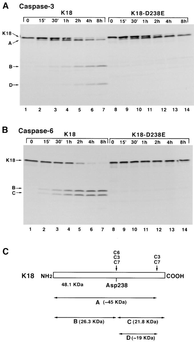
Keratins 8 (K8) and 18 (K18) are major components of intermediate filaments (IFs) of simple epithelial cells and tumors derived from such cells. Structural cell changes during apoptosis are mediated by proteases of the caspase family. During apoptosis, K18 IFs reorganize into granular structures enriched for K18 phosphorylated on serine 53. K18, but not K8, generates a proteolytic fragment during drug- and UV light-induced apoptosis; this fragment comigrates with K18 cleaved in vitro by caspase-6, -3, and -7. K18 is cleaved by caspase-6 into NH2-terminal, 26-kD and COOH-terminal, 22-kD fragments; caspase-3 and -7 additionally cleave the 22-kD fragment into a 19-kD fragment. The cleavage site common for the three caspases was the sequence VEVD/A, located in the conserved L1-2 linker region of K18. The additional site for caspases-3 and -7 that is not cleaved efficiently by caspase-6 is located in the COOH-terminal tail domain of K18. Expression of K18 with alanine instead of serine at position 53 demonstrated that cleavage during apoptosis does not require phosphorylation of serine 53. However, K18 with a glutamate instead of aspartate at position 238 was resistant to proteolysis during apoptosis. Furthermore, this cleavage site mutant appears to cause keratin filament reorganization in stably transfected clones. The identification of the L1-2 caspase cleavage site, and the conservation of the same or very similar sites in multiple other intermediate filament proteins, suggests that the processing of IFs during apoptosis may be initiated by a similar caspase cleavage.


Figures

Figure 1
Figure 1
Induction of apoptosis in SNG-M cells results in reorganization of K18 IF. SNG-M cells were treated with etoposide (250 μg/ml) for 24 h and stained by indirect immunofluorescence for K18 with monoclonal antibody CK5 (A, C, and E) and with propidium iodide for DNA (B, D, and F). Images were analyzed with a confocal microscope. Note the granular K18 staining associated with altered nuclear morphology.
Figure 2
Figure 2
K18 granular structures are preferentially phosphorylated. Etoposide-treated SNG-M cells were fixed and stained by indirect immunofluorescence for phospho-ser53 K18 with antibody 3055 (A, C, and E) and with propidium iodide for DNA (B, D, and F). Note the granular keratin staining of some cells with normal nuclear morphology in A, the staining of cell–cell junctions in A and C, and advanced keratin disorganization with nuclear fragmentation in C and E.
Figure 3
Figure 3
Treatment of SNG-M cells with etoposide results in specific cleavage of K18. (A) SNG-M cells were treated with etoposide (250 μg/ml) for 0, 12, 24, 36, 48, 60, 72, and 84 h. Adherent and floating cells were combined, and whole-cell lysates were analyzed by Western blot with polyclonal antibody anti-K18 1589. This membrane was stripped and probed consecutively with monoclonal anti-K8 M20 (C), anti–phospho ser53-K18 antibodies (B), and monoclonal anti-K18 CK5 (D). (E) HR-9 cells were treated with daunomycin for 0, 12, 24, 36, 48, and 60 h. Adherent and floating cells were combined, and whole-cell lysates were analyzed by Western blot with anti-K18 polyclonal antibody, 1589.
Figure 4
Figure 4
(A) Kinetics of induction of apoptosis in etoposide-treated SNG-M cells. In parallel experiments to the ones described in Fig. 3 A, SNG-M cells were treated with etoposide (250 μg/ml) for 0, 12, 24, 36, 48, 60, 72, and 84 h. Adherent and floating cells were combined, fixed in methanol, and stained with DAPI. Nuclei fragmentation was used as the criterion of apoptosis. (B) Comparison of the kinetics of appearance of the K18 23-kD fragment (K18 B) and its phosphorylated form (P-K18 B) by densitometric analysis of the signal detected in Western blotting shown in Fig. 3, A and B. The y-axis represents the percentage of the maximum signal. Maximum signal for P-K18 B was obtained after 36 h and for K18 B after 60 h. (C) Comparison of the kinetics of disappearance of K18 and P-K18 by densitometric analysis of the signal detected in Fig. 3, A and B.
Figure 5
Figure 5
Different apoptotic stimuli results in the cleavage of K18 and EndoB. (A) Western blot analysis of HR-9 (lanes 1–7) and SNG-M (lanes 8–14) cells subjected to different apoptotic stimuli. Adherent (A) and floating (F) cells were separately collected after etoposide (ETP) and daunomycin (DNM) treatment and UV exposure, and whole-cell lysates were analyzed by Western blot with polyclonal anti-K18. Lanes 1 and 9 represent control untreated cells. (B) Low molecular weight genomic DNA was analyzed from cells represented in A except for lane 1, which contained size markers. (C) Comparison of K18 found in apoptotic cells and purified K18 cleaved in vitro by caspase-6. Cell lysates or purified protein preparation was analyzed by Western blotting with antibody 3055. Untreated SNG-M cells (lane 1), etoposide (ETP) treated floating SNG-M cells (lane 7), and mouse HR-9 cells (lane 2) or apoptotic HR-9 cells (lane 6) were compared with purified K18 (lane 4), purified K18 mixed with HR-9 cell lysate (lane 3), or K18 cleaved with caspase-6 (C6) in vitro and mixed with HR-9 cell lysate (lane 5). Note the comigration of caspase-6–cleaved K18 (lane 5) and K18 B found in apoptotic SNG-M cells (lane 7).
Figure 6
Figure 6
Specific cleavage in vitro of K18, but not K8, by caspase-6, -3, and -7. (A) [35S]methionine-labeled K8 and K18 were incubated for 2 h at 37°C with decreasing concentrations of caspase-6 (Cas-6) (lanes 2–10), caspase-3 (Cas-3) (lanes 11–18), and caspase-7 (Cas-7) (lanes 19–26), and subsequently analyzed by SDS-PAGE and fluorography. Protease was decreased by serial 1:10 dilutions. (B) Comparison of the proteolytic pattern of K18 and EndoB cleavage generated by caspase-6 (C6), caspase-3 (C3), and caspase-7 (C7). A–D represent the proteolytic fragments obtained by cleavage of K18. a–d represent the proteolytic fragments obtained by cleavage of EndoB.
Figure 7
Figure 7
Mapping of the caspase cleavage sites for K18 and EndoB. (A) Schematic representation of the deletion mutants of K18 obtained by in vitro transcription and translation of K18 cDNA digested with HindIII (K18), BamHI (K18-339), and ScaI (K18-269). (B) [35S]methionine-labeled K18 and K18 mutants were separately incubated for 2 h at 37°C with caspase-6 (C6), caspase-3 (C3), and caspase-7 (C7) and subsequently analyzed by SDS-PAGE and fluorography. (C) Schematic representation of the deletion mutants of EndoB obtained by in vitro transcription and translation of EndoB cDNA digested with BamHI (EndoB), AvaI (EndoB-316), ScaI (EndoB-262), and PvuII (EndoB-191). (D) [35S]methionine-labeled EndoB and EndoB mutants were separately incubated for 2 h at 37°C with caspase-6 (C6), caspase-3 (C3), and caspase-7 (C7) and subsequently analyzed by SDS-PAGE and fluorography.
Figure 8
Figure 8
Site-directed mutagenesis to K18-D238E abolishes cleavage by caspases. (A) Time-dependent cleavage of K18 and K18-D238E with caspase-3. Equal amounts of [35S]methionine-labeled K18 and K18-D238E were incubated with caspase-3. The reaction was stopped at 0, 15 min, 30 min, 1 h, 2 h, 4 h, and 8 h, and samples were analyzed by SDS-PAGE and fluorography. (B) Time-dependent cleavage of K18 and K18-D238E with caspase-6. Equal amounts of [35S]methionine-labeled K18 and K18-D238E were incubated with caspase-6. The reaction was stopped at the indicated times, and samples were analyzed by SDS-PAGE and fluorography. (C) Schematic representation of the different proteolytic fragments generated by cleavage of K18 by caspases.
Figure 9
Figure 9
Western blot analysis of K18, K18-D238E, and K18-S53A proteins expressed in HR-9 normal and apoptotic cells. (A) Whole cell lysates of stably transfected HR-9 cells expressing the indicated proteins were prepared from control cultures (C) and after treatment with daunomycin for 24 h. Attached cells (A) and floating cells (F) were analyzed separately. After SDS-PAGE and transfer to nitrocellulose, the proteins were probed with the 1589 polyclonal EndoB antibody. (B) The same filter was reprobed with 3055 polyclonal antibody specific for K18 phosphorylated on ser53.
Figure 10
Figure 10
Immunofluorescent localization of K18, P-K18, and EndoA in HR-9 cells expressing K18 (A and D), K18-S53A (B and E) or K18-D238E (C and F–L). Cells were stained for K18 with CK5 monoclonal antibody (A–C) and with 3055 antibody for P-K18 (D–F). Note the presence of keratin aggregates (arrows) that are preferentially phosphorylated in HR-9 K18-D238E cells. Double staining of HR-9 K18-D238E cells with 3055 antibody (G–I) and monoclonal antibody anti-EndoA TROMA1 (J–L) show colocalization of EndoA with P-K18 in the granular structures.
Figure 11
Figure 11
Amino acid sequences of the linker L1-2 region of the central rod domain of the different members of the IF family. Potential caspase cleavage sites are highlighted. Sequences were obtained from Swiss-Prot database.

References

    1. Albers K, Fuchs E. Expression of mutant keratin cDNAs in epithelial cells reveals possible mechanisms for initiation and assembly of intermediate filaments. J Cell Biol. 1989;108:1477–1493. - PMC - PubMed
    1. Alnemri ES, Livingston DJ, Nicholson DW, Salvesen G, Thornberry NA, Wong WW, Yuan J. Human ICE/CED-3 protease nomenclature. Cell. 1996;87:171. - PubMed
    1. Anderson JM, Heindl LM, Bauman PA, Ludi CW, Dalton WS, Cress AE. Cytokeratin expression results in a drug-resistant phenotype to six different chemotherapeutic agents. Clin Cancer Res. 1996;2:97–105. - PubMed
    1. Baribault H, Price J, Miyai K, Oshima RG. Mid-gestational lethality in mice lacking keratin 8. Genes Dev. 1993;7:1191–1202. - PubMed
    1. Baribault H, Penner J, Iozzo RV, Wilson-Heiner M. Colorectal hyperplasia and inflammation in keratin 8-deficient FVB/N mice. Genes Dev. 1994;8:2964–2973. - PubMed

Publication types

MeSH terms