

Increased expression of transforming growth factor alpha precursors in acute experimental colitis in rats
- PMID: 9301498
- PMCID: PMC1891469
- DOI: 10.1136/gut.41.2.195
Increased expression of transforming growth factor alpha precursors in acute experimental colitis in rats
Abstract
Background and aim: Epidermal growth factor (EGF) and transforming growth factor alpha (TGF-alpha), members of the EGF family of growth factors, protect rat gastric and colonic mucosa against injury. Having shown previously that exogenously applied EGF protects rat colonic mucosa against injury, the aim of the present study was to evaluate the endogenously expressed ligand mediating the protective effect of EGF/TGF-alpha in vivo.
Methods: In an experimental model of trinitrobenzene sulphonic acid (TNBS)/ethanol induced colitis in rats EGF and TGF-alpha expression was evaluated using a ribonuclease protection assay, northern blot analysis, western blot analysis, and immunohistochemistry.
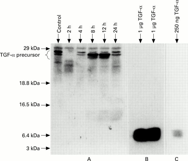
Results: TGF-alpha mRNA increased 3-4 times at 4-8 hours after induction of colitis and returned to control levels within 24 hours. TGF-alpha immunoreactive protein with a molecular size of about 28 kDa representing TGF-alpha precursors increased markedly after induction of colitis with a peak at 8-12 hours. No fully processed 5.6 kDa TGF-alpha protein was detected in normal or inflamed colon tissue. Only a weak signal for EGF mRNA expression was detected in the rat colon and no EGF protein was observed by immunohistochemistry or western blot analysis.
Conclusions: TGF-alpha precursors are the main ligands for the EGF receptor in acute colitis. It is hypothesised that TGF-alpha precursors convey the biological activity of endogenous TGF-alpha peptides during mucosal defence and repair.
Figures


Similar articles
-
Increased expression of epidermal growth factor-receptor in an experimental model of colitis in rats.Scand J Gastroenterol. 2000 Nov;35(11):1174-80. doi: 10.1080/003655200750056655. Scand J Gastroenterol. 2000. PMID: 11145289
-
Increased duodenal expression of transforming growth factor-alpha and epidermal growth factor during experimental colitis in rats.Eur J Gastroenterol Hepatol. 2008 Oct;20(10):989-94. doi: 10.1097/MEG.0b013e3283036758. Eur J Gastroenterol Hepatol. 2008. PMID: 18787466
-
Control of the epidermal growth factor receptor and its ligands during renal injury.Nephron. 2001 May;88(1):71-9. doi: 10.1159/000045962. Nephron. 2001. PMID: 11340354
-
Structure-function relationships for the EGF/TGF-alpha family of mitogens.Growth Factors. 1994;11(4):235-57. doi: 10.3109/08977199409010997. Growth Factors. 1994. PMID: 7779404 Review.
-
From wavy hair to naked proteins: the role of transforming growth factor alpha in health and disease.Semin Cell Dev Biol. 2014 Apr;28:12-21. doi: 10.1016/j.semcdb.2014.03.003. Epub 2014 Mar 12. Semin Cell Dev Biol. 2014. PMID: 24631356 Free PMC article. Review.
Cited by
-
Establishment of Intestinal Organoid from Rousettus leschenaultii and the Susceptibility to Bat-Associated Viruses, SARS-CoV-2 and Pteropine Orthoreovirus.Int J Mol Sci. 2021 Oct 5;22(19):10763. doi: 10.3390/ijms221910763. Int J Mol Sci. 2021. PMID: 34639103 Free PMC article.
-
ErbB receptors and their growth factor ligands in pediatric intestinal inflammation.Pediatr Res. 2014 Jan;75(1-2):127-32. doi: 10.1038/pr.2013.210. Epub 2013 Nov 6. Pediatr Res. 2014. PMID: 24402051 Free PMC article. Review.
-
Long-term effects of transient chemically induced colitis on the visceromotor response to mechanical colorectal distension.Dig Dis Sci. 2004 Jan;49(1):96-101. doi: 10.1023/b:ddas.0000011609.68882.3a. Dig Dis Sci. 2004. PMID: 14992442
-
Interactions between stromal cell--derived keratinocyte growth factor and epithelial transforming growth factor in immune-mediated crypt cell hyperplasia.J Clin Invest. 1998 Oct 15;102(8):1473-80. doi: 10.1172/JCI2792. J Clin Invest. 1998. PMID: 9788959 Free PMC article.
-
Use of mouse models to evaluate the persistence, safety, and immune modulation capacities of lactic acid bacteria.Clin Diagn Lab Immunol. 2003 Jul;10(4):696-701. doi: 10.1128/cdli.10.4.696-701.2003. Clin Diagn Lab Immunol. 2003. PMID: 12853407 Free PMC article.
References
Publication types
MeSH terms
Substances
LinkOut - more resources
Full Text Sources