

Induction of intestinal lesions in nu/nu mice induced by transfer of lymphocytes from syngeneic mice infected with murine retrovirus
- PMID: 9301502
- PMCID: PMC1891452
- DOI: 10.1136/gut.41.2.221
Induction of intestinal lesions in nu/nu mice induced by transfer of lymphocytes from syngeneic mice infected with murine retrovirus
Abstract
Background: Murine leukemia virus, LP-BM5, induces severe immunodeficiency with abnormal lymphoproliferation in susceptible C57BL/6 mice. In a previous study, it was shown that a Sjögren's syndrome-like systemic exocrinopathy is induced in the virus infected mice.
Aims: To examine lymphocyte functions of the virus infected mice.
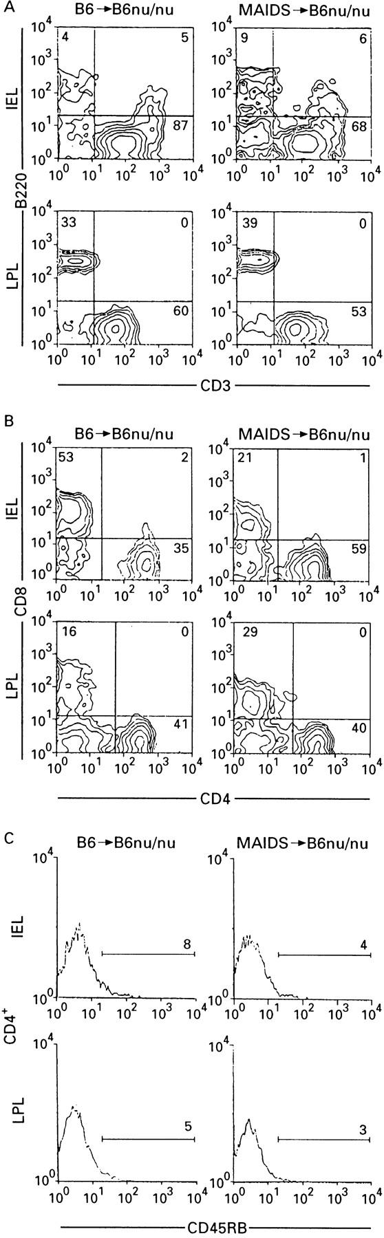
Methods: Four-week old mice were inoculated with the virus and their spleen cells were transferred into syngeneic nu/nu mice. Their organs were examined by light and electron microscopy. Phenotypes of the colon infiltrating cells were examined by flow cytometry.
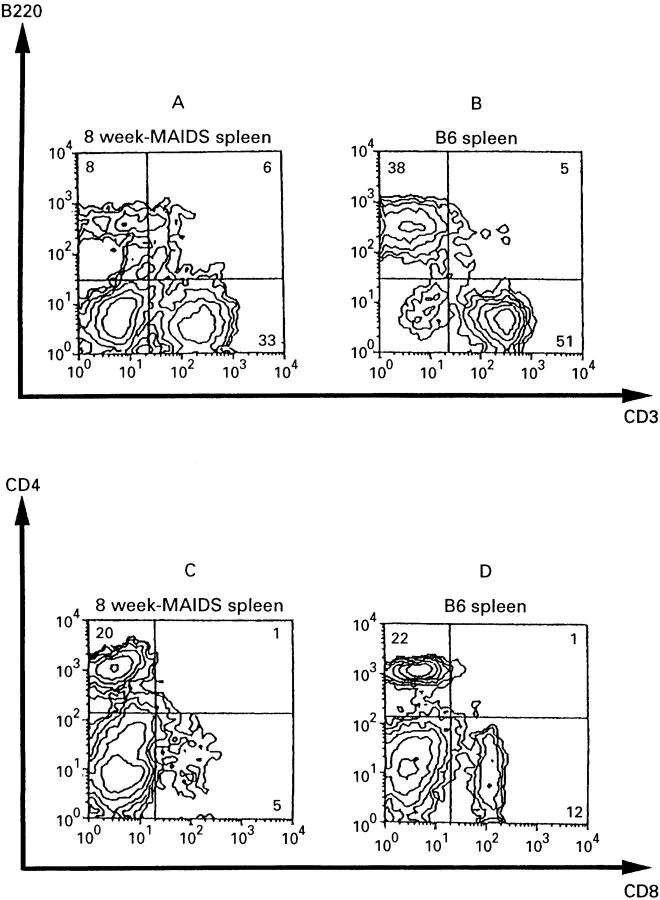
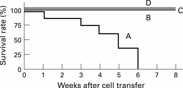
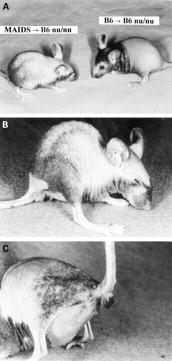
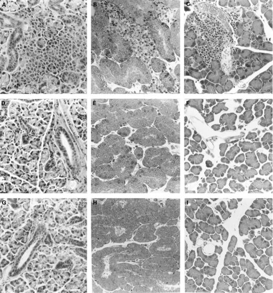
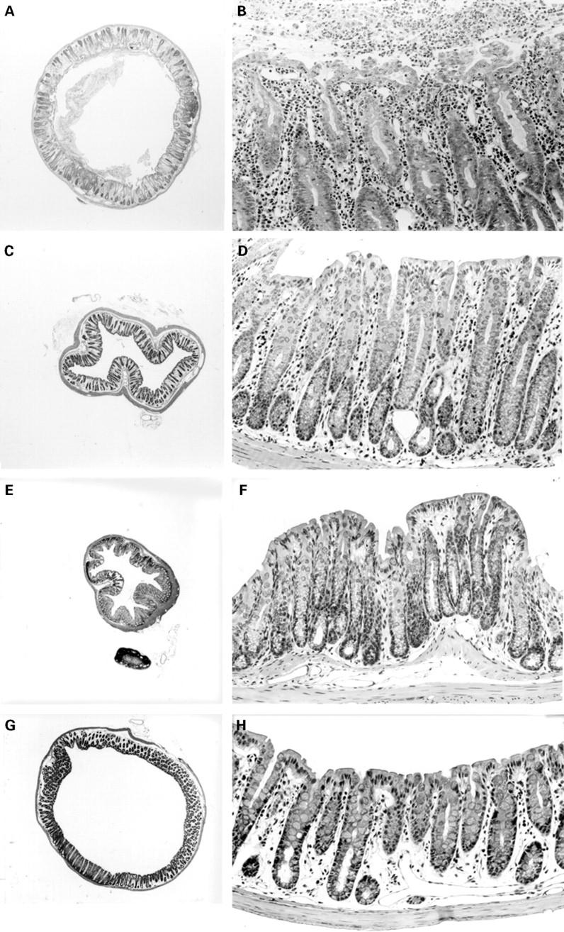
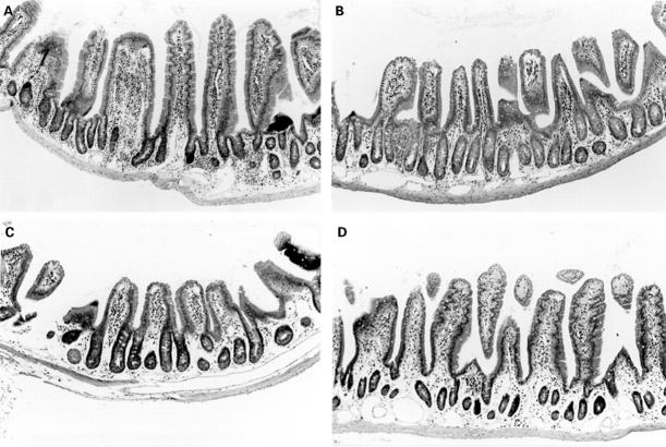
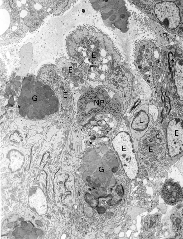
Results: All nu/nu recipients had died by six weeks after cell transfer, showing runting disease like cachexia with diarrhoea and anal bleeding. Histopathological examination revealed that systemic exocrinopathy was adoptively transferable and that the colon became thickened due to mononuclear cell infiltration into the mucosal and submucosal layer with hyperplasia of intestinal epithelial cells. No virus particles were found in the colon. Flow cytometric analyses revealed that most of the infiltrating CD4+ T cells showed CD45RBlow. No intestinal lesions were observed in the virus infected mice nor in nu/nu mice inoculated with normal lymphocytes.
Conclusion: Lymphocytes of the virus infected mice induced colitis and hyperplasia of intestinal epithelial cells as well as systemic exocrinopathy in nu/nu mice. Our experimental system may give some insight into intestinal lesions associated with virus infection.
Figures
Similar articles
-
Analysis of cytokine production in the colon of nude mice with experimental colitis induced by adoptive transfer of immunocompetent cells from mice infected with a murine retrovirus.Clin Immunol. 2000 Oct;97(1):33-42. doi: 10.1006/clim.2000.4909. Clin Immunol. 2000. PMID: 10998315
-
Exocrinopathy resembling Sjögren's syndrome induced by a murine retrovirus.Lab Invest. 1993 Oct;69(4):430-5. Lab Invest. 1993. PMID: 8231110
-
The drug monosodium luminol (GVT) preserves crypt-villus epithelial organization and allows survival of intestinal T cells in mice infected with the ts1 retrovirus.Immunol Lett. 2009 Feb 21;122(2):150-8. doi: 10.1016/j.imlet.2008.12.012. Epub 2009 Jan 30. Immunol Lett. 2009. PMID: 19186189
-
A murine model of retroviral encephalopathy.Curr Protoc Neurosci. 2002 Feb;Chapter 9:Unit9.8. doi: 10.1002/0471142301.ns0908s17. Curr Protoc Neurosci. 2002. PMID: 18428573 Review.
-
Murine models for acquired immune deficiency syndrome.Life Sci. 1989;44(3):iii-xv. doi: 10.1016/0024-3205(89)90592-4. Life Sci. 1989. PMID: 2536876 Review.
Cited by
-
Citrobacter-induced colitis in mice with murine acquired immunodeficiency syndrome.Vet Pathol. 2010 Mar;47(2):312-7. doi: 10.1177/0300985809358605. Epub 2010 Jan 29. Vet Pathol. 2010. PMID: 20118320 Free PMC article.
-
Mouse mammary tumor virus is implicated in severity of colitis and dysbiosis in the IL-10-/- mouse model of inflammatory bowel disease.Microbiome. 2023 Mar 3;11(1):39. doi: 10.1186/s40168-023-01483-4. Microbiome. 2023. PMID: 36869359 Free PMC article.
-
Cyclophilin C-associated protein (CyCAP) knock-out mice spontaneously develop colonic mucosal hyperplasia and exaggerated tumorigenesis after treatment with carcinogen azoxymethane.BMC Cancer. 2009 Jul 24;9:251. doi: 10.1186/1471-2407-9-251. BMC Cancer. 2009. PMID: 19627619 Free PMC article.
References
Publication types
MeSH terms
LinkOut - more resources
Full Text Sources
Research Materials