Pathology of idiopathic megarectum and megacolon
- PMID: 9301507
- PMCID: PMC1891468
- DOI: 10.1136/gut.41.2.252
Pathology of idiopathic megarectum and megacolon
Abstract
Background: The aetiology and pathology of both idiopathic megarectum and idiopathic megacolon are unknown. In particular, it is unknown whether there are abnormalities involving enteric nerves or smooth muscle.
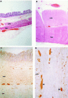
Methods: Resected tissue was examined from 24 patients who underwent surgery for idiopathic megarectum, from six patients who had tissue resected for idiopathic megacolon, and 17 control patients who had surgery for non-obstructing large bowel cancer. Qualitative and quantitative histological examination was performed after staining with haematoxylin and eosin, periodic acid Schiff (PAS), Martius scarlet blue (MSB), and phosphotungstic acid haematoxylin (PTAH). Neural and glial tissue were examined after immuno-staining with S100 and PGP9.5.
Results: Compared with controls, patients with idiopathic megarectum had significant thickening of their muscularis mucosae (median 78 v 33 microns, p < 0.005), circular muscle (1000 v 633 microns, p < 0.005), and longitudinal muscle (1083 v 303 microns, p < 0.005), despite rectal dilatation. This thickening was relatively greater in the longitudinal than in the circular muscle. Fibrosis of the longitudinal muscle was seen, using MSB staining, in 58%, of circular muscle in 38%, and of muscularis mucosae in 29% of patients. The relation between muscle thickening and fibrosis was variable. The density of neural tissue in the longitudinal muscle seemed to be reduced in patients with idiopathic megarectum. There was no thickening of enteric muscle or alteration in the density of innervation in patients with idiopathic megacolon.
Conclusion: There is notable thickening of the enteric smooth muscle in patients with idiopathic megarectum, but the architecture of the enteric innervation seems to be intact. Functional abnormalities of the latter remain a possible cause of the smooth muscle hypertrophy.
Figures

Similar articles
-
Enteric innervation in idiopathic megarectum and megacolon.Int J Colorectal Dis. 1996;11(6):264-71. doi: 10.1007/s003840050059. Int J Colorectal Dis. 1996. PMID: 9007620
-
Altered contractile proteins and neural innervation in idiopathic megarectum and megacolon.Histopathology. 1998 Jul;33(1):34-8. doi: 10.1046/j.1365-2559.1998.00438.x. Histopathology. 1998. PMID: 9726046
-
Clinical features of idiopathic megarectum and idiopathic megacolon.Gut. 1997 Jul;41(1):93-9. doi: 10.1136/gut.41.1.93. Gut. 1997. PMID: 9274479 Free PMC article.
-
Surgery for idiopathic megarectum and megacolon.Int J Colorectal Dis. 1991 Aug;6(3):171-4. doi: 10.1007/BF00341241. Int J Colorectal Dis. 1991. PMID: 1744491 Review. No abstract available.
-
Hirschsprung's disease: a review of the morphology and physiology.Postgrad Med J. 1972 Aug;48(562):471-7. doi: 10.1136/pgmj.48.562.471. Postgrad Med J. 1972. PMID: 4116711 Free PMC article. Review. No abstract available.
Cited by
-
Idiopathic proximal hemimegacolon in an adult woman.J Neurogastroenterol Motil. 2010 Apr;16(2):203-6. doi: 10.5056/jnm.2010.16.2.203. Epub 2010 Apr 27. J Neurogastroenterol Motil. 2010. PMID: 20535353 Free PMC article.
-
Idiopathic Megacolon-Short Review.Diagnostics (Basel). 2021 Nov 15;11(11):2112. doi: 10.3390/diagnostics11112112. Diagnostics (Basel). 2021. PMID: 34829459 Free PMC article. Review.
-
A Rare Case of Idiopathic Megacolon and Megarectum.Cureus. 2024 Dec 6;16(12):e75229. doi: 10.7759/cureus.75229. eCollection 2024 Dec. Cureus. 2024. PMID: 39759630 Free PMC article.
-
Symptoms and diagnostic criteria of acquired Megacolon - a systematic literature review.BMC Gastroenterol. 2018 Jan 31;18(1):25. doi: 10.1186/s12876-018-0753-7. BMC Gastroenterol. 2018. PMID: 29385992 Free PMC article.
-
Use of radiographic and histologic scores to evaluate cats with idiopathic megacolon grouped based on the duration of their clinical signs.Front Vet Sci. 2022 Dec 16;9:1033090. doi: 10.3389/fvets.2022.1033090. eCollection 2022. Front Vet Sci. 2022. PMID: 36590806 Free PMC article.
References
Publication types
MeSH terms
LinkOut - more resources
Full Text Sources
Medical
