

Medial temporal atrophy on MRI in normal aging and very mild Alzheimer's disease
- PMID: 9305341
- PMCID: PMC2730601
- DOI: 10.1212/wnl.49.3.786
Medial temporal atrophy on MRI in normal aging and very mild Alzheimer's disease
Abstract
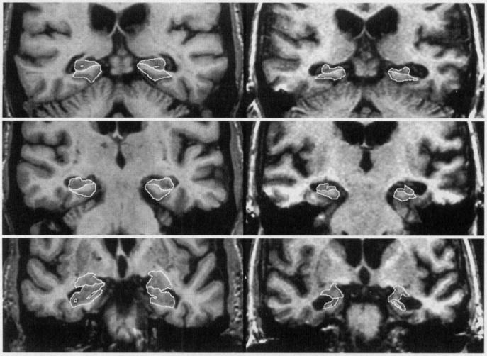
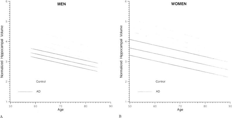
Magnetic resonance imaging (MRI)-based volumetric measurements of medial temporal lobe (MTL) structures can discriminate between normal elderly control subjects and patients with Alzheimer's disease (AD) of moderate to advanced severity. In terms of clinical utility, however, a more important issue concerns the ability of the technique to differentiate between normal elderly control subjects and AD patients with the very mildest form of the disease. We performed MRI-based volumetric measurements of the hippocampus, parahippocampal gyrus, and amygdala in 126 cognitively normal elderly control subjects and 94 patients with probable AD. The diagnosis of AD was made according to NINDS/ADRDA criteria, and disease severity was categorized by Clinical Dementia Rating (CDR) scores. Patients with CDR 0.5 were classified as very mild, CDR 1 as mild, and CDR 2 as moderate disease severity. Volumes of each structure declined with increasing age in control subjects and did so in parallel for men and women. The volume of each measured MTL structure also declined with age in patients with AD. The volume of each MTL structure was significantly smaller in AD patients than control subjects (p < 0.001). Of the several MTL measures, the total hippocampal volumetric measurements were best at discriminating control subjects from AD patients. The mean hippocampal volumes for AD patients relative to control subjects by severity of disease were as follows: very mild AD (CDR 0.5) -1.75 SD below the control mean, mild AD (CDR 1) -1.99 SD, and moderate AD (CDR 2) -2.22 SD. Age- and gender-adjusted, normalized MRI-based hippocampal volumetric measurements provide a sensitive marker of the MTL neuroanatomic degeneration in AD early in the disease process.
Figures
Comment in
-
The added value of MRI measures of atrophy in the diagnosis of AD.Neurology. 1998 Oct;51(4):1238. doi: 10.1212/wnl.51.4.1238. Neurology. 1998. PMID: 9781589 No abstract available.
References
-
- McKhann G, Drachman D, Folstein M, Katzman R, Price D, Stadlan EM. Clinical diagnosis of Alzheimer's disease: report of the NINCDS-ADRDA work group under the auspices of Department of Health and Human Services Task Force on Alzheimer's disease. Neurology. 1984;34:939–944. - PubMed
-
- Katzman R. Alzheimer's Disease. NEJM. 1986;314:964–973. - PubMed
-
- Kokmen E, Beard CM, O'Brien PC, Offord KP, et al. Is the incidence of dementing illness changing? A 25-year-time-trend study in Rochester, MN (1960-1984). Neurology. 1993;43:1887–1892. - PubMed
-
- DeCarli C, Kaye JA, Horwitz B, Rapoport SI. Critical analysis of the use of computer-assisted transverse axial tomography to study human brain in aging and dementia of the Alzheimer type. Neurology. 1990;40:872–883. - PubMed
-
- Squire LR. memory and the hippocampus: a synthesis from findings with rats, monkeys, and humans. Psychological Review. 1992;99:195–231. - PubMed
Publication types
MeSH terms
Grants and funding
LinkOut - more resources
Full Text Sources
Other Literature Sources
Medical