Molecular damage in the bronchial epithelium of current and former smokers
- PMID: 9308707
- PMCID: PMC5193483
- DOI: 10.1093/jnci/89.18.1366
Molecular damage in the bronchial epithelium of current and former smokers
Abstract
Background: Most lung cancers are attributed to smoking. These cancers have been associated with multiple genetic alterations and with the presence of preneoplastic bronchial lesions. In view of such associations, we evaluated the status of specific chromosomal loci in histologically normal and abnormal bronchial biopsy specimens from current and former smokers and specimens from nonsmokers.
Methods: Multiple biopsy specimens were obtained from 18 current smokers, 24 former smokers, and 21 nonsmokers. Polymerase chain reaction-based assays involving 15 polymorphic microsatellite DNA markers were used to examine eight chromosomal regions for genetic changes (loss of heterozygosity [LOH] and microsatellite alterations).
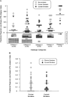
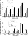
Results: LOH and microsatellite alterations were observed in biopsy specimens from both current and former smokers, but no statistically significant differences were observed between the two groups. Among individuals with a history of smoking, 86% demonstrated LOH in one or more biopsy specimens, and 24% showed LOH in all biopsy specimens. About half of the histologically normal specimens from smokers showed LOH, but the frequency of LOH and the severity of histologic change did not correspond until the carcinoma in situ stage. A subset of biopsy specimens from smokers that exhibited either normal or preneoplastic histology showed LOH at multiple chromosomal sites, a phenomenon frequently observed in carcinoma in situ and invasive cancer. LOH on chromosomes 3p and 9p was more frequent than LOH on chromosomes 5q, 17p (17p13; TP53 gene), and 13q (13q14; retinoblastoma gene). Microsatellite alterations were detected in 64% of the smokers. No genetic alterations were detected in nonsmokers.
Conclusions: Genetic changes similar to those found in lung cancers can be detected in the nonmalignant bronchial epithelium of current and former smokers and may persist for many years after smoking cessation.
Figures


References
-
- Parker SL, Tong T, Bolden S, Wingo PA. Cancer statistics, 1996. CA Cancer J Clin. 1996;46:5–27. - PubMed
-
- Parkin DM, Pisani P, Lopez AD, Masuyer E. At least one in seven cases of cancer is caused by smoking. Global estimates for 1985. Int J Cancer. 1994;5904:494–504. - PubMed
-
- Saccomanno G, Archer VE, Auerbach O, Saunders RP, Brennan LM. Development of carcinoma of the lung as reflected in exfoliated cells. Cancer. 1974;33:256–270. - PubMed
-
- Auerbach O, Stout AP, Hammond EC, Garfinkel L. Changes in bronchial epithelium in relation to smoking and cancer of the lung. N Engl J Med. 1961;265:253–267. - PubMed
-
- Auerbach O, Hammond EC, Garfinkel L. Changes in bronchial epithelium in relation to cigarette smoking, 1955–1960 vs. 1970–1977. N Engl J Med. 1979;300:381–385. - PubMed
Publication types
MeSH terms
Grants and funding
LinkOut - more resources
Full Text Sources
Other Literature Sources
Research Materials
Miscellaneous

