. 1997 Sep 15;11(18):2347-58.
doi: 10.1101/gad.11.18.2347.

PARP is important for genomic stability but dispensable in apoptosis

Affiliations

PARP is important for genomic stability but dispensable in apoptosis

Z Q Wang et al. Genes Dev. .

Abstract

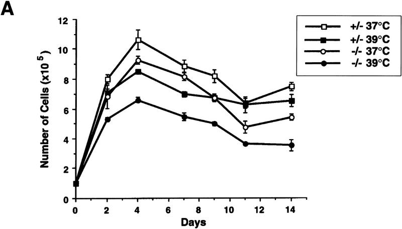
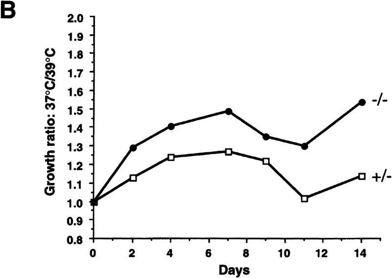
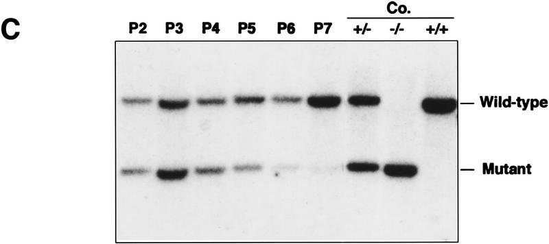
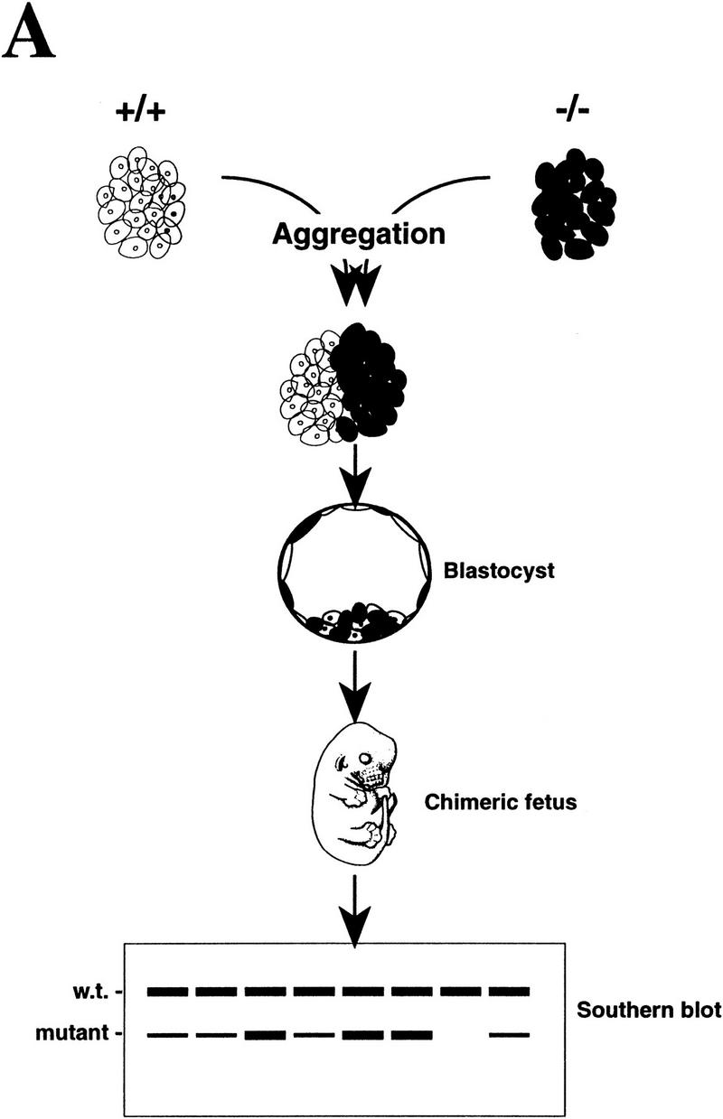
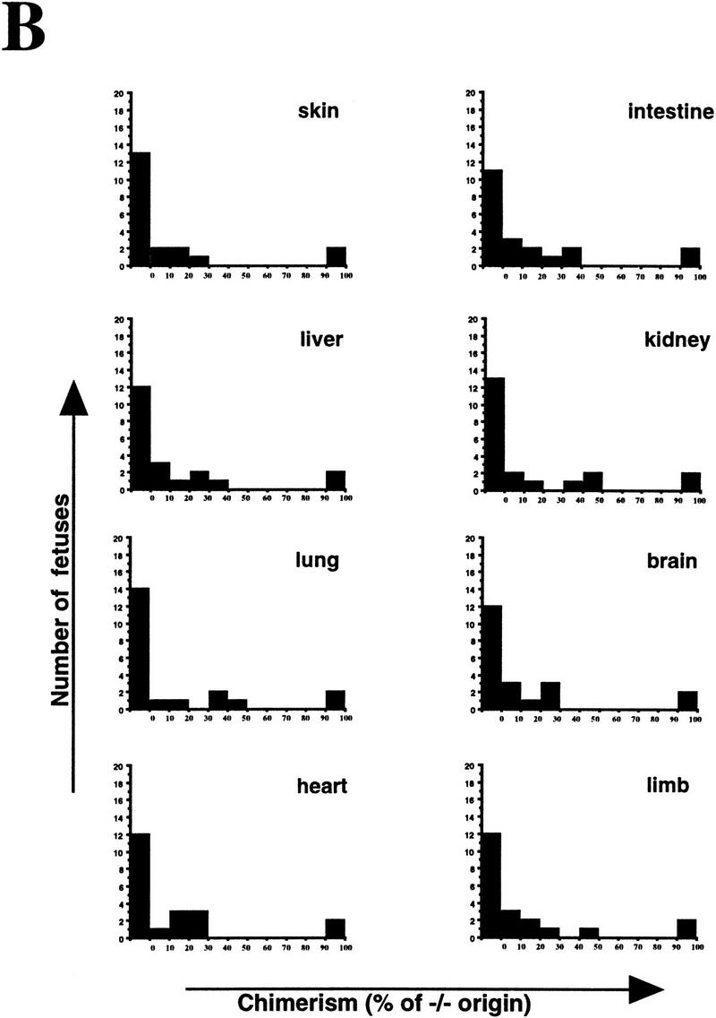
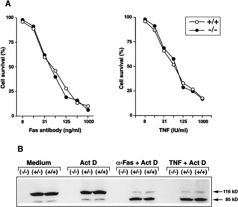
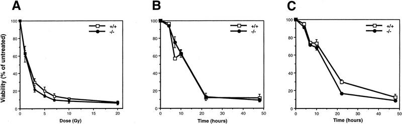
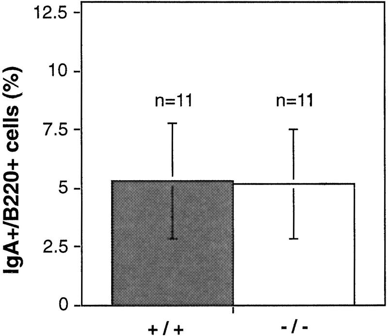
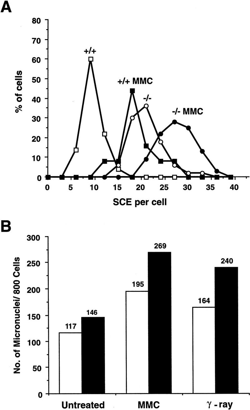
Mice lacking the gene encoding poly(ADP-ribosyl) transferase (PARP or ADPRT) display no phenotypic abnormalities, although aged mice are susceptible to epidermal hyperplasia and obesity in a mixed genetic background. Whereas embryonic fibroblasts lacking PARP exhibit normal DNA excision repair, they grow more slowly in vitro. Here we investigated the putative roles of PARP in cell proliferation, cell death, radiosensitivity, and DNA recombination, as well as chromosomal stability. We show that the proliferation deficiency in vitro and in vivo is most likely caused by a hypersensitive response to environmental stress. Although PARP is specifically cleaved during apoptosis, cells lacking this molecule apoptosed normally in response to treatment with anti-Fas, tumor neurosis factor alpha, gamma-irradiation, and dexamethasone, indicating that PARP is dispensable in apoptosis and that PARP-/- thymocytes are not hypersensitive to ionizing radiation. Furthermore, the capacity of mutant cells to carry out immunoglobulin class switching and V(D)J recombination is normal. Finally, primary PARP mutant fibroblasts and splenocytes exhibited an elevated frequency of spontaneous sister chromatid exchanges and elevated micronuclei formation after treatment with genotoxic agents, establishing an important role for PARP in the maintenance of genomic integrity.


Figures

Figure 1
Figure 1
Proliferation of PARP mutant cells in response to experimental stress. (A) PARP heterozygous (+/−) and homozygous (−/−) primary fibroblasts were cultured at 37°C and 39°C for 14 days. At each time point the number of cells was counted from triplicate culture. (B) The ratio of number of cells at 37°C vs. 39°C indicates the magnitude of proliferation reduction of each genotype. (C) Southern blot analysis of genomic DNA extracted from the co-cultures of PARP−/− (mutant) and wild-type cells at different passages from passage 2 (P2) through 7 (P7). DNA was digested by PvuII and hybridized with a specific probe (see Materials and Methods). Control DNAs (Co.) are from wild-type (+/+), heterozygous (+/−) and homozygous (−/−) mouse tail biopsies respectively. The presence of mutant cells in cocultured mixtures is depicted by the intensity of the bands on the Southern blot.
Figure 1
Figure 1
Proliferation of PARP mutant cells in response to experimental stress. (A) PARP heterozygous (+/−) and homozygous (−/−) primary fibroblasts were cultured at 37°C and 39°C for 14 days. At each time point the number of cells was counted from triplicate culture. (B) The ratio of number of cells at 37°C vs. 39°C indicates the magnitude of proliferation reduction of each genotype. (C) Southern blot analysis of genomic DNA extracted from the co-cultures of PARP−/− (mutant) and wild-type cells at different passages from passage 2 (P2) through 7 (P7). DNA was digested by PvuII and hybridized with a specific probe (see Materials and Methods). Control DNAs (Co.) are from wild-type (+/+), heterozygous (+/−) and homozygous (−/−) mouse tail biopsies respectively. The presence of mutant cells in cocultured mixtures is depicted by the intensity of the bands on the Southern blot.
Figure 2
Figure 2
Proliferation of PARP mutant cells in vivo. (A) The diagram shows the experimental approach to examine the proliferation capacity and competence of PARP mutant cells. Wild-type (w.t., +/+) and PARP mutant (−/−) embryos were aggregated at the morula stage. The proliferation status of mutant cells in chimeric embryos was monitored by the contribution of each origin, which was determined by quantitative Southern blot analysis using DNA isolated from different organs (see also Materials and Methods). (B) A total of 21 E17.5 fetuses was obtained and eight tissues from each were analyzed by Southern blotting. The contribution from PARP−/− origin is represented by the percentage along with the number of fetuses that contained the same range of contribution in a given organ.
Figure 2
Figure 2
Proliferation of PARP mutant cells in vivo. (A) The diagram shows the experimental approach to examine the proliferation capacity and competence of PARP mutant cells. Wild-type (w.t., +/+) and PARP mutant (−/−) embryos were aggregated at the morula stage. The proliferation status of mutant cells in chimeric embryos was monitored by the contribution of each origin, which was determined by quantitative Southern blot analysis using DNA isolated from different organs (see also Materials and Methods). (B) A total of 21 E17.5 fetuses was obtained and eight tissues from each were analyzed by Southern blotting. The contribution from PARP−/− origin is represented by the percentage along with the number of fetuses that contained the same range of contribution in a given organ.
Figure 3
Figure 3
Apoptosis of PARP mutant cells. (A) Primary embryonic fibroblasts were treated for 18 hr with different concentrations of TNF or anti-Fas antibody and the viability was monitored by the MTT assay. (B) Detection of PARP cleavage: Primary embryonic fibroblasts of wild-type (+/+), heterozygous (+/−), and homozygous (−/−) mice were incubated for 8 hr with anti-Fas (α-Fas) or TNF in the presence of actinomycin D (Act D). Total cell extracts were prepared, subjected to immunoprecipitation with an PARP-specific antiserum, and analyzed by SDS-PAGE and immunoblotting. Cleavage of PARP (116 kD) into the characteristic 85-kD fragment was visualized with anti-PARP antibodies. Note the absence of PARP in homozygous (−/−) cells.
Figure 4
Figure 4
Apoptosis of thymocytes from wild-type (+/+) and PARP mutant (−/−) mice following the treatment with γ-irradiation and dexamethasone. (A) Cells were irradiated with different doses of γ-ray and their viability was measured after 24 hr. (B) After treatment with 5 Gy of γ-irradiation, cell viability was monitored at different time points. (C) Induction of cell death following treatment of thymocytes with dexamethasone.
Figure 5
Figure 5
IgA switching in wild-type (+/+) and PARP mutant (−/−) mice. Splenocytes were treated with TGFβ to induce expression of surface IgA. After 4 days, IgA-positive B lymphocytes were determined by costaining using anti-IgA and anti-B220 antibodies. The results from 11 mice per genotype are depicted.
Figure 6
Figure 6
(A) SCE rate in splenocytes freshly isolated from wild-type (+/+) and PARP mutant (−/−) mice. Cells treated and nontreated with MMC were counted for SCE. Whereas 50 chromosomal spreads were counted for the untreated groups, 25 spreads were counted after MMC treatment. (B) Micronuclei formation in primary embryonic fibroblasts isolated from wild-type (open bars) and mutant (solid bars) mice. A total of 800 cells was counted for the presence of micronuclei with or without treatment with γ-irradiation or MMC. Although untreated cells showed no significant difference in the number of micronuclei between +/+ and −/− cells, after treatment the number of micronuclei in −/− cells was significantly higher than in +/+ cells (P  0.01).

Similar articles

Cited by

References

    1. Agarwal, M., A. Agarwal, W.R. Taylor, Z.-Q. Wang, E.F. Wagner, and G. Stark. 1997. Defective induction but normal activation and function of p53 in mouse cells lacking poly-ADP-ribose polymerase. Oncogene (in press). - PubMed
    1. Althaus FR, Richter C. ADP-ribosylation of proteins—Enzymology and biological significance. Mol Biol Biochem Biophys. 1987;37:1–125. - PubMed
    1. Casciola-Rosen L, Nicholson DW, Chong T, Rowan KR, Thornberry NA, Miller DK, Rosen A. Apopain/CPP32 cleaves proteins that are essential for cellular repair: A fundamental principle of apoptotic death. J Exp Med. 1996;183:1957–1964. - PMC - PubMed
    1. Catena C, Villani P, Conti D, Righi E. Micronuclei and 3AB index in X-irradiated human lymphocytes in G0 and G1 phases. Mutat Res. 1994;311:231–237. - PubMed
    1. Cleaver JE, Mitchell DL, Feeney L, Afzal V. Chromatid exchanges may be induced by damage in sites of transcriptional activity. Mutagenesis. 1996;11:183–187. - PubMed

Publication types

Substances