. 1997 Sep 15;11(18):2383-95.
doi: 10.1101/gad.11.18.2383.

DNA methylation inhibits elongation but not initiation of transcription in Neurospora crassa

Affiliations

DNA methylation inhibits elongation but not initiation of transcription in Neurospora crassa

M R Rountree et al. Genes Dev. .

Abstract

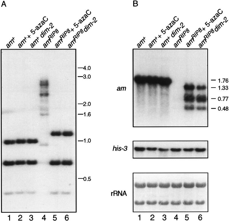
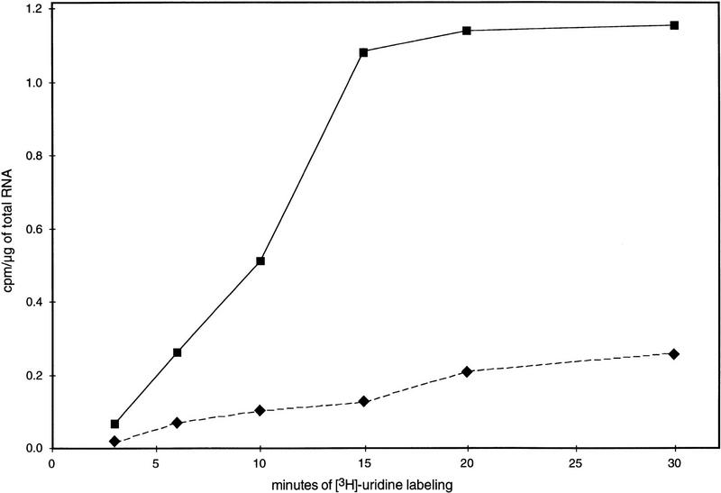
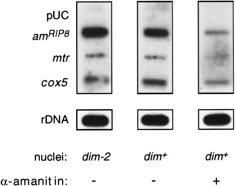
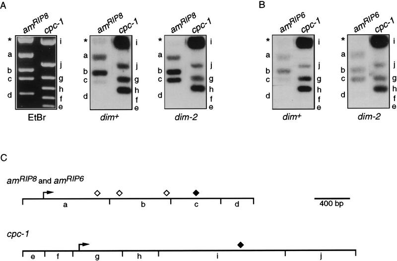
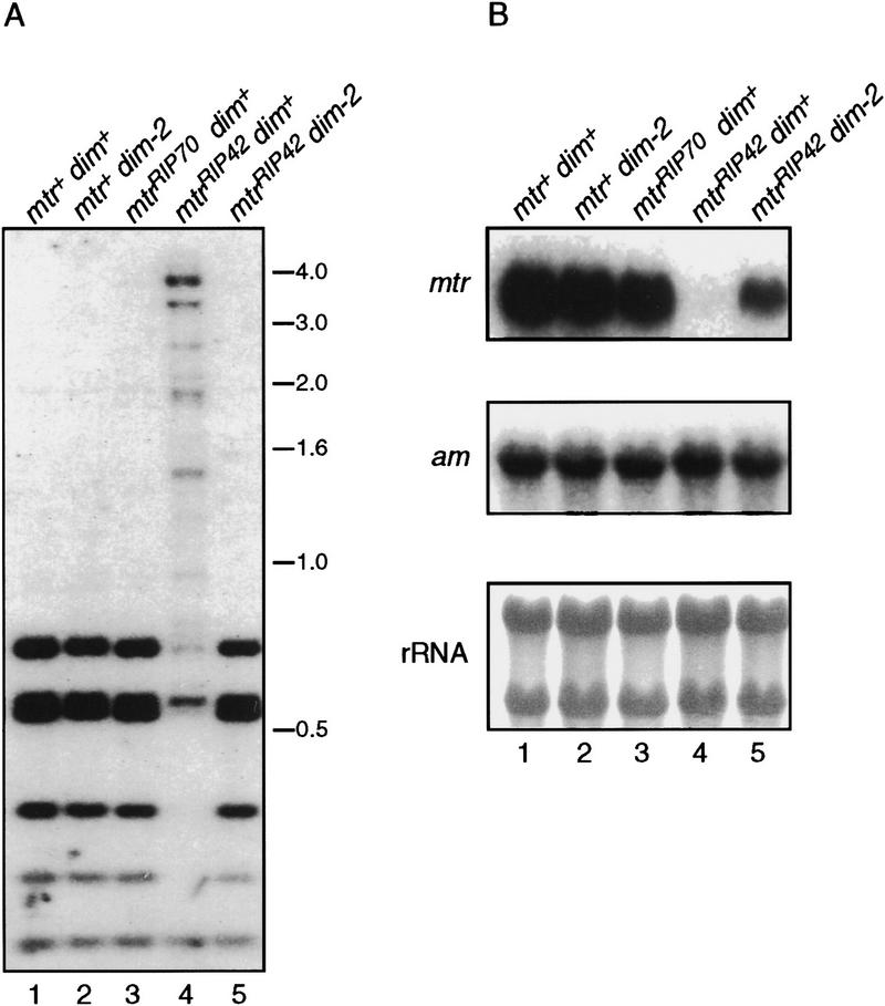
In plants, animals, and fungi, DNA methylation is frequently associated with gene silencing, yet little is known about the role of the methylation in silencing. In Neurospora crassa, repeated sequences are silenced by repeat-induced point mutation (RIP) and genes that have suffered numerous GC --> AT mutations by RIP are typically methylated at remaining cytosines. We investigated possible effects on transcription from methylation associated with RIP by taking advantage of 5-azacytidine, which prevents most methylation in Neurospora and a dim-2 mutation that abolishes all detectable methylation. Northern analyses revealed that methylation prevents the accumulation of transcripts from genes mutated by RIP. Measurements of transcription rates in vivo showed that methylation inhibits transcription severely but does not influence mRNA stability. Results of nuclear run-on experiments demonstrated that transcription initiation was not significantly inhibited by the dense methylation in the promoter sequences. In contrast, methylation blocked transcription elongation in vivo.


Figures

Figure 1
Figure 1
Treatment with 5-azaC results in an accumulation of transcripts from the methylated amRIP alleles. DNA and RNA were isolated from strains grown in the presence (+) or absence (−) of 24 μm 5-azaC. (A) 5-AzaC treatment results in demethylation of the methylated amRIP alleles. Genomic DNA (750 ng) from strains N150 [am+ (lanes 1,2)], N665 [amRIP2 (lanes 3,4)], N673 [amRIP5 (lanes 5,6)], N675 [amRIP6 (lanes 7,8)], and N617 [amRIP8 (lanes 9,10)] was digested with the methylation-sensitive restriction enzyme Sau3A1, subjected to Southern blotting, and hybridized to an am probe. Size standards (kb) are indicated at left. The Southern blot was stripped and reprobed with the unmethylated mtr gene to confirm complete digestion of the DNA (data not shown). (B) Corresponding Northern analyses. The blot at top was hybridized to a wild-type am probe, whereas the blot in the middle was hybridized to a his-3 probe. (Bottom) Methylene blue staining of the rRNA of the blot from the top. (C) Map of the am gene. The am locus (horizontal line) with the exons (thick bars), transcription start sites (arrow), Sau3A1 restriction sites (ticks below the horizontal line), and scale bar are indicated. The mutations (vertical lines) for the amRIP2, amRIP5, amRIP6, and amRIP8 alleles are depicted below the diagram of the am locus. The asterisk (*) marks the Sau3A1 site that is mutated in the amRIP2 and amRIP8 alleles.
Figure 2
Figure 2
Prevention of methylation by the dim-2 mutation increases amRIP8 transcript levels. (A) Southern analysis of genomic DNA from the strains N150 [am+; dim+ (lane 1)], N150 treated with 5-azaC (lane 2), N540 [am+; dim-2 (lane 3)], N617 [amRIP8; dim+ (lane 4)], N617 treated with 5-azaC (lane 5), and N618 [amRIP8; dim-2 (lane 6)]. Genomic DNA (750 ng) was digested with the methylation-sensitive restriction enzyme Sau3A1, subjected to Southern blotting, and hybridized to an am probe. Size standards (kb) are indicated at right. The Southern blot was stripped and reprobed with the unmethylated mtr gene to ensure complete digestion of the DNA (data not shown). (B) Northern analysis of total RNA (10 μg) from the strains depicted in the Southern blot. The blot at top was hybridized to a wild-type am probe. The size (kb) of the wild-type am transcript and of the three amRIP8 transcripts is indicated right of the top panel. The blot in the middle was hybridized to a his-3 probe. (Bottom) The methylene blue staining of the rRNA of the blot from the top.
Figure 3
Figure 3
Methylation analysis of amRIP8. The amRIP8 allele is represented by the horizontal line. The upstream enhancer-like sequences (α and β) are represented by open boxes. The start sites of transcription are indicated by the arrow. The mutations in amRIP8 are indicated by the ticks above the horizontal line. The approximate ends of the three amRIP8 transcripts are indicated by the open diamonds, whereas the end of the wild-type am transcript is indicated by the black diamond. The recognition sites for the methylation-sensitive restriction enzymes BamHI (B), BglII (G), Bsp106-I (S), NaeI (N), NarI (A), NsiI (I), PstI (P), PvuII (V), XbaI (X), and XhoI (H) are indicated below the horizontal line. The approximate level of methylation at each restriction enzyme site is depicted in black in a pie chart below each site.
Figure 4
Figure 4
In vivo labeling analysis of amRIP8 transcription in dim+ and dim-2 backgrounds. Time course of [3H]uridine incorporation into amRIP8 mRNA from N617 [amRIP8; dim+ (♦)] and N618 [amRIP8; dim-2 (▪)]. The transcription rates of the amRIP8 allele in the methylated (N617) and unmethylated (N618) states was estimated from the slope of the best-fit line over the first three time points. Transcription rates of the unmethylated histone H4 gene were very similar in the sibling strains, N1223 (0.194 cpm/μg of total RNA/min) and N1224 (0.188 cpm/μg of total RNA/min).
Figure 5
Figure 5
Analysis of amRIP8 transcription in dim+ and dim-2 backgrounds by the nuclear run-on assay. Slot–blots containing 5 μg of linearized denatured plasmids or 5 μg of a denatured rDNA fragment were probed with [32P]UTP-labeled transcripts isolated from nuclear run-on reactions. The pUC19 plasmid was included as a negative control, whereas the rDNA fragment and the plasmids containing the mtr and cox5 genes served as positive controls. The first two panels show representative blots probed with labeled RNA from nuclear run-on reactions conducted with nuclei isolated from N617 (amRIP8; dim+) and N618 (amRIP8; dim-2). The right panel shows a blot probed with labeled RNA from a run-on reaction performed with N617 nuclei in the presence of 1 mg/ml of α-amanitin.
Figure 6
Figure 6
Nuclear run-on analysis of amRIP8 and amRIP6 transcription elongation. Plasmids (5 μg) containing amRIP8 (pBM7), wild-type am (pMS2), or cpc-1 (pCPC1-2) were digested with the appropriate enzymes: pBM7 and pMS2 with BamHI and XmnI and pCPC1-2 with Bsu36I and PstI. Fragments were separated on 1.5% agarose gels, transferred to nylon membranes, and probed with [32P]UTP-labeled transcripts from nuclear run-on reactions. (A) A representative ethidium bromide-stained gel is shown at left with the restriction fragments of amRIP8 and cpc-1 labeled with lowercase letters corresponding to the fragments indicated in the restriction maps (C). Blots probed with [32P]UTP-labeled transcripts from nuclear run-on reactions performed with dim+ (amRIP8) nuclei (middle) or dim-2 (amRIP8) nuclei (right). (B) Blots containing separated wild-type am and cpc-1 fragments were probed with [32P]UTP-labeled transcripts from nuclear run-on reactions performed with dim+ (amRIP6) nuclei (left) or dim-2 (amRIP6) nuclei (right). The weaker signals obtained for amRIP6, when compared with amRIP8, were most likely attributable to hybridization of the amRIP6 transcripts to wild-type am sequences. (C) Maps of amRIP8, amRIP6, and cpc-1. (⋄) The approximate ends of the amRIP8 transcripts; (♦) the ends of the amRIP6 and cpc-1 transcripts.
Figure 7
Figure 7
The effect of methylation on mtrRIP42 transcript accumulation. (A) Southern analysis of genomic DNA from strains N1164 [mtr+; dim+ (lane 1)], N540 [mtr+; dim-2 (lane 2)], N1165 [mtrRIP70; dim+ (lane 3)], N1185 [mtrRIP42; dim+ (lane 4)], and N1183 [mtrRIP42; dim-2 (lane 5)]. Genomic DNA (750 ng) was digested with the methylation-sensitive restriction enzyme Sau3A1 and hybridized to an mtr probe. Size standards (kb) are indicated at right. The Southern blot was stripped and reprobed with the unmethylated am gene to ensure complete digestion of the DNA (data not shown). (B) Corresponding Northern analysis of total RNA (10 μg) from the strains depicted in the Southern blot. The blot at top was probed with mtr, whereas the equivalent blot in the middle was probed with am. The blot at bottom is a methylene blue staining of the rRNA from the blot at top.

Similar articles

Cited by

References

    1. Barry C, Faugeron G, Rossignol J-L. Methylation induced premeiotically in Ascobolus: Coextension with DNA repeat lengths and effect on transcript elongation. Proc Natl Acad Sci. 1993;90:4557–4561. - PMC - PubMed
    1. Bartolomei MS, Webber AL, Brunkow ME, Tilghman SM. Epigenetic mechanisms underlying the imprinting of the mouse H19 gene. Genes & Dev. 1993;7:1663–1673. - PubMed
    1. Bednarik DP, Duckett C, Kim SU, Perez VL, Griffis K, Guenther PC, Folks TM. DNA CpG methylation inhibits binding of NF-kB proteins to the HIV-1 long terminal repeat cognate DNA motifs. New Biol. 1991;3:969–976. - PubMed
    1. Beelman CA, Parker R. Degradation of mRNA in eukaryotes. Cell. 1995;81:179–183. - PubMed
    1. Ben-Hattar J, Beard P, Jiricny J. Methylation in CTF and Sp1 recognition sites of an HSV tk promoter: Effects on transcription in vivo and on factor binding in vitro. Nucleic Acids Res. 1989;17:10179–10190. - PMC - PubMed

Publication types

MeSH terms

LinkOut - more resources