. 1997 Sep 15;11(18):2396-413.
doi: 10.1101/gad.11.18.2396.

GTP hydrolysis controls stringent selection of the AUG start codon during translation initiation in Saccharomyces cerevisiae

Affiliations

GTP hydrolysis controls stringent selection of the AUG start codon during translation initiation in Saccharomyces cerevisiae

H K Huang et al. Genes Dev. .

Abstract

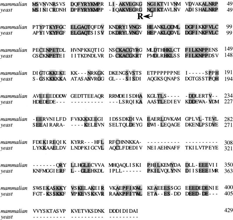
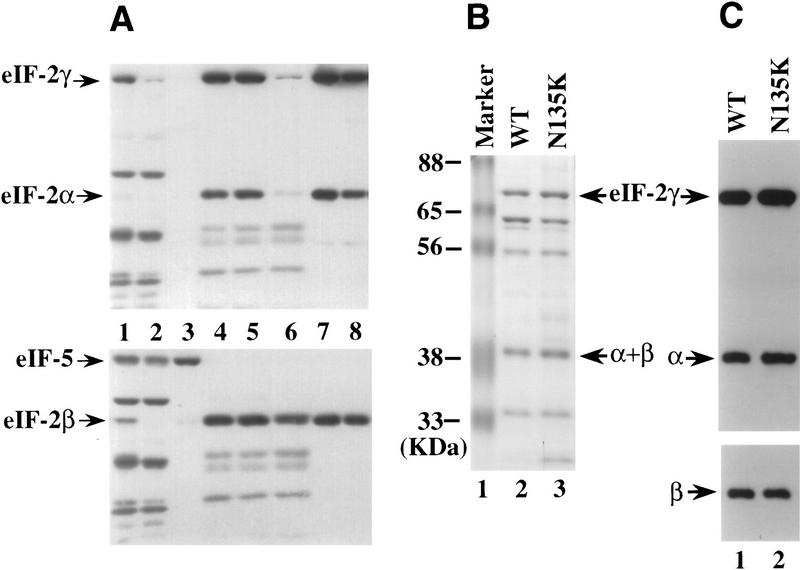
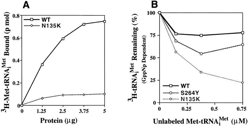
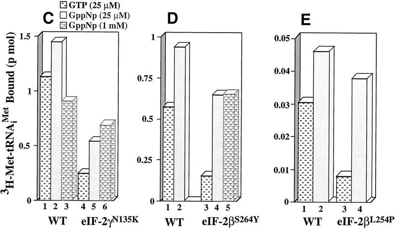
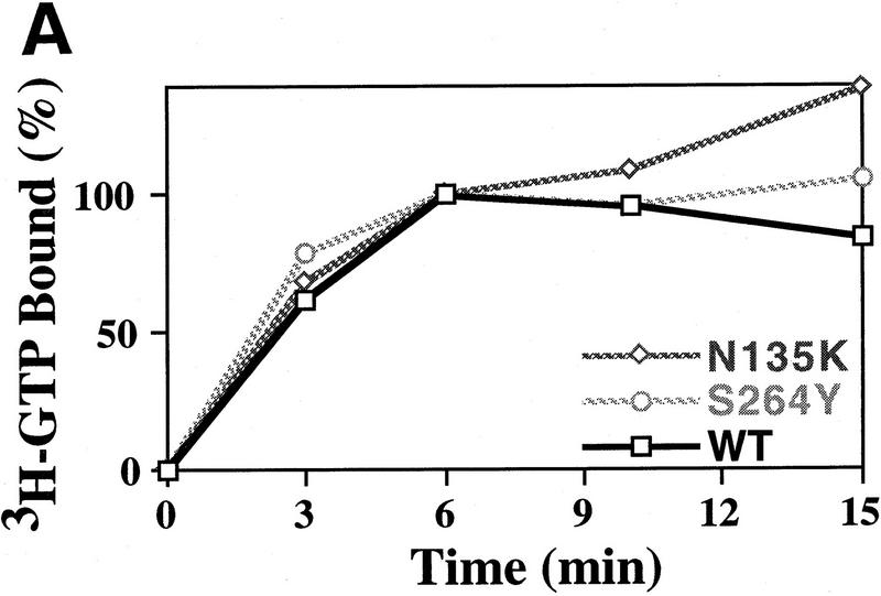
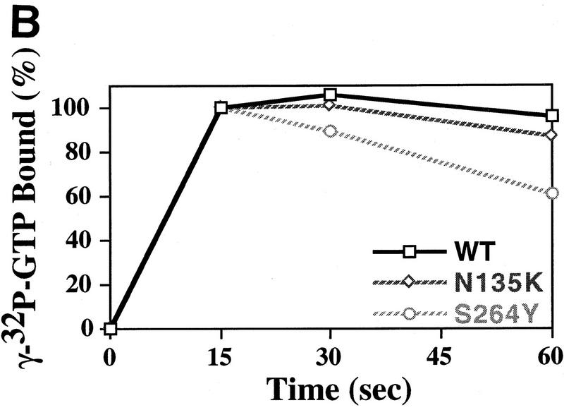
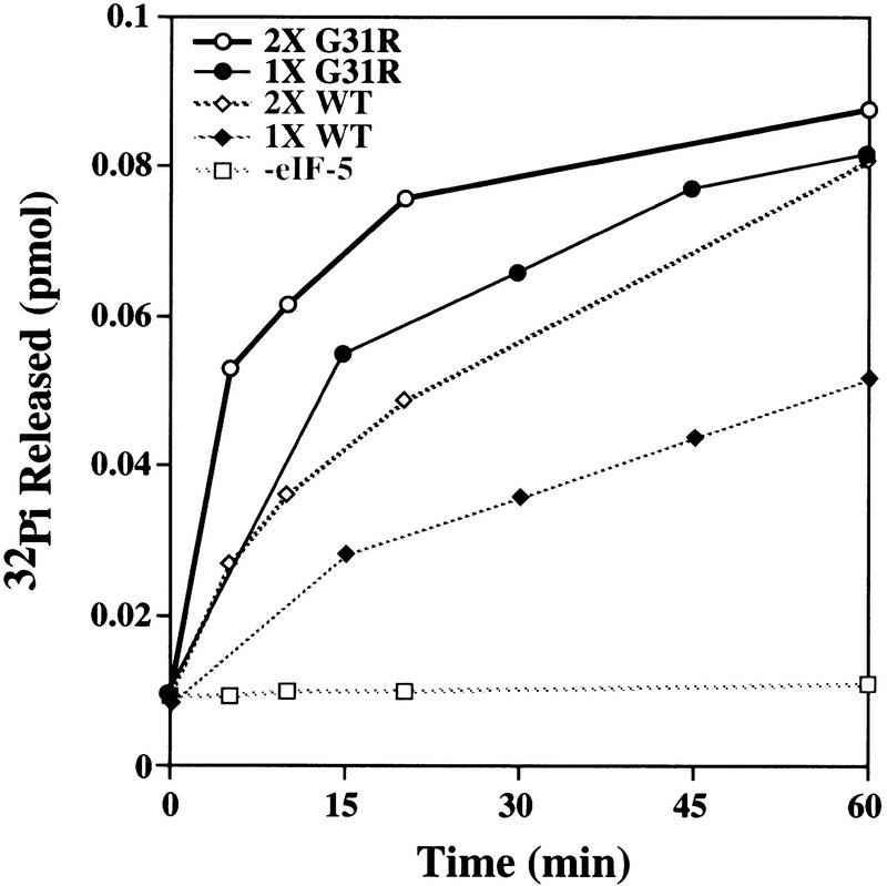
We have isolated and characterized two suppressor genes, SUI4 and SUI5, that can initiate translation in the absence of an AUG start codon at the HIS4 locus in Saccharomyces cerevisiae. Both suppressor genes are dominant in diploid cells and lethal in haploid cells. The SUI4 suppressor gene is identical to the GCD11 gene, which encodes the gamma subunit of the eIF-2 complex and contains a mutation in the G2 motif, one of the four signature motifs that characterizes this subunit to be a G-protein. The SUI5 suppressor gene is identical to the TIF5 gene that encodes eIF-5, a translation initiation factor known to stimulate the hydrolysis of GTP bound to eIF-2 as part of the 43S preinitiation complex. Purified mutant eIF-5 is more active in stimulating GTP hydrolysis in vitro than wild-type eIF-5, suggesting that an alteration of the hydrolysis rate of GTP bound to the 43S preinitiation complex during ribosomal scanning allows translation initiation at a non-AUG codon. Purified mutant eIF-2gamma complex is defective in ternary complex formation and this defect correlates with a higher rate of dissociation from charged initiator-tRNA in the absence of GTP hydrolysis. Biochemical characterization of SUI3 suppressor alleles that encode mutant forms of the beta subunit of eIF-2 revealed that these mutant eIF-2 complexes have a higher intrinsic rate of GTP hydrolysis, which is eIF-5 independent. All of these biochemical defects result in initiation at a UUG codon at the his4 gene in yeast. These studies in light of other analyses indicate that GTP hydrolysis that leads to dissociation of eIF-2 x GDP from the initiator-tRNA in the 43S preinitiation complex serves as a checkpoint for a 3-bp codon/anticodon interaction between the AUG start codon and the initiator-tRNA during the ribosomal scanning process.


Figures

Figure 1
Figure 1
Protein sequence alignment of mammalian (top) and yeast (bottom) eIF-5. The amino acid sequences of mammalian and yeast eIF-5 are compared for maximal alignment. The shaded regions indicate identical amino acid residues among the two proteins. The SUI5 suppressor allele contained a point mutation (GGT → CGT) that altered Gly-31 to Arg as indicated by the arrow.
Figure 2
Figure 2
Purification of the His-tagged wild-type eIF-2 and the eIF-2γN135K complexes. (A) Crude extracts and fractions from P-11 and Ni chromatography columns were resolved by 10% SDS-PAGE, transferred to a nitrocellulose membrane, and analyzed by Western blot analysis using antisera directed against eIF2-α and eIF2-γ (top) or eIF2-β and eIF-5 (bottom). Purification of wild-type eIF-2: (Lane 1) 50 μg of crude extract; (lane 2) 50 μg of protein from the flowthrough fraction of the P-11 column; (lane 3) 20 μg of protein from the 450 mm KCl wash fraction of the P-11 column; (lane 4) 10 μg of protein from the 750 mm KCl elution fraction of the P-11 column (predialysis); (lane 5) 10 μg of protein from the 750 mm KCl elution fraction of the P-11 column (after dialysis); (lane 6) 10 μg of protein from the flowthrough fraction from the nickel affinitiy column; (lane 7) 5 μg of protein from the pooled elution fraction from the nickel affinity column; (lane 8) 5 μg of wild-type eIF-2 after concentration of the pooled elution fraction by centricon-30. (B) Coomassie blue staining of purified wild-type eIF-2 and eIF-2γN135K complexes. Fifteen micrograms of protein from the concentration step after Ni affinity chromatography were resolved on a 10% SDS–polyacrylamide gel. (Lane 1) Molecular mass markers with positions noted as (kD); (lane 2) wild-type eIF-2 complex; and (lane 3) mutant eIF-2γN135K complex. Arrows point to the position of each of the eIF-2 subunits as determined by Western blot analysis. (C) Western blot analysis of purified wild-type eIF-2 and eIF-2γN135K complexes. Five micrograms of protein was resolved by 10% SDS-polyacrylamide gel and probed for the presence of the α, β, and γ subunits of eIF-2 by Western blot analysis. The position of each is noted by an arrow. (Lane 1) Wild-type eIF-2, (lane 2) mutant eIF-2γN135K.
Figure 3
Figure 3
Ternary complex formation by purified wild-type and mutant eIF-2 complexes. (A) Purified wild-type eIF-2 (□) and mutant eIF-2γN135K (⋄) were each assayed for the ability to promote GTP-dependent binding to the [3H] methionine charged initiator-tRNA [3H]Met–tRNAiMet, 60,000 cpm/pmole, 0.075 μm) as a function of protein concentration (μg). Identical reactions without GTP were performed as a control for nonspecific GTP-independent binding activity. The number of picomoles of [3H]Met–tRNAiMet bound in the absence of GTP was subtracted from the number of pimoles of [3H]Met–tRNAiMet] bound in the presence of GTP to determine the GTP-dependent-binding activity, for each respective eIF-2 complex. (B) Wild-type eIF-2 (□), the mutant eIF-2γN135K complex (⋄), and the mutant eIF-2βS264Y complex (○) were each assayed for their ability to dissociate from the [3H]Met–tRNAiMet in the presence of the nonhydrolyzable GTP analog, GppNp. For the wild-type eIF-2 and eIF-2βS264Y complexes, ternary complex formed after 5 min of incubation with GppNp and [3H]Met–tRNAiMet (0.075 μm) was competed with various concentrations of unlabeled, charged initiator-tRNA for an additional 5 min and the level of labeled ternary complex determined by the filter-binding assay. The same reaction conditions were used to assay the eIF-2γN135K complex with the exception that a 10-min incubation time was used in the initial step to enhance the level of labeled ternary complex. Ternary complex without addition of unlabeled charged initiator-tRNA was stable at 37°C for up to 15 min (data not shown). Identical reactions without GppNp were performed as a control for nonspecific binding of the labeled tRNA and subtracted from the respective assays as background. The amount of eIF-2 preparation in each reaction was adjusted to compensate for similar initial levels of ternary complex formation in the presence of GppNp. Total protein in each reaction was 1.25 μg for the wild-type eIF-2 and 5 μg for each of the mutant complexes. (C) The eIF-2γN135K complex (5 μg of total protein) was assayed for its ability to bind [3H]Met–tRNAiMet (60,000 cpm/pmole) in the presence of GppNp as compared with to the wild-type eIF-2 (5 μg of total protein). (Lanes 1–3) The amount of ternary complex formed by wild-type eIF-2 in the presence of GTP (25 μm), and 25 μm, and 1 mm GppNp, respectively, in a 5-min reaction. (Lanes 4–6) The amount of ternary complex formed by mutant eIF-2γN135K complex in the presence of GTP (25 μm); and 25 μm, and 1 mm GppNp, respectively, in a 10-min reaction. The 10-min time point used for the mutant complex analysis was to maximize the amount of initiator-tRNA binding. However, only a 10% increase in binding is observed by using a 10-min incubation vs. a 5-min incubation period. (D) Same as C except using purified mutant eIF-2βS264Y complex (5 μg of total protein) compared with wild-type eIF-2 complex (2.5 μg of total protein). The different levels of total protein added to each reaction adjusts for the lower yields of eIF-2 in the former preparation as determined by Western blot analysis using antibodies directed against the α subunit of eIF-2. (Lanes 1,2) The amount of ternary complex formed by wild-type eIF-2 in the presence of GTP (25 μm); and 25 μm GppNp, respectively, in a 5-min reaction. (Lanes 3–5) The amount of ternary complex formed by mutant eIF-2βS264Y complex in the presence of GTP (25 μm), and 25 μm, and 1 mm GppNp, respectively, in a 5-min reaction. (E) Same as C except using purified mutant eIF-2βL254P complex (5 μg of total protein) compared with wild-type eIF-2 complex (0.17 μg of total protein). The different levels of total protein added to each reaction adjusts for the lower yields of eIF-2 in the former preparation as determined by Western blot analysis using antibodies directed against the α subunit of eIF-2. (Lanes 1,2) The amount of ternary complex formed by wild-type eIF-2 in the presence of GTP (25 μm), and 25 μm GppNp, respectively in a 5-min reaction. (Lanes 3,4) The amount of ternary complex formed by mutant eIF-2βL254P complex in the presence of GTP (25 μm); and 25 μm GppNp, respectively, in a 5-min reaction. The data in panels B–E represent the average of two independent experiments with a standard deviation <15%.
Figure 3
Figure 3
Ternary complex formation by purified wild-type and mutant eIF-2 complexes. (A) Purified wild-type eIF-2 (□) and mutant eIF-2γN135K (⋄) were each assayed for the ability to promote GTP-dependent binding to the [3H] methionine charged initiator-tRNA [3H]Met–tRNAiMet, 60,000 cpm/pmole, 0.075 μm) as a function of protein concentration (μg). Identical reactions without GTP were performed as a control for nonspecific GTP-independent binding activity. The number of picomoles of [3H]Met–tRNAiMet bound in the absence of GTP was subtracted from the number of pimoles of [3H]Met–tRNAiMet] bound in the presence of GTP to determine the GTP-dependent-binding activity, for each respective eIF-2 complex. (B) Wild-type eIF-2 (□), the mutant eIF-2γN135K complex (⋄), and the mutant eIF-2βS264Y complex (○) were each assayed for their ability to dissociate from the [3H]Met–tRNAiMet in the presence of the nonhydrolyzable GTP analog, GppNp. For the wild-type eIF-2 and eIF-2βS264Y complexes, ternary complex formed after 5 min of incubation with GppNp and [3H]Met–tRNAiMet (0.075 μm) was competed with various concentrations of unlabeled, charged initiator-tRNA for an additional 5 min and the level of labeled ternary complex determined by the filter-binding assay. The same reaction conditions were used to assay the eIF-2γN135K complex with the exception that a 10-min incubation time was used in the initial step to enhance the level of labeled ternary complex. Ternary complex without addition of unlabeled charged initiator-tRNA was stable at 37°C for up to 15 min (data not shown). Identical reactions without GppNp were performed as a control for nonspecific binding of the labeled tRNA and subtracted from the respective assays as background. The amount of eIF-2 preparation in each reaction was adjusted to compensate for similar initial levels of ternary complex formation in the presence of GppNp. Total protein in each reaction was 1.25 μg for the wild-type eIF-2 and 5 μg for each of the mutant complexes. (C) The eIF-2γN135K complex (5 μg of total protein) was assayed for its ability to bind [3H]Met–tRNAiMet (60,000 cpm/pmole) in the presence of GppNp as compared with to the wild-type eIF-2 (5 μg of total protein). (Lanes 1–3) The amount of ternary complex formed by wild-type eIF-2 in the presence of GTP (25 μm), and 25 μm, and 1 mm GppNp, respectively, in a 5-min reaction. (Lanes 4–6) The amount of ternary complex formed by mutant eIF-2γN135K complex in the presence of GTP (25 μm); and 25 μm, and 1 mm GppNp, respectively, in a 10-min reaction. The 10-min time point used for the mutant complex analysis was to maximize the amount of initiator-tRNA binding. However, only a 10% increase in binding is observed by using a 10-min incubation vs. a 5-min incubation period. (D) Same as C except using purified mutant eIF-2βS264Y complex (5 μg of total protein) compared with wild-type eIF-2 complex (2.5 μg of total protein). The different levels of total protein added to each reaction adjusts for the lower yields of eIF-2 in the former preparation as determined by Western blot analysis using antibodies directed against the α subunit of eIF-2. (Lanes 1,2) The amount of ternary complex formed by wild-type eIF-2 in the presence of GTP (25 μm); and 25 μm GppNp, respectively, in a 5-min reaction. (Lanes 3–5) The amount of ternary complex formed by mutant eIF-2βS264Y complex in the presence of GTP (25 μm), and 25 μm, and 1 mm GppNp, respectively, in a 5-min reaction. (E) Same as C except using purified mutant eIF-2βL254P complex (5 μg of total protein) compared with wild-type eIF-2 complex (0.17 μg of total protein). The different levels of total protein added to each reaction adjusts for the lower yields of eIF-2 in the former preparation as determined by Western blot analysis using antibodies directed against the α subunit of eIF-2. (Lanes 1,2) The amount of ternary complex formed by wild-type eIF-2 in the presence of GTP (25 μm), and 25 μm GppNp, respectively in a 5-min reaction. (Lanes 3,4) The amount of ternary complex formed by mutant eIF-2βL254P complex in the presence of GTP (25 μm); and 25 μm GppNp, respectively, in a 5-min reaction. The data in panels B–E represent the average of two independent experiments with a standard deviation <15%.
Figure 4
Figure 4
GTP-binding activity of wild-type and mutant eIF-2 complexes. (A) Purified wild-type eIF-2 complex (□; 1.25 μg of total protein), mutant eIF-2γN135K complex (⋄; 1.25 μg of total protein) and mutant eIF-2βS264Y (○; 2.5 μg of total protein) were assayed for their ability to bind [3H]GTP (1 μm final concentration) using a filter-binding assay. The different levels of total protein added to each reaction adjusts for the lower yields of eIF-2 in the latter preparation as determined by Western blot analysis using antibodies directed against the α subunit of eIF-2. The number of picomoles of [3H]GTP bound in the absence of protein was subtracted from the number of picomoles of [3H]GTP bound in the presence of protein to determine the binding activity. The amount of [3H]GTP bound at the 6-min timepoint was arbitrarily chosen as the 100% level for comparative purposes. (B) Same as A except using [γ-32P]GTP. For this reaction the specific activity of [γ-32P]GTP was adjusted with [3H]GTP, using a [3H]GTP:[γ-32P]GTP ratio of 1000:1 (10 μm final concentration), as [3H]GTP is purer than unlabeled GTP (see Materials and Methods). The protein–[γ-32P]GTP complex was quantitated using a scintillation counter but without scintillation fluid to avoid interference with [3H]GTP counts. The data represent the average of two independent experiments with a standard deviation <7%. (C) Purified wild-type eIF-2 complex (□; 1.25 μg of total protein), mutant eIF-2γN135K complex (⋄; 1.25 μg of total protein), and mutant eIF-2βS264Y (○; 2.5 μg of total protein) were assayed for their ability to dissociate from [α-32P]GTP (1 μm final concentration) using a filter-binding assay. The different levels of total protein added to each reaction adjusts for the lower yields of eIF-2 in the latter preparations as determined by Western blot analysis using antibodies directed against the α subunit of eIF-2. For this reaction the specific activity of [α-32P]GTP was adjusted with [3H]GTP, using a [3H]GTP:[α-32P]GTP ratio of 100:1 (1 μm final concentration), as [3H]GTP is purer than unlabeled GTP (see Material and Methods). The protein–[α-32P]GTP complex was quantitated using a scintillation counter but without scintillation fluid to avoid interference with [3H]GTP counts.
Figure 4
Figure 4
GTP-binding activity of wild-type and mutant eIF-2 complexes. (A) Purified wild-type eIF-2 complex (□; 1.25 μg of total protein), mutant eIF-2γN135K complex (⋄; 1.25 μg of total protein) and mutant eIF-2βS264Y (○; 2.5 μg of total protein) were assayed for their ability to bind [3H]GTP (1 μm final concentration) using a filter-binding assay. The different levels of total protein added to each reaction adjusts for the lower yields of eIF-2 in the latter preparation as determined by Western blot analysis using antibodies directed against the α subunit of eIF-2. The number of picomoles of [3H]GTP bound in the absence of protein was subtracted from the number of picomoles of [3H]GTP bound in the presence of protein to determine the binding activity. The amount of [3H]GTP bound at the 6-min timepoint was arbitrarily chosen as the 100% level for comparative purposes. (B) Same as A except using [γ-32P]GTP. For this reaction the specific activity of [γ-32P]GTP was adjusted with [3H]GTP, using a [3H]GTP:[γ-32P]GTP ratio of 1000:1 (10 μm final concentration), as [3H]GTP is purer than unlabeled GTP (see Materials and Methods). The protein–[γ-32P]GTP complex was quantitated using a scintillation counter but without scintillation fluid to avoid interference with [3H]GTP counts. The data represent the average of two independent experiments with a standard deviation <7%. (C) Purified wild-type eIF-2 complex (□; 1.25 μg of total protein), mutant eIF-2γN135K complex (⋄; 1.25 μg of total protein), and mutant eIF-2βS264Y (○; 2.5 μg of total protein) were assayed for their ability to dissociate from [α-32P]GTP (1 μm final concentration) using a filter-binding assay. The different levels of total protein added to each reaction adjusts for the lower yields of eIF-2 in the latter preparations as determined by Western blot analysis using antibodies directed against the α subunit of eIF-2. For this reaction the specific activity of [α-32P]GTP was adjusted with [3H]GTP, using a [3H]GTP:[α-32P]GTP ratio of 100:1 (1 μm final concentration), as [3H]GTP is purer than unlabeled GTP (see Material and Methods). The protein–[α-32P]GTP complex was quantitated using a scintillation counter but without scintillation fluid to avoid interference with [3H]GTP counts.
Figure 5
Figure 5
Purification and quantitation of wild-type and mutant eIF-5. (A) Coomassie blue staining of purified wild-type and mutant eIF-5 protein. Protein was resolved by 10% SDS-acrylamide gel electrophoresis. (Lane 1) Molecular mass markers, the positions of which are noted as kD; (lanes 2,3) 15 and 30 μg, respectively, of concentrated wild-type eIF-5 from the Ni affinity column; (lanes 4,5) 15 and 30 μg, respectively, of concentrated mutant eIF-5G31R from the Ni affinity column. (B) Quantitation of the level of wild-type eIF-5 and mutant eIF-5G31R by Western blot analysis. Five different amounts of total protein in both the wild-type (⋄) and mutant eIF-5 (□) purified preparations were resolved on a 10% SDS-acrylamide gel and transferred to a nitrocellulose membrane and detected by Western blot analysis (bottom) using antiserum directed against eIF-5 protein (1:25,000). 125I-labeled protein A (Amersham, 30 mCi/mg) was used as the secondary probe (1:4000). The membrane was also exposed to a PhosphorImager screen (Molecular Dynamics) to determine levels of radioactivity. Data were plotted using a linear-regression program (CA-Cricket Graph III; Computer Associates) in a Macintosh computer (top). The ratio of the two slopes (WT:G31R = 2.67:1) was used as the difference in eIF-5 protein levels in the two preparations.
Figure 6
Figure 6
Comparison of the rate of hydrolysis of GTP bound to eIF-2 by purified wild-type eIF-5 and mutant eIF-5G31R protein. Wild-type eIF-5 and mutant eIF-5G31R were each assayed for the ability to promote hydrolysis of GTP bound to eIF-2 when part of the preinitiation complex. Formation and purification of the 43S preinitiation complex is described in Material and Methods. The amount of total protein added to each reaction was adjusted to compensate for the lower yields of eIF-5G31R in final purified preparations (i.e., 2.67 times more mutant total protein than wild-type total protein as described in Fig. 5B). Identical results were determined with independent preparations of both wild-type and mutant eIF-5. (♦) Three micrograms of wild-type eIF-5; (•) 8 μg of mutant eIF-5G31R; (⋄) 6 μg of wild-type eIF-5; (○) 16 μg of mutant eIF-5G31R; (□) no eIF-5.
Figure 7
Figure 7
Schematic diagram of how GTP hydrolysis controls AUG selection during ribosomal scanning in wild-type cells and SUI suppressor mutants. (A,B) Translation initiation in wild-type cells. When the 43S preinitiation complex (including eIF-2 ⋅ GTP, eIF-3, 40S ribosomal subunit, and the initiator-tRNA) pauses at a non-AUG codon, such as UUG, GTP hydrolysis is inhibited or not induced attributable to the absence of the stringent 3-bp codon/anticodon signal and no translation initiation occurs. Therefore, the ribosome continues to scan the leader and encounters an AUG start codon (B). A 3-bp codon/anticodon interaction occurs, which signals the eIF-5-dependent hydrolysis of GTP bound to eIF-2. Translation initiation factors are released leaving the initiator-tRNA in the P site and the 60S ribosome joins such that elongation can begin. (C,D) Translation initiation at a non-AUG codon in SUI3, SUI4, and SUI5 strains. The 43S preinitiation complex pauses at a UUG codon, a mutation in eIF-5 or eIF-2β allows GTP to be hydrolyzed without a 3-bp codon/anticodon interaction between the UUG and the initiator-tRNA (C). Alternatively, when the 43S preinitiation complex pauses at UUG, the mutation in eIF-2γ results in dissociation of eIF-2 from the initiator-tRNA (D). Either event results in leaving the initiator-tRNA at the P site mismatched base-paired with the UUG codon. The 60S ribosome joins and the ribosome is committed to the elongation phase of translation. (formula image
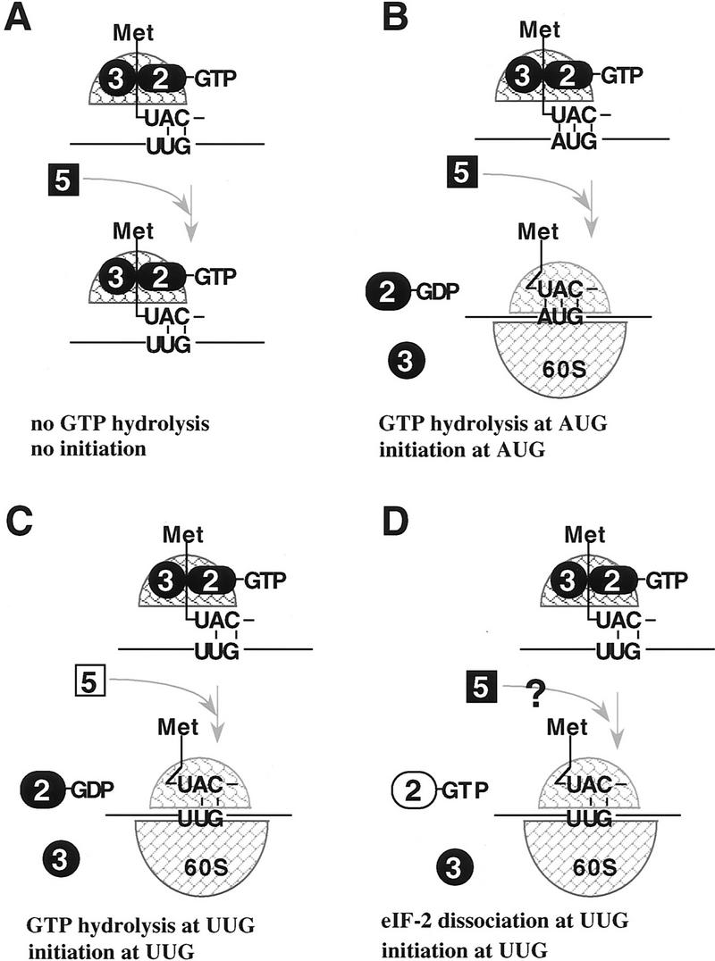
) Initiator tRNA; (➋) eIF-2; (➌) eIF-3; (formula image) eIF-5; (formula image) the 40S ribosomal subunit; (formula image) the 60S ribosomal subunit.

Similar articles

Cited by

References

    1. Balch WE. Small GTP-binding proteins in vesicular transport. Trends Biochem Sci. 1990;15:473–477. - PubMed
    1. Bourne HR, Sanders DA, McCormick F. The GTPase superfamily: Conserved structure and molecular mechanism. Nature. 1991;349:117–127. - PubMed
    1. Bushman JL, Asuru AI, Matts RL, Hinnebusch AG. Evidence that GCD6 and GCD7, translational regulator of GCN4, are subunits of guanine nucleotide exchange factor for eIF-2 in Saccharomyces cerevisiae. Mol Cell Biol. 1993a;13:1920–1932. - PMC - PubMed
    1. Bushman JL, Foiani M, Cigan AM, Paddon CJ, Hinnebusch AG. Guanine nucleotide exchange factor for eukaryotic translation initiation factor 2 in Saccharomyces cerevisiae: Interactions between the essential subunits GCD2, GCD6 and GCD7 and the regulatory subunit GCN3. Mol Cell Biol. 1993b;13:4618–4631. - PMC - PubMed
    1. Castilho-Valavacius B, Yoon H, Donahue TF. Genetic characterization of the Saccharomyces cerevisiae translation initiation suppressors sui1, sui2, and SUI3 and their effects on HIS4 expression. Genetics. 1990;24:483–495. - PMC - PubMed

Publication types

MeSH terms

Substances