. 1997 Sep 15;11(18):2414-25.
doi: 10.1101/gad.11.18.2414.

Rpp1, an essential protein subunit of nuclear RNase P required for processing of precursor tRNA and 35S precursor rRNA in Saccharomyces cerevisiae

Affiliations

Rpp1, an essential protein subunit of nuclear RNase P required for processing of precursor tRNA and 35S precursor rRNA in Saccharomyces cerevisiae

V Stolc et al. Genes Dev. .

Corrected and republished in

Abstract

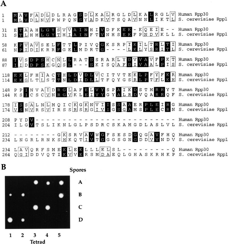
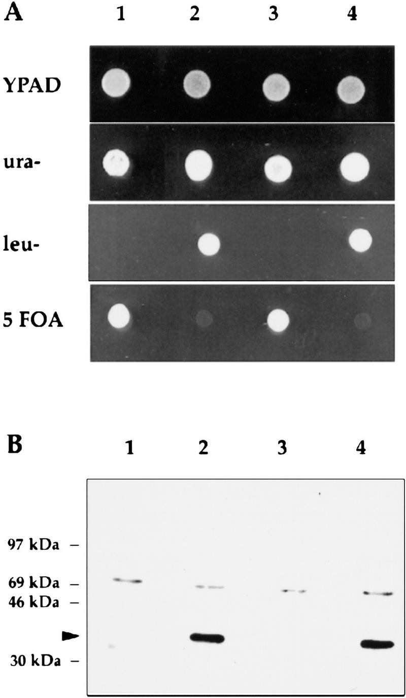
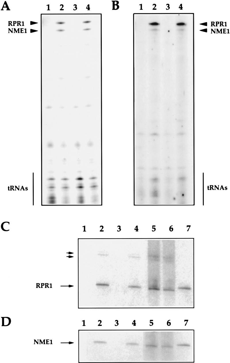
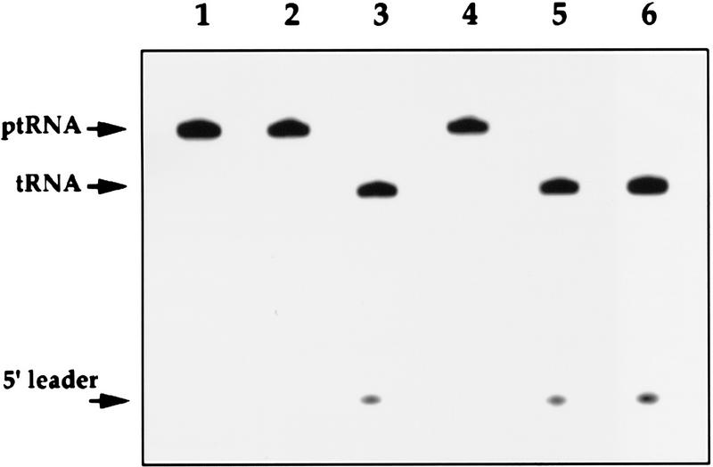
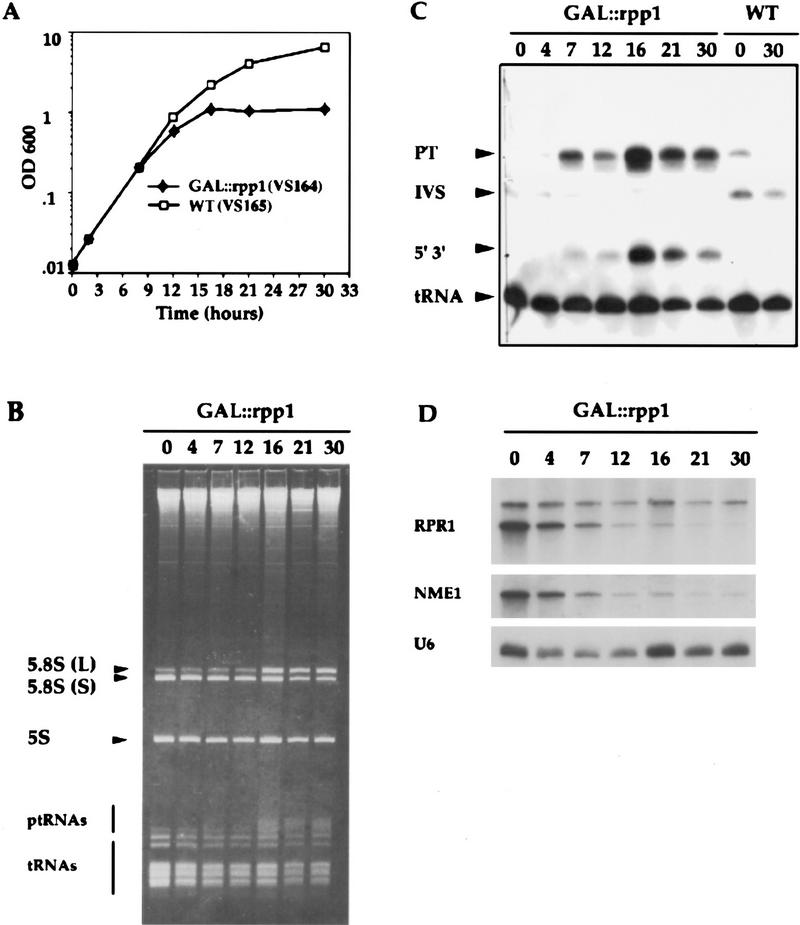
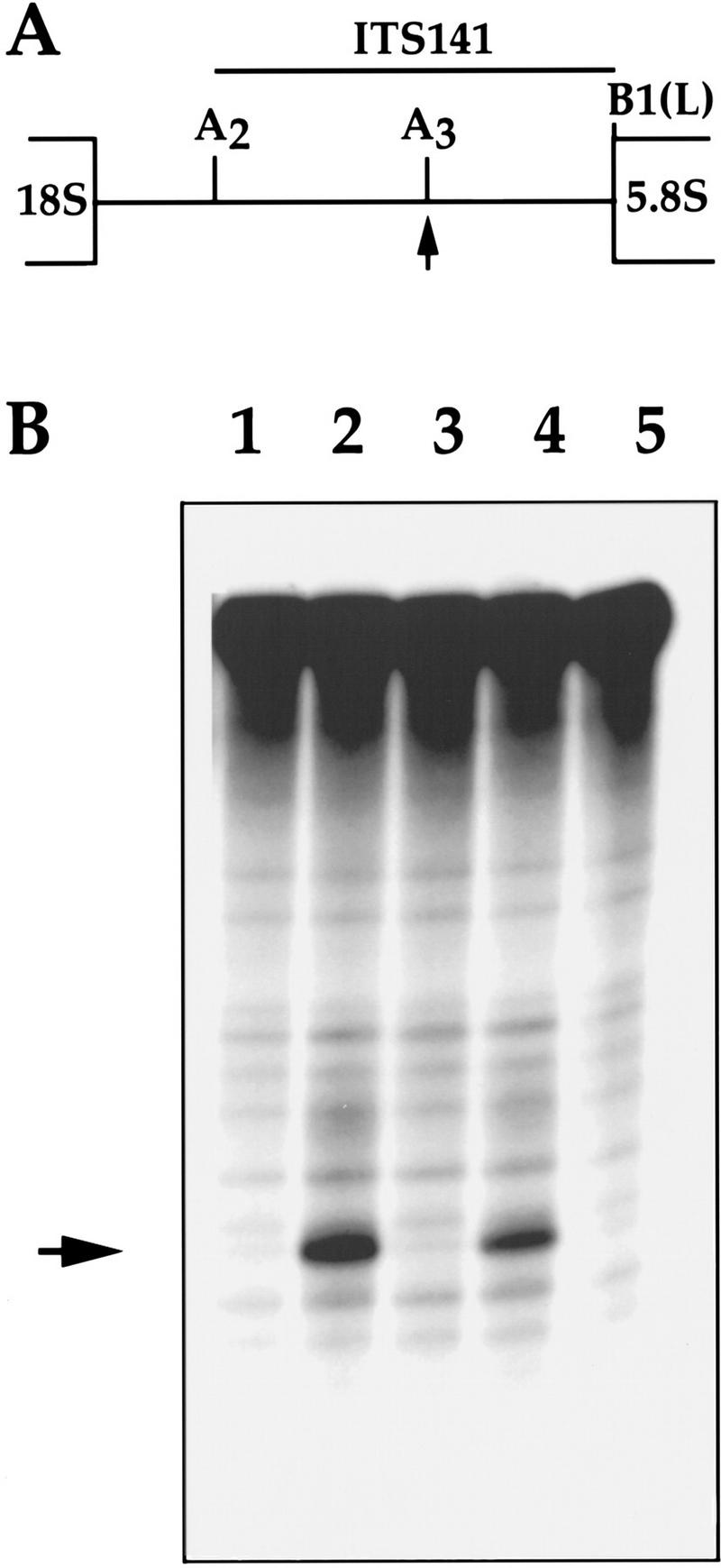
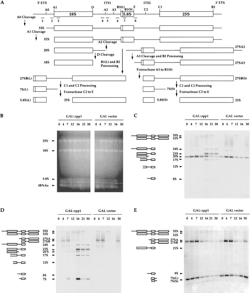
The gene for an essential protein subunit of nuclear RNase P from Saccharomyces cerevisiae has been cloned. The gene for this protein, RPP1, was identified by virtue of its homology with a human scleroderma autoimmune antigen, Rpp30, which copurifies with human RNase P. Epitope-tagged Rpp1 can be found in association with both RNase P RNA and a related endoribonuclease, RNase MRP RNA, in immunoprecipitates from crude extracts of cells. Depletion of Rpp1 in vivo leads to the accumulation of precursor tRNAs with unprocessed 5' and 3' termini and reveals rRNA processing defects that have not been described previously for proteins associated with RNase P or RNase MRP. Immunoprecipitated complexes cleave both yeast precursor tRNAs and precursor rRNAs.


Figures

Figure 1
Figure 1
RPP1, an essential yeast gene encodes a 32.2-kD protein ortholog of the human scleroderma autoimmune antigen, Rpp30. (A) Predicted amino acid sequence of S. cerevisiae Rpp1. The protein is encoded by ORF YHR062c on chromosome VIII and its alignment with the human scleroderma autoimune antigen Rpp30 is shown. The amino acid sequences are numbered from the first methionine residue of each protein. Identical amino acids are shaded and similar amino acids are boxed. (B) The heterozygous diploid strain VS161, RPP1/rpp1::LEU2, was sporulated and dissections were performed on 20 tetrads. The four spores (A–D) derived from each of five tetrads are shown in vertical rows.
Figure 2
Figure 2
3 × myc–Rpp1 is functional and recognized by 9E10 antibody. (A) The four spores (VS162 A–D; lanes 1–4, respectively) derived from the diploid strain VS162, were dissected on rich medium plates (YPAD), and then replica-plated onto plates that contained synthetic complete medium that lacked either leucine (leu) or uracil (ura), or that contained 5-FOA. (B) Immunoblot of protein extracts prepared from the four spores VS162A′, VS162B, VS162C′, and VS162D, derived from the diploid strain VS162. All four spores are isogenic except the VS162A′ and VS162C′ spores do not have the pRS316–3 × myc–Rpp1 plasmid. (Lanes 1–4) VS162A′, VS162B, VS162C′, and VS162D, respectively. The arrowhead points to the 36-kD 3 × myc–Rpp1 fusion protein. The upper band is a nonspecifically reacting protein.
Figure 3
Figure 3
The RNA subunits of RNase P (RPR1) and RNase MRP (NME1) coprecipitate with 3 × myc–Rpp1. (A) Immunoprecipitated RNAs extracted from the 9E10 Ab–IgG–agarose beads that were incubated with protein extracts from the four spores (VS162A′, VS162B, VS162C′, and VS162D; lanes 1–4, respectively). RNA was extracted from the immunoprecipitated beads that were washed with 150 mm KCl (see Materials and Methods). The RNA was 3′ end-labeled with [5′-32P]pCp, and fractionated on a 8% polyacrylamide/7 m urea gel. (B) Immunoprecipitated RNAs derived from the same immunoprecipitated beads as in A (lanes 1–4, respectively) except that the immunoprecipitated beads were washed with 600 mm KCl prior to 3′ end-labeling of the RNA. (C) Immunoprecipitated RNAs derived from the same immunoprecipitated beads as in A were transferred to a positively charged nylon membrane (Boehringer Mannheim) by electroblotting and hybridized with a uniformly labeled DNA probe complementary to the RPR1 gene (see Materials and Methods). (Lanes 1–4) Immunoprecipitated RNA from spores VS162A′, VS162B, VS162C′, and VS162D, respectively; (lane 5) RNA from supernatant of the immunoprecipitated extract derived from spore VS162A′ after centrifugation of beads; (lane 6) RNA from supernatant of the immunoprecipitated extract derived from spore VS162B after centrifugation of beads; (lane 7) RNase P (RPR1) and RNase MRP (NME1) RNAs (0.001 pmoles each) transcribed in vitro. Two small arrows point to extended species, which may be precursors to mature RNase P RNA, RPR1 (Lee et al. 1991). The larger arrows indicate RNase P (RPR1) and RNase MRP (NME1) RNAs. (D) Same as in C, except the membrane was hybridized with a uniformly labeled DNA probe complementary to the NME1 gene (Schmitt and Clayton 1992).
Figure 4
Figure 4
RNase P activity coprecipitates with 3 × myc–Rpp1. (A) IgG–agarose pellets, to which is bound immunoprecipitated RNase P RNA, that were derived from immunoprecipitates from spores VS162A′, VS162B, VS162C′, and VS162D (as in Fig. 3B), were incubated with a uniformly labeled precursor tRNASer for 30 min at 37°C, and then fractionated on a 8% polyacrylamide/7 m urea gel (see Materials and Methods). (Lane 1) Precursor tRNASer; (lanes 2–5) immunoprecipitated RNase P from the four spores VS162A′, VS162B, VS162C′, and V6S162D, respectively; (lane 6) glycerol gradient-purified human RNase P, fraction F29 (Eder et al. 1997). Arrows indicate ptRNA, accurately processed mature tRNA, and 5′ leader sequence.
Figure 5
Figure 5
RNase P is required for processing of ptRNA and 5.8S rRNA in vivo. (A) Growth of the strain VS164 (▪), and the wild-type, isogenic strain VS165 (□), after transfer from galactose-containing to glucose-containing medium at T = 0. Cell density was measured at the times indicated and the cultures were diluted with glucose-containing medium at each time point to prevent nutritional depravation and to maintain exponetial growth. (B) RNA was extracted from VS164 following growth in glucose-containing medium at the indicated times, and was fractionated in an 8% polyacrylamide/7 m urea gel, and stained with ethidium bromide. The positions of 5.8S (L) RNA, 5.8S (S) RNA, 5S RNA, ptRNA, and mature tRNA are indicated. (C) Total RNA extracted from VS164 and VS165 was transferred to a positively charged nylon membrane (Boehringer Mannheim) by electroblotting and hybridized with a γ-32P-labeled oligonucleotide complementary to mature tRNALeu (see Materials and Methods). The position of the three processing intermediates of the ptRNA are indicated. PT is the primary transcript, which has extra sequences at both of its termini and contains an intron; IVS is the ptRNA that contains the intron but has been processed at both the 5′ and 3′ ends; 5′ 3′ is the spliced ptRNA that is unprocessed at both termini. (D) Steady-state levels of RNase P RNA (RPR1) and RNase MRP RNA (NME1) in VS164 after transfer from galactose-containing to glucose-containing medium. The upper band in the RPR1 panel is the putative precursor RPR1 RNA that has extra sequences at both termini (Lee et al. 1991). U6 snRNA levels were detected with oligo U6 (Table 2), as an internal control for RNA levels (Brow and Guthrie 1990).
Figure 6
Figure 6
Processing of prRNA by Rpp1 immunoprecipitates. (A) Position of the ITS141 rRNA substrate relative to the 35S rRNA precursor [5′ end is at A2 site and 3′ end is at B1(L) site]. (B) Uniformly labeled rRNA transcript, ITS141 (see Materials and Methods), was incubated with 3 × myc–Rpp1 immunoprecipitates derived from the same immunoprecipitates as in Fig. 3A for 2 hr at 37°C, and then fractionated on a 8% polyacrylamide/7 m urea gel. (Lanes 1–4) ITS141 rRNA, plus IgG–agarose pellets to which are bound immunoprecipitated RNase P and RNase MRP RNAs, respectively, as in Fig. 3A. (Lane 5) ITS141 rRNA. The arrow indicates the position of two cleavage products of almost identical size from the ITS1 rRNA transcripts. The site of cleavage corresponds to the region of the A3 site in the internal transcribed sequence 1 (ITS1) (Lygerou et al. 1996).
Figure 7
Figure 7
Effects of Rpp1 depletion on the processing of 35S prRNA. (A) Schematic plan of the rRNA processing pathway (Venema and Tollervey 1995). Steady-state levels of rRNA, prRNA intermediates, and aberrant rRNA species were detected by ethidium bromide staining and by Northern hybridization with oligonucleotide probes 1–7. (B) Total RNA extracted from VS164 and VS165 was obtained after transfer from galactose-containing to glucose-containing medium at the times indicated, and separated in a l.2% agarose gel stained with ethidium bromide. Arrows indicate 25S rRNA, 18S rRNA, 5.8S rRNA, and tRNAs. (C–E) The gel shown in B was transferred to a positively charged nylon membrane (Boehringer Mannheim) by capillary diffusion and hybridized with γ-32-labeled oligonucleotide probes 4–6 (see Materials and Methods). Oligonucleotide 4 (C) is complementary to the ITS1, between 3′ end of 18S rRNA and the A2 site (position 157–180), oligonucleotide 5 (D) is complementary to the ITS1, between A2 and A3 sites (position 119–236), and oligonucleotide 8 (E) is complementary to the ITS2, between the 3′ end of 5.8S rRNA and C2 sites (position −3 to +21). The 24S precursor rRNA is predicted to extend from the 5′ end of the ETS to the 3′ end of 5.8S rRNA, and represents a product of the 35S rRNA precursor that is cleaved in ITS2 in the absence of cleavage at the A0, A1, A2, and A3 sites. The 21S precursor rRNA appears to be comprised of rRNA species that are predicted to extend from the 5′ end of 18S rRNA to the 3′ end of 5.8S rRNA, and also have 3′ ends that extend 3′ to the 3′ end of 5.8S rRNA, and may be heterogeneous. The 20′S rRNA species is predicted to extend from the 5′ end of 18S rRNA to the region of the A3 site in the ITS1. The 17S′ and 12S′ aberrant rRNA species represent stable degradation intermediates, which may have fragmented 5′ ends that correspond to sequences within 18S rRNA and 3′ ends close to the 3′ end of 5.8S rRNA. The 8S rRNA species is predicted to extend from the 5′ end of ITS1 to the 3′ extended ends of 5.8S rRNA. The 7S RNA is the precursor of the 5.8S rRNA, which is predicted to extend from the region of the A2 site in the ITS1 to the 3′ end of 5.8S rRNA.

Similar articles

Cited by

References

    1. Alifano P, Rivellini F, Piscitelli C, Arraiano CM, Bruni CB, Carlomagno MS. Ribonuclease E provides substrates for ribonuclease P-dependent processing of a polycistronic mRNA. Genes & Dev. 1994;8:3021–3031. - PubMed
    1. Allmang C, Henry Y, Morrissey JP, Wood H, Petfalski E, Tollervey D. Processing of the yeast pre-rRNA at sites A(2) and A(3) is linked. RNA. 1996a;2:63–73. - PMC - PubMed
    1. Allmang C, Henry Y, Wood H, Morrissey JP, Petfalski E, Tollervey D. Recognition of cleavage site A(2) in the yeast pre-rRNA. RNA. 1996b;2:51–62. - PMC - PubMed
    1. Altman S, Kirseborm L, Talbot S. Recent studies of ribonuclease P. FASEB J. 1993;7:7–14. - PubMed
    1. ————— . Recent studies of RNase P. In: Soll D, RajBhandary UL, editors. tRNA: Structure biosynthesis and function. Washington, DC.: American Society for Microbiology; 1995. pp. 67–78.

Publication types

Associated data