. 1997 Sep;7(9):932-41.
doi: 10.1101/gr.7.9.932.

Cloning, characterization, and mapping of a murine promiscuous chemokine receptor gene: homolog of the human Duffy gene

Cloning, characterization, and mapping of a murine promiscuous chemokine receptor gene: homolog of the human Duffy gene

H Luo et al. Genome Res. 1997 Sep.

Abstract

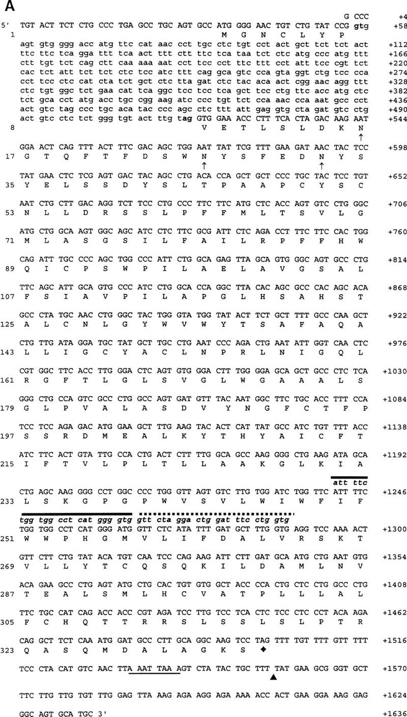
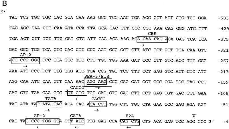
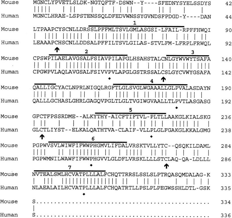
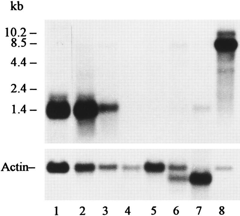
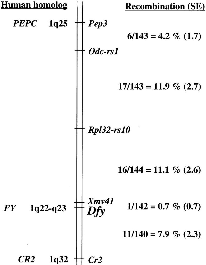
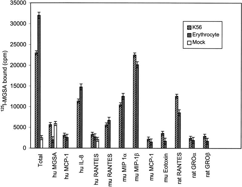
We report here the isolation and genomic organization of the orthologous mouse Duffy gene, named Dfy. It is a single copy gene located in chromosome 1 in a region homologous to the human Duffy gene (FY). Sequence analyses indicate that Dfy consists of two exons: exon 1 of 55 nucleotides, which encodes 7 amino acid residues; and exon 2 of 1038 nucleotides, which encodes 327 residues. The single intron consists of 462 nucleotides. The 5'-end promoter region contains motifs involved in vertebrate development in addition to potential binding sites of factors for globin transcription. The open reading frame (ORF) shows 60% homology with the human Duffy protein. However, mouse erythrocytes are serologically Duffy-negative and mouse erythrocyte membrane proteins do not cross-react with two Duffy-specific rabbit polyclonal antibodies. The deduced protein predicts a M(r) of 36,692 and carries three potential N-glycosylation sites to asparagine residues. Hydropathy analysis predicts an exocellular amino-terminal domain of 57 residues, seven transmembrane alpha-helices, and an endocellular carboxy-terminal domain of 29 residues. In bone marrow and spleen, Dfy expresses a major 1.4-kb and a minor 1.8-kb mRNA. Contrary to humans, Dfy is expressed in liver, synthesizing a 1.4-kb mRNA, and is repressed in kidney. Dfy is highly expressed in mouse brain and produces a major 8.5-kb and a minor 10.2-kb mRNA. The human erythroleukemia K562 cells, transfected with cDNA encoding the mouse Duffy-like protein and mouse erythrocytes, have the same chemokine binding profiles indicating that they contain the same protein.


Figures

Figure 1
Figure 1
(A) Genomic DNA and cDNA structure of Dfy and deduced amino acid sequence of mouse Duffy-like protein. Amino acid residues are numbered on left; nucleotide positions are numbered on right. The exon and intron sequences are shown in uppercase and lowercase, respectively. The consensus sequence at the intron splice site is indicated in boldface letters. The three potential carbohydrate-binding sites to asparagine residues are shown in up arrows. The primers’ nucleotide sequences are shown in lowercase, italic, and boldface letters. P2 nucleotides are shown by a solid line and P1 by a dotted line. The underline is the polyadenylation signal. (▴) The polyadenylation site; (♦) the stop codon. (B) 5′-end promoter flanking sequence of Dfy. (▿) The position (+1) of the major transcription initiation site. Consensus sequences for the transcription factor binding sites are boxed, and the name of the corresponding factors are shown. The arrows indicate consensus sequences in sense strand (right) and antisense strand (left).
Figure 1
Figure 1
(A) Genomic DNA and cDNA structure of Dfy and deduced amino acid sequence of mouse Duffy-like protein. Amino acid residues are numbered on left; nucleotide positions are numbered on right. The exon and intron sequences are shown in uppercase and lowercase, respectively. The consensus sequence at the intron splice site is indicated in boldface letters. The three potential carbohydrate-binding sites to asparagine residues are shown in up arrows. The primers’ nucleotide sequences are shown in lowercase, italic, and boldface letters. P2 nucleotides are shown by a solid line and P1 by a dotted line. The underline is the polyadenylation signal. (▴) The polyadenylation site; (♦) the stop codon. (B) 5′-end promoter flanking sequence of Dfy. (▿) The position (+1) of the major transcription initiation site. Consensus sequences for the transcription factor binding sites are boxed, and the name of the corresponding factors are shown. The arrows indicate consensus sequences in sense strand (right) and antisense strand (left).
Figure 2
Figure 2
Alignment of the amino acid sequences of the proteins codified by mouse and human spliced mRNAs. Amino acid residues are at right. Horizontal lines indicate seven transmembrane spanning α-helices. Vertical lines indicate identical residues. Arrows and ♦ indicate invariant cysteines and prolines, respectively.
Figure 3
Figure 3
RNA blot analysis of mRNA from mouse tissues probed with exon 2 of Dfy. (Lane 1) 2 μg of bone marrow mRNA; (lane 2) 5 μg of spleen mRNA; (lane 3) 5 μg of liver mRNA; (lane 4) 5 μg of lung mRNA; (lane 5) 10 μg of kidney mRNA; (lane 6) 10 μg of heart mRNA; (lane 7) 5 μg of skeletal muscle mRNA; (lane 8) 1 μg of brain mRNA. Actin probe was used to test the integrity of the mRNA fraction and to determine the relative abundance of poly(A)+ RNA. RNAs were resolved on a 1.5% denaturing agarose gel hybridized and autoradiographed for 5 days. The relative amount of mRNA was determined with the Eagle Eye II Still Video System (Stratagene). The Duffy-like mRNA/actin mRNA ratios were as follows: (lane 1) 1.80; (lane 2) 2.30; (lane 3) 0.78; (lane 4) −; (lane 5) −; (lane 6) 0.09; (lane 7) 0.09; (lane 8) 10.72.
Figure 4
Figure 4
Location of Dfy on mouse chromosome 1. The genetic map was derived from segregation analysis of 144 interspecific backcross progeny. The number of observed recombinant chromosomes over the total number of analyzed chromosomes was used to estimate the recombination frequency between adjacent loci. These interlocus recombination estimates are shown to the right of the chromosome along with their associated standard errors (shown in parentheses). Map locations for known human homologs are shown at left.
Figure 5
Figure 5
Inhibition of 125I-labeled MGSA binding to K562 cells stably expressing the mouse Duffy-like protein and mouse erythrocytes. K562 cells and mouse erythrocytes were incubated for 30 min at 37°C with 125I-labeled MGSA (0.5 nm) in the absence or presence of 0.25 μm of unlabeled MGSA, MCP-1, IL-8, RANTES, MIP-1α, MIP-1β, Eotaxin, GROα, and GROβ. The binding reactions were stopped as described (Neote et al. 1993; Chaudhuri et al. 1994). The data are expressed as cpm of 125I-labeled MGSA bound (per 106 cells) for K562 cells and (per 107 cells) for mouse erythrocytes. Abbreviations for human and murine chemokines are hu and mu, respectively.

References

    1. Chaudhuri A, Polyakova J, Zbrzezna V, Williams K, Gulati S, Pogo AO. Cloning of glycoprotein D cDNA, which encodes the major subunit of the Duffy blood group system and the receptor for the Plasmodium vivax malaria parasite. Proc Natl Acad Sci. 1993;90:10793–10797. - PMC - PubMed
    1. Chaudhuri A, Zbrzezna V, Polyakova J, Pogo AO, Hesselgesser J, Horuk R. Expression of the Duffy antigen in K562 cells. J Biol Chem. 1994;269:7835–7838. - PubMed
    1. Chaudhuri A, Polyakova J, Zbrzezna V, Pogo AO. The coding sequence of Duffy blood group gene in humans and simians: Restriction fragment length polymorphism, antibody and malarial parasite specificities, and expression in nonerythroid tissues in Duffy-negative individuals. Blood. 1995;85:615–621. - PubMed
    1. Chaudhuri A, Nielsen S, Elkjaer M-L, Zbrzezna V, Fang F, Pogo AO. Detection of Duffy antigen in the plasma membranes and caveolae of vascular endothelial and epithelial cells of nonerythroid organs. Blood. 1997;89:701–712. - PubMed
    1. Collins A, Keast BJ, Dracopoli N, Shield DC, Morton NE. Integration of gene maps: Chromosome 1. Proc Natl Acad Sci. 1992;89:4598–4602. - PMC - PubMed

Publication types

Associated data