. 1997 Oct 6;139(1):23-36.
doi: 10.1083/jcb.139.1.23.

SOI1 encodes a novel, conserved protein that promotes TGN-endosomal cycling of Kex2p and other membrane proteins by modulating the function of two TGN localization signals

Affiliations

SOI1 encodes a novel, conserved protein that promotes TGN-endosomal cycling of Kex2p and other membrane proteins by modulating the function of two TGN localization signals

J H Brickner et al. J Cell Biol. .

Abstract

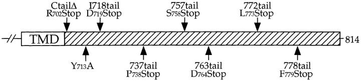
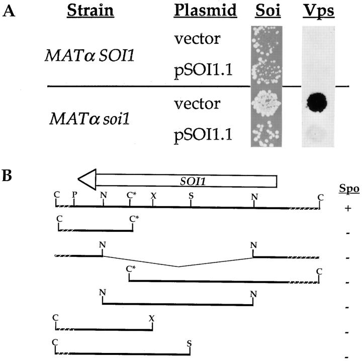
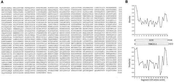
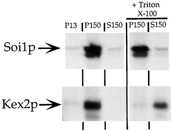
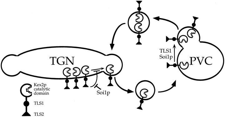
Localization of yeast Kex2 protease to the TGN requires a signal (TLS1) in its cytosolic tail (C-tail). Mutation of TLS1 results in rapid transit of Kex2p to the vacuole. Isolation of suppressors of the Tyr713Ala mutation in TLS1 previously identified three SOI genes. SOI1, cloned by complementation of a sporulation defect, encodes a novel, hydrophilic 3,144-residue protein with homologues in Caenorhabditis elegans, Drosophila melanogaster, and humans. Epitope-tagged Soi1p existed in a detergent-insensitive, sedimentable form. Deletion of SOI1 impaired TGN localization of wild-type Kex2p and a fusion protein containing the C-tail of Ste13p, and also caused missorting of carboxypeptidase Y and accelerated vacuolar degradation of the Vps10p sorting receptor. Deletion of SOI1 improved retention of Tyr713Ala Kex2p in the pro-alpha-factor processing compartment but, unlike the original soi1 alleles, did not increase the half-life of Tyr713Ala Kex2p. These results suggested that Soi1p functions at two steps in the cycling of Kex2p and other proteins between the TGN and prevacuolar compartment (PVC). This hypothesis was confirmed in several ways. Soi1p was shown to be required for optimal function of TLS1. Suppression of the Tyr713Ala mutation by mutation of SOI1 was shown to be caused by activation of a second signal (TLS2) in the Kex2p C-tail. TLS2 delayed exit of Kex2p from the TGN, whereas TLS1 did not affect this step. We propose that Soi1p promotes cycling of TGN membrane proteins between the TGN and PVC by antagonizing a TGN retention signal (TLS2) and facilitating the function of a retrieval signal (TLS1) that acts at the PVC.


Figures

Figure 1
Figure 1
Schematic of mutations in the Kex2p cytosolic tail (C-tail). The positions of mutations in various forms of Kex2p that were used in this study are indicated. The Y713A (55) and I718tail (34) mutations have been described. TMD, transmembrane domain.
Figure 2
Figure 2
Isolation of SOI1. (A) KRY18-1A (MATα kex2Δ) and JBY11-r1 (MATα kex2Δ soi1-2), expressing Y713A Kex2p under the control of the GAL1 promoter from plasmid pCWKX21 (55), were transformed with either pBS32 (vector control) or pSOI1.1, and were analyzed for their Soi and Vps phenotypes, as described in Materials and Methods. For the mating assay, strains were shifted to glucose for 6 h before testing mating competence. (B) Restriction map of pSOI1.1 and analysis of subcloned fragments. At the top is shown a restriction map of the insert from pSOI1.1. Indicated above the map is the extent and direction of the SOI1 open reading frame. Restriction fragments from pSOI1.1 were subcloned into either pBS32 or pRS313, transformed into JBY96 (MATa/MATα, soi1-2/soi1-2), and tested for complementation of the Spo phenotype (scores are shown to the right of each subclone). Restriction sites are indicated as follows: C, ClaI; P, PstI; N, NcoI; S, SalI; X, XhoI. The ClaI site indicated by an asterisk is not unique, but is the only one within the insert not blocked by dam methylation.
Figure 3
Figure 3
Sequence of Soi1p and analysis of conserved sequences. (A) The deduced amino acid sequence of Soi1p (GenBank/EMBL/ DDBJ accession number AF001317). (B) Schematic representation of Soi1p and the deduced amino acid sequence from C. elegans open reading frame T08G11.1. Percent identity and similarity (using a PAM250 substitution matrix) were calculated using Clustal W (49) and plotted for segments of 120 amino acids (the COOH-terminal segment was 87 amino acids). Upper plot, percent identity; lower plot, percent similarity. A second potential C. elegans homologue of ∼3,200 codons (not shown) can be formed by joining two adjacent, predicted open reading frames from cosmid C25H3 (sequences C25H3.8 and C25H3.9, GenBank/EMBL/DDBJ accession No. U29535).
Figure 4
Figure 4
Soi1p is a high molecular weight protein that is found in a detergent-insensitive, sedimentable fraction. Labeled cell extracts were produced and processed as described in Materials and Methods. After a clearing spin, lysates were spun at ∼13,000 g to produce a pellet (P13). Triton X-100 (0.1%) was added to half of the supernatant fraction from this spin and the detergent-treated and untreated samples were then centrifuged at ∼150,000 g to produce a pellet (P150) and a supernatant fraction (S150). Kex2p and Soi1p-HA were immunoprecipitated from SDS-denatured samples and were separated by SDS-PAGE (see Materials and Methods).
Figure 5
Figure 5
Indirect immunofluorescence analysis of Kex2p in SOI1 and soi1Δ-2 strains. JBY154-2A (kex2Δ soi1Δ-2) transformed with either pCWKX20 (a and b, WT Kex2p under GAL1 promoter control; reference 55) or pCWKX21 (c and d, Y713A Kex2p under GAL1 promoter control; reference 55), and either pSOI1.1 (a and c) or a vector control (b and d) were processed for immunofluorescence as described (35). Expression of Kex2p under GAL1 promoter control from a CEN plasmid results in ∼15-fold overexpression that is not thought to alter localization significantly (35). Vacuolar staining, apparent from bright field viewing, is indicated with narrow arrows. Wide arrows indicate larger structures present at a higher frequency in the soi1Δ strain.
Figure 6
Figure 6
SOI1 is required for TGN localization of Kex2p, Vps10p, and Ste13p, and for vacuolar targeting of proCPY. (A) Pulse-chase/immunoprecipitation of Kex2p. BFY106-4D (kex2Δ SOI1) and JBY134.1 (kex2Δ soi1Δ-1), transformed with CEN plasmids expressing either WT Kex2p (pCWKX10) or Y713A Kex2p (pCWKX11) from the KEX2 promoter (55), were pulse labeled with [35S]H2SO4 for 10 min before addition of chase. Cells were collected at the indicated times after addition of chase, lysed, and processed for immunoprecipitation of Kex2p. (B) BFY106-4D and JBY134.1 were labeled as described in A and chased. Samples were collected at the indicated times, separated into cells (I, intracellular) and medium (E, extracellular), and processed for immunoprecipitation of CPY. Forms of CPY are as follows: p1CPY, core-glycosylated ER form; p2CPY, Golgi form; mCPY, mature vacuolar form (47). (C and D) JBY135-1A (soi1Δ-1) and JBY135-2D (SOI1) were labeled with [35S]H2SO4 for 10 min and then chased. Samples were collected, lysed, and immunoprecipitated using anti-Vps10p (C) or anti-ALP (D) sera. Vps10p (C) and precursor A-ALP (D) were quantified after SDS-PAGE and plotted relative to initial band intensity. Half-life (C) or half-time of processing (D) were determined by linear regression (Table II).
Figure 7
Figure 7
Deletion of SOI1 reveals a second TLS in the C-tail of Kex2p. (A) BFY106-4D (SOI1 kex2Δ) and JBY134.1 (soi1Δ-1 kex2Δ) expressing WT Kex2p, Y713A Kex2p or I718tail Kex2p, under the control of the GAL1 promoter, were shifted from galactose to glucose for the indicated times before testing mating competence. (B and D) JBY135-1A (soi1Δ-1) and JBY135-2D (SOI1) carried CEN plasmids expressing either I718tail Kex2p (pCWKX10-I718tail; B) or C-tailΔ Kex2p (pCWKX17; D) from the KEX2 promoter (34, 55). (C) JBY173 (soi1Δ-2 pep4 prb1 prc1) and CB018 (SOI1 pep4 prb1 prc1) carried pCWKX10-I718tail. Strains were labeled for 10 min (B and C) or 5 min (D), chased, and samples were collected at the indicated times after the addition of chase. Cells were lysed, and Kex2p was immunoprecipitated using antiserum raised against the Kex2p lumenal domain (55). WT Kex2p is indicated by closed arrows. I718tail Kex2p (B and C) and C-tailΔ Kex2p (D) are indicated by open arrows.
Figure 8
Figure 8
(A) Suppression of the localization defect of Y713A Kex2p in the soi1Δ strain requires TLS2. JBY154-1A (MATα soi1Δ-2 kex2Δ) and JBY154-2A (MATα SOI1 kex2Δ) expressing either Y713A Kex2p (pCWKX21), Y713A I718tail Kex2p (pCWKX21-I718), or C-tailΔ Kex2p (pCWKX27) under the control of the GAL1 promoter on CEN plasmids were shifted from galactose to glucose for 5 h before testing mating competence. (B) Optimal TLS1 function requires Soi1p, but TLS1 exhibits residual function in the absence of Soi1p. Strains JBY154-1A and JBY154-2A expressing either I718tail Kex2p (pCWKX20-I718) or Y713A I718tail Kex2p under the control of the GAL1 promoter on CEN plasmids were shifted from galactose to glucose medium for the indicated times before testing mating competence. Indicated to the left of each row of patches is the form of Kex2p expressed. The relevant SOI1 allele is indicated above the columns.
Figure 9
Figure 9
(A) Improved TGN localization of F87A A-ALP in a soi1 mutant strain. Strains JBY135-1A (soi1Δ-1) and JBY135-2D (SOI1), transformed with pSN98 (expressing F87A A-ALP; reference 29), were labeled for 10 min with [35S]H2SO4 and chased for 120 min. Samples were collected after 10, 20, 30, 60, and 120 min of chase and processed for immunoprecipitation using anti-ALP antiserum. Band intensity of pro-A-ALP was quantified after SDS-PAGE (see Materials and Methods) and plotted relative to initial band intensity. (B) Deletion of SOI1 does not affect transport through early secretory compartments. Strains JBY154-1D (KEX2 soi1Δ-2) and JBY154-8B (KEX2 SOI1) were labeled with EXPRE35S35S label for 2 min before addition of chase. Cells were collected at the indicated times after addition of chase. Indicated beside the top panel are precursor forms of Kex2p, I1 (pro-Kex2p possessing core glycosyl modifications) and I2 (mature, core glycosylated Kex2p), and Golgi-modified, mature Kex2p (J; reference 54). Indicated beside the bottom panel are proALP and mature ALP (21).
Figure 10
Figure 10
TLS2 slows delivery of Kex2p to the PVC. (A) Mutation of TLS1 does not affect the rate of delivery to the PVC. Strain 0472-28 (vps28 KEX2) expressing either I718tail Kex2p (pCWKX10-I718tail; top panel) or Y713A I718tail Kex2p (pCWKX11-I718tail; bottom panel) on a CEN plasmid was pulse-labeled for 10 min at 30°C in SDC-Met-Ura and chased for 80 min (see Materials and Methods). At the indicated times after addition of chase, cells were collected, lysed, and immunoprecipitated using antisera against the Kex2p lumenal domain. After SDS PAGE, WT Kex2p, I718tail Kex2p, or Y713A I718tail Kex2p were quantified, and t 1/2 values were obtained by linear regression. Indicated are the positions of WT Kex2p (Full length; filled arrows), I718tail Kex2p, or Y713A I718tail Kex2p (open arrows). (B) Strain 0472-28 containing pCWKX10-I718tail was grown at 23°C in SD + Ade, His, Leu, Lys, and Trp, labeled, chased, and processed for immunoprecipitation as described in A. Note that under otherwise identical conditions, the rate of degradation of Kex2p in the vps28 mutant is slower in SDC-Met-Ura than in SD + Ade, His, Leu, Lys, and Trp (data not shown). This may be caused by increased proteolytic activity in the PVC caused by amino acid limitation.
Figure 11
Figure 11
Mapping TLS2. A series of Kex2p truncation mutants (shown schematically in Fig. 1) was expressed under the control of the GAL1 promoter in JBY154-1A (MATα kex2Δ soi1Δ-2) and JBY154-2A (MATα kex2Δ SOI1), and was tested for mating competence in the onset of impotence assay after 10 h on glucose. Sequence analysis of the 778tail Kex2p used in this experiment revealed that this protein had a second mutation at the carboxy terminus (P778S). This second mutation was inconsequential in that other isolates of this truncation that lacked this second mutation behaved identically to the one shown (data not shown).
Figure 12
Figure 12
Model for role of Soi1p, TLS1, and TLS2 in the cycling of Kex2p between the TGN and PVC. TLS2 inhibits/delays entrance of Kex2p into the PVC transport vesicle at the TGN. Soi1p inhibits this function of TLS2. TLS1 and Soi1p together promote entry of Kex2p into newly forming TGN transport vesicle at PVC.

References

    1. Ausubel, F.M., R. Brent, R.E. Kingston, D.D. Moore, J.G. Seidman, J.A. Smith, and K. Struhl. 1989. Mutagenesis of cloned DNA. In Current Protocols in Molecular Biology. Vol. 1. John Wiley and Sons, New York. 8.0.1–8.5.9.
    1. Bordier C. Phase separation of integral membrane proteins in Triton X-114 solution. J Biol Chem. 1981;256:1604–1607. - PubMed
    1. Bosshart H, Humphrey J, Deignan E, Davidson J, Drazba J, Yuan L, Oorschot V, Peters PJ, Bonifacino JS. The cytoplasmic domain mediates localization of furin to the trans-Golgi network en route to the endosomal/lysosomal system. J Cell Biol. 1994;126:1157–1172. - PMC - PubMed
    1. Brenner C, Bevan A, Fuller RS. One-step site-directed mutagenesis of the Kex2 protease oxyanion hole. Curr Biol. 1993;3:498–506. - PubMed
    1. Bryant NJ, Stevens TH. Two separate signals act independently to localize a yeast late Golgi membrane protein through a combination of retrieval and retention. J Cell Biol. 1997;136:287–297. - PMC - PubMed

Publication types

MeSH terms

Associated data