. 1997 Oct 6;139(1):63-74.
doi: 10.1083/jcb.139.1.63.

Large plasma membrane disruptions are rapidly resealed by Ca2+-dependent vesicle-vesicle fusion events

Affiliations

Large plasma membrane disruptions are rapidly resealed by Ca2+-dependent vesicle-vesicle fusion events

M Terasaki et al. J Cell Biol. .

Abstract

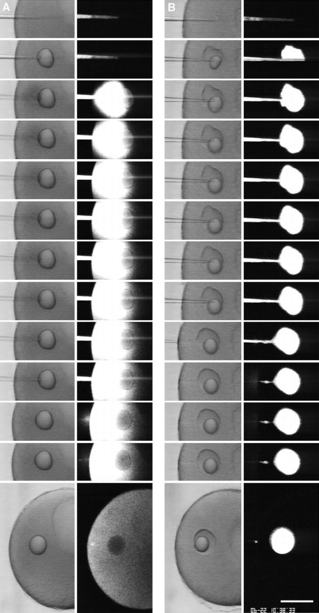
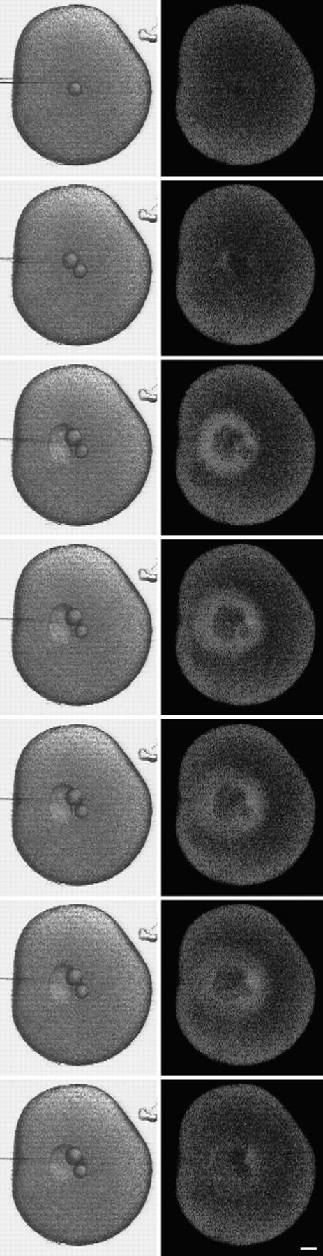
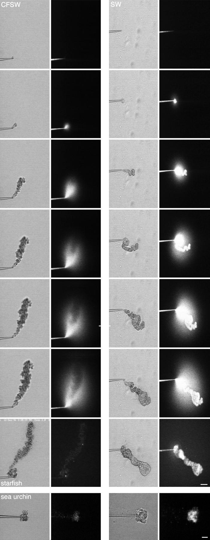
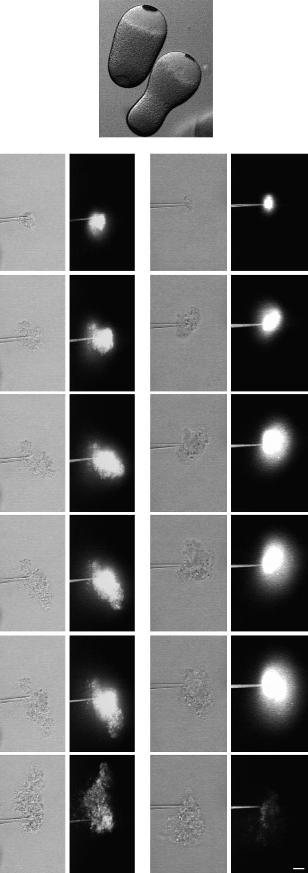
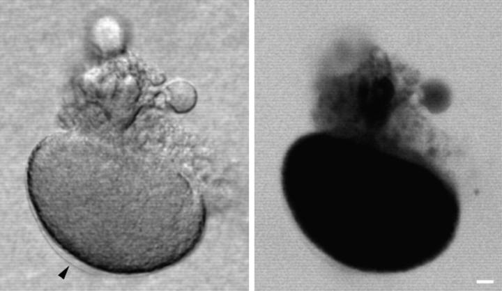
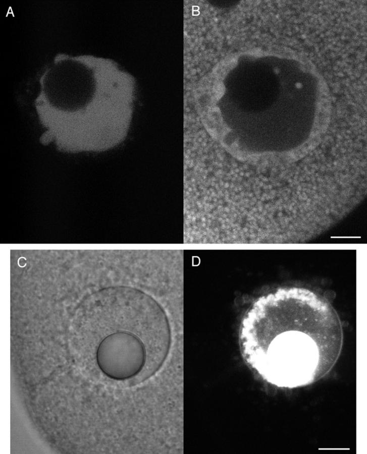
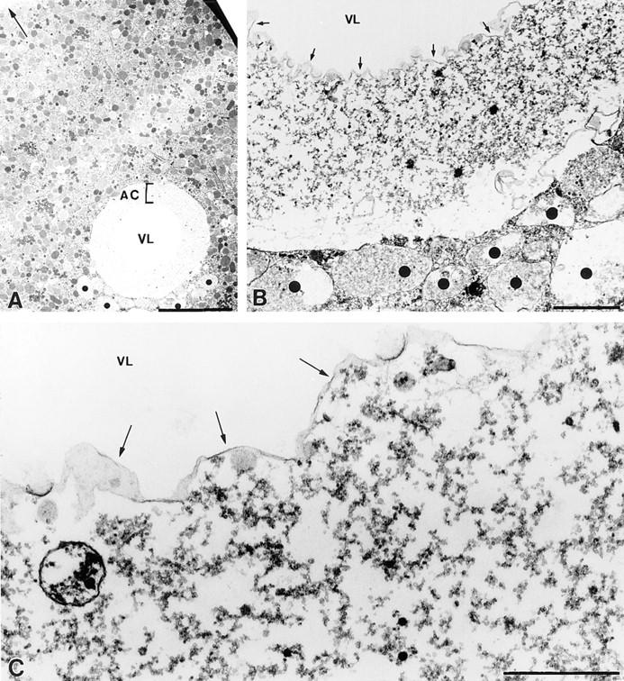
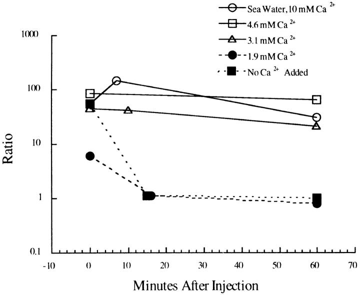
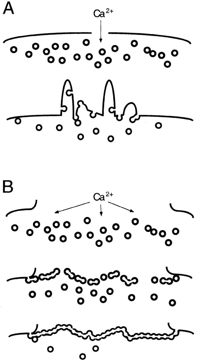
A microneedle puncture of the fibroblast or sea urchin egg surface rapidly evokes a localized exocytotic reaction that may be required for the rapid resealing that follows this breach in plasma membrane integrity (Steinhardt, R.A,. G. Bi, and J.M. Alderton. 1994. Science (Wash. DC). 263:390-393). How this exocytotic reaction facilitates the resealing process is unknown. We found that starfish oocytes and sea urchin eggs rapidly reseal much larger disruptions than those produced with a microneedle. When an approximately 40 by 10 microm surface patch was torn off, entry of fluorescein stachyose (FS; 1, 000 mol wt) or fluorescein dextran (FDx; 10,000 mol wt) from extracellular sea water (SW) was not detected by confocal microscopy. Moreover, only a brief (approximately 5-10 s) rise in cytosolic Ca2+ was detected at the wound site. Several lines of evidence indicate that intracellular membranes are the primary source of the membrane recruited for this massive resealing event. When we injected FS-containing SW deep into the cells, a vesicle formed immediately, entrapping within its confines most of the FS. DiI staining and EM confirmed that the barrier delimiting injected SW was a membrane bilayer. The threshold for vesicle formation was approximately 3 mM Ca2+ (SW is approximately 10 mM Ca2+). The capacity of intracellular membranes for sealing off SW was further demonstrated by extruding egg cytoplasm from a micropipet into SW. A boundary immediately formed around such cytoplasm, entrapping FDx or FS dissolved in it. This entrapment did not occur in Ca2+ -free SW (CFSW). When egg cytoplasm stratified by centrifugation was exposed to SW, only the yolk platelet-rich domain formed a membrane, suggesting that the yolk platelet is a critical element in this response and that the ER is not required. We propose that plasma membrane disruption evokes Ca2+ regulated vesicle-vesicle (including endocytic compartments but possibly excluding ER) fusion reactions. The function in resealing of this cytoplasmic fusion reaction is to form a replacement bilayer patch. This patch is added to the discontinuous surface bilayer by exocytotic fusion events.


Figures

Figure 1
Figure 1
Survival and rapid healing of a large wound in the plasma membrane. A polylysine-coated microneedle was maneuvered so that it contacted the surface of an immobilized sea urchin egg and then was rapidly moved away from the surface (the movement was in the plane of focus of the microscope). This ripped off the contacted surface and created a wound ∼40 by 10 μm. (Top) (A) an apparent sharp boundary formed immediately (within 1–2 s) at the interface of cytoplasm and SW produced by the rip-off. After addition of sperm, the wounded egg became fertilized (B; arrow denotes fertilization envelope) and underwent several rounds of division (C; shown 2.5 h after fertilization). This shows that the wounded egg healed successfully after the rip-off. (Bottom) A sea urchin egg was immersed in 100 μg/ml FS in SW and observed by confocal microscopy during the rip-off wound procedure. No entry of FS was observed after a rip-off, indicating that the wound had resealed rapidly. Images were obtained at 1-s intervals; consecutive images are shown except the last image, which was 40 s after wounding. Bars: (top) 20 μm; (bottom) 10 μm.
Figure 2
Figure 2
Cytosolic Ca2+ during the healing of a wound. A sea urchin egg was injected with the fluorescent Ca2+ indicator calcium green dextran (10 kD; 20 μM final concentration). As in Fig. 1, a polylysine-coated microneedle was used to create a wound. There is a local rise in Ca2+ during wounding, but the rise diminishes rapidly. In view of the large gradient of Ca2+ from SW to cytosol, this is further indication of a rapid healing reaction. Images were obtained at 1-s intervals; consecutive images are shown except the last image, which was 15 s after wounding. The small dark circle near the center of the egg is an oil drop resulting from the calcium green dextran injection. Bar, 10 μm.
Figure 4
Figure 4
Injected SW is contained at the injection site. FDx (10 mg/ml; 70 kD) dissolved in CFSW or in SW was injected into a starfish oocyte. The micropipet contained an oil cap at the tip to prevent mixing of the injectate with chamber SW. The oil cap comes out first during the injection and forms a spherical droplet in the cytoplasm. The injections were observed by simultaneous scanning transmission and fluorescence imaging. (A) When CFSW containing FDx was injected, the fluorescence spread throughout the cytoplasm. (B) When SW containing FDx was injected, the fluorescence did not spread from the injection site and was contained within a wound vesicle that is visible in the transmitted light image. These results suggest that high Ca2+ causes fusion of intracellular membranes, creating a boundary that prevents spreading of high Ca2+ throughout the cytoplasm. Images were obtained at 1-s intervals; consecutive images are shown except that last image of the CFSW sequence was 125 s after injection, and the last image of the SW sequence was 306 s after injection. Bar, 20 μm.
Figure 5
Figure 5
Cytosolic Ca2+ during SW injection rises only briefly. A mature starfish egg was injected with the fluorescent Ca2+ indicator calcium green dextran (20 μM final concentration) then was imaged by simultaneous scanning transmission and fluorescence confocal microscopy during an injection of SW. There was a brief rise in fluorescence around the injection site which then decreased rapidly. The oil drop in the first transmitted light image is from the calcium green dextran injection. Images were obtained every 0.5 s; consecutive images are shown except the last frame which is 6 s after the beginning of the sequence. Bar, 20 μm.
Figure 9
Figure 9
Cytoplasm extruded into SW retains a fluorescent marker in the cytosol. Starfish oocytes were injected with a final concentration of 0.2 mg/ml FS. After allowing the FS to diffuse throughout the oocyte, cytoplasm was removed by micropipet (2% of the oocyte volume). The cytoplasm was then extruded while observing by simultaneous scanning transmission and fluorescence confocal microscopy. When extruded into CFSW (left), the fluorescent marker diffused away, indicating lack of a boundary formation. When extruded into SW (right), the fluorescent marker was retained, indicating that the Ca2+ caused fusion of intracellular membranes, trapping the marker. Images were obtained at 1-s intervals; consecutive images are shown except the last image, which was 30 s after the sequence began. (bottom) Sea urchin eggs were injected as described above with FS and their cytoplasm was then extruded into CFSW (bottom left) or SW (bottom right). As was the case for starfish, when sea urchin egg cytoplasm was extruded into CFSW, the FS diffused away whereas the FS became trapped when cytoplasm was extruded into SW. Both images were taken 45 s after extrusion. Bars, 10 μm.
Figure 10
Figure 10
Behavior of extruded cytoplasm from centrifuged eggs. Sea urchin eggs were centrifuged to stratify the cytoplasm (top); the clear region is devoid of yolk granules. Centrifuged eggs were injected with a final concentration of 0.2 mg/ml 70-kD FDx. Cytoplasm was removed by micropipet (2% of the total cytoplasmic volume) and then extruded into SW and observed by simultaneous scanning transmission and fluorescence microscopy. (bottom left) When yolk granule-containing cytoplasm was extruded, the fluorescence was retained. (bottom right) When clear cytoplasm was extruded, the fluorescence diffused away. These results suggest that yolk granule membranes are involved in the Ca2+-dependent fusion of intracellular membranes. Images were obtained at 1-s intervals; consecutive images are shown except for the last images, which were both taken at 40 s. Bar, 10 μm.
Figure 3
Figure 3
Sea urchin eggs are able to heal very large wounds in their surface. Eggs were attached to a polylysine-coated coverslip and then sheared in SW. This process leaves a ∼50-μm patch of cortex on the coverslip (Vacquier, 1975), and creates an equivalently sized wound in the sheared egg surface. After 3 min, the sheared eggs were transferred to SW containing 0.3 mg/ml 10-kD FDx and then were imaged by scanning transmission and fluorescence confocal microscopy. The fluorescence image shows that this very large wound has healed, since the FDx is excluded from the egg interior. Part of a low fertilization envelope is seen in the transmitted light image (arrow), showing that the wound healing was not able to prevent a partial activation of the egg by Ca2+ entry. Bar, 10 μm.
Figure 6
Figure 6
Yolk platelet and membrane staining near the wound vesicle. SW containing 0.5 mg/ml FS was injected into a sea urchin egg whose yolk platelets were stained by Nile red. The egg was imaged using dual channel fluorescence confocal microscopy. (A) Fluorescein channel imaging, showing that the FS had been contained within the wound vesicle. (B) Rhodamine channel imaging showing the fluorescence from Nile red. Unaltered yolk platelets are seen throughout most of the cytoplasm. A more uniformly stained region surrounds the SW injectate that is marked by the FS seen in A. This domain seems to represent yolk platelets that have fused together or disintegrated, and corresponds with the region surrounding the SW in the electron micrographs in Fig. 7. By transmitted light, the wound vesicle often was seen to have a sharp inner boundary and a finer, less distinct outer boundary. The inner boundary corresponds to the sea water injectate interface, and the outer boundary corresponds to the boundary between the disrupted and normal appearing yolk platelets. (C and D) Composition of the wound vesicle boundary. A SW injection was made using DiI-saturated oil for the oil cap in the microinjection needle. DiI spreads in membranes contacted by the oil drop (Terasaki and Jaffe, 1991). 15 min after injection, the wound vesicle was viewed by (C) scanning transmission and (D) fluorescence confocal imaging. The fluorescence image shows that DiI has labeled the wound vesicle boundary, providing strong evidence that the boundary is a membrane. Also labeled are free-floating vesicles that are often observed in the wound vesicle lumen. Bar, 10 μm.
Figure 7
Figure 7
Electron micrographs of the SW injection site. (A) Low magnification view of a section through the SW injection site. An empty central region or vesicle lumen (VL) at the injection site is surrounded by a shell of abnormal cytoplasm (AC) and then, abruptly, by normal appearing cytoplasm (Arrow points to plasma membrane). (B) At higher magnification, the shell of abnormal cytoplasm surrounding the vesicle lumen (VL; arrows indicate boundary to SW) is seen to be devoid of organelles, and appears to consist predominantly of a course granular material. Vesicles (dots) smaller than the wound vesicle but larger than any normally seen in egg cytoplasm, presumably also formed during the SW injection, are common in the immediate vicinity of the injection site. (C) At the interface of the VL with the abnormal cytoplasm is a continuous electron-dense boundary (arrows), suggesting that this is the site of SW vesicle's permeability barrier. These images were from eggs fixed in glutaraldehyde ∼10–20 min after the SW injection. Bars, (A) 10 μm; (B and C) 1 μm.
Figure 8
Figure 8
Ca2+ dependence for forming a wound vesicle. SW containing FS and with varying Ca2+ concentration was injected into starfish oocytes and imaged by confocal microscopy. The ratio of fluorescence at the injection site compared with cytoplasmic fluorescence far from the injection site was determined from measurements of the average fluorescence brightness in a small region at these two sites. A high ratio indicates containment of the SW in a wound vesicle whereas a ratio of 1.0 indicates uniform spreading throughout the cytoplasm. Under these conditions, there is a threshold concentration of ∼3 mM Ca2+ for forming stable wound vesicles.
Figure 11
Figure 11
Mechanisms for rapid resealing. (A) Small disruptions evoke vesicle transport to the breached site, followed by an exocytotic reaction at this site; vesicle–plasma membrane fusion predominates. (B) A large plasma membrane disruption evokes the rapid formation of a large membrane sheet across the breach site, followed by exocytotic joining of this sheet with the plasma membrane; vesicle– vesicle fusion predominates. See text for further discussion of this model.

References

    1. Bi G-Q, Alderton JM, Steinhardt RA. Calcium-regulated exocytosis is required for cell membrane resealing. J Cell Biol. 1995;131:1747–1758. - PMC - PubMed
    1. Blinc A, Francis CE. Transport processes in fibrinolysis and fibrinolytic therapy. Thromb Haemostasis. 1996;76:481–491. - PubMed
    1. Casademont J, Carpenter S, Karpati G. Vacuolation of muscle fibers near sarcolemmal breaks represents T tubule dilatation secondary to enhanced sodium pump activity. J Neuropathol Exp Neurol. 1988;47:618–628. - PubMed
    1. Chambers, R., and E.L. Chambers. 1961. Explorations into the Nature of the Living Cell. Harvard University Press, Cambridge, MA. 352 pp.
    1. Chandler DE, Heuser J. The vitelline layer of the sea urchin egg and its modification during fertilization. A freeze-fracture study using quick-freezing and deep-etching. J Cell Biol. 1980;84:618–632. - PMC - PubMed

Publication types