. 1997 Oct 6;139(1):129-44.
doi: 10.1083/jcb.139.1.129.

Desmin is essential for the tensile strength and integrity of myofibrils but not for myogenic commitment, differentiation, and fusion of skeletal muscle

Affiliations

Desmin is essential for the tensile strength and integrity of myofibrils but not for myogenic commitment, differentiation, and fusion of skeletal muscle

Z Li et al. J Cell Biol. .

Abstract

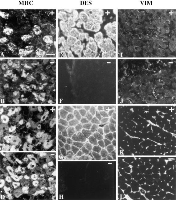
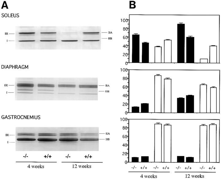
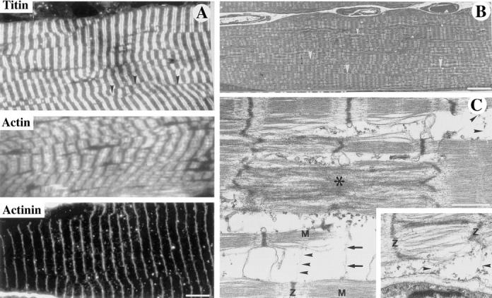
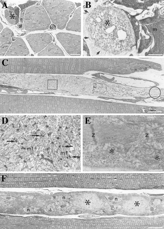
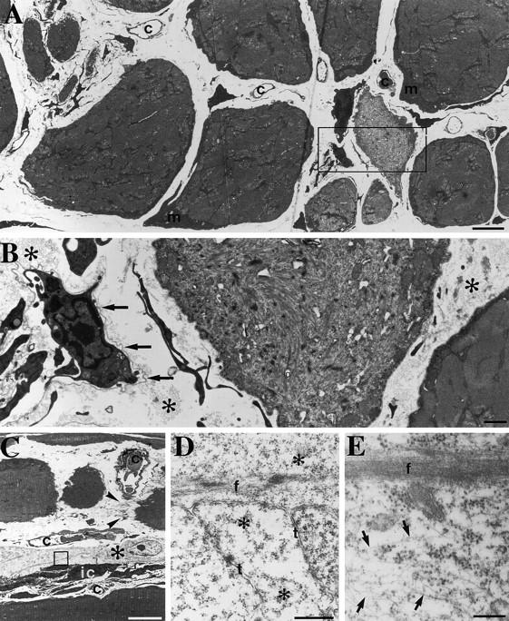
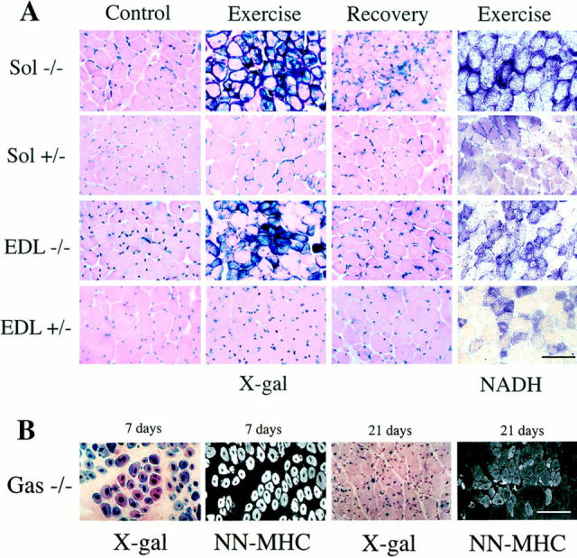
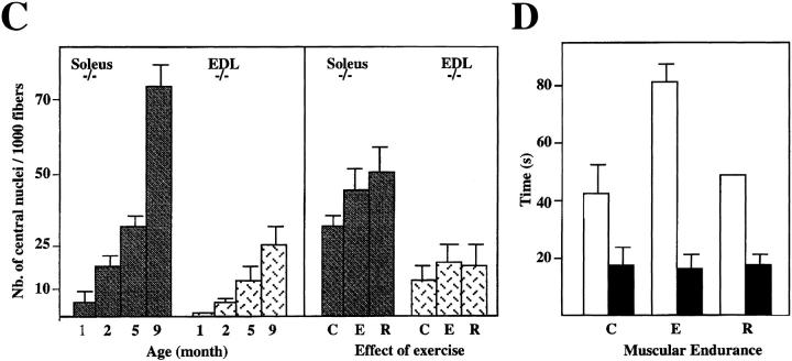
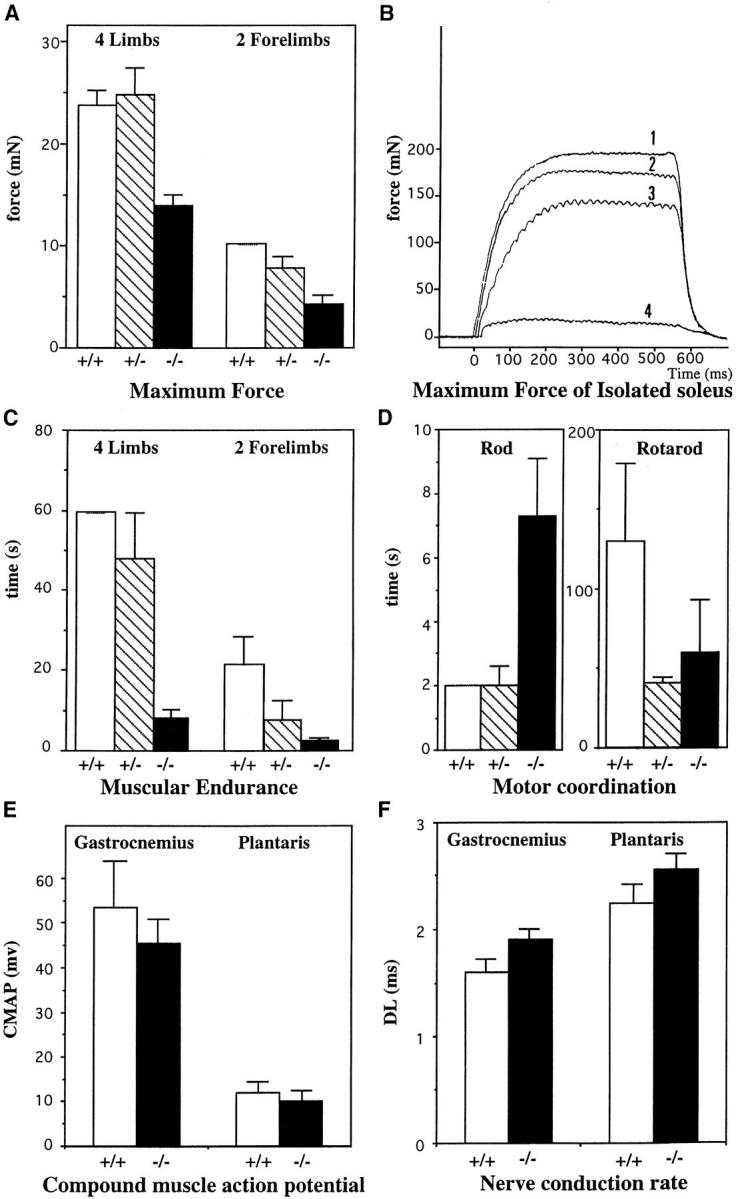
A null mutation was introduced into the mouse desmin gene by homologous recombination. The desmin knockout mice (Des -/-) develop normally and are fertile. However, defects were observed after birth in skeletal, smooth, and cardiac muscles (Li, Z., E. Colucci-Guyon, M. Pincon-Raymond, M. Mericskay, S. Pournin, D. Paulin, and C. Babinet. 1996. Dev. Biol. 175:362-366; Milner, D.J., G. Weitzer, D. Tran, A. Bradley, and Y. Capetanaki. 1996. J. Cell Biol. 134:1255- 1270). In the present study we have carried out a detailed analysis of somitogenesis, muscle formation, maturation, degeneration, and regeneration in Des -/- mice. Our results demonstrate that all early stages of muscle differentiation and cell fusion occur normally. However, after birth, modifications were observed essentially in weight-bearing muscles such as the soleus or continually used muscles such as the diaphragm and the heart. In the absence of desmin, mice were weaker and fatigued more easily. The lack of desmin renders these fibers more susceptible to damage during contraction. We observed a process of degeneration of myofibers, accompanied by macrophage infiltration, and followed by a process of regeneration. These cycles of degeneration and regeneration resulted in a relative increase in slow myosin heavy chain (MHC) and decrease in fast MHC. Interestingly, this second wave of myofibrillogenesis during regeneration was often aberrant and showed signs of disorganization. Subsarcolemmal accumulation of mitochondria were also observed in these muscles. The lack of desmin was not compensated by an upregulation of vimentin in these mice either during development or regeneration. Absence of desmin filaments within the sarcomere does not interfere with primary muscle formation or regeneration. However, myofibrillogenesis in regenerating fibers is often abortive, indicating that desmin may be implicated in this repair process. The results presented here show that desmin is essential to maintain the structural integrity of highly solicited skeletal muscle.


Figures

Figure 1
Figure 1
Skeletal muscle formation in mouse embryos. Myotome formation in somites of 11-d.p.c. embryos: Des −/+ (A) and Des −/− (B) embryos were stained for β-galactosidase activity. All myogenic cells appeared blue with a higher intensity in the homozygous (two LacZ copies) than in the heterozygous (one LacZ copy). We observed a normal dermomyotome ventral extension at the thoracic level. h, heart; dm, dermomyotome; hg, hindgut; hl, hindlimb. Muscle growth in 13.5–15-d.p.c embryos in the absence of desmin: Des −/− 13.5-d.p.c. (C) and 15-d.p.c. (D) embryos were stained for β-galactosidase activity. All myofibers are stained blue. We observed a normal morphogenesis and growth of the skeletal muscles. ld, latissimus dorsi; cm, cutaneous maximus; d, deltoidus; t, trapezius. Formation of head and limb musculature at 16 d.p.c. in Des −/− fetuses: β-galactosidase activity in head muscles (E), tongue (F), forelimb (G), and hindlimb musculature (H). bb, biceps brachii; tb, triceps brachii; ad, acromiodeltoidus; sd, spinodeltoidus; e, extensor; f, flexor; vl, vastus lateralis; ta, tibialis anterior; s, soleus.
Figure 2
Figure 2
Myotome differentiation and migration of myogenic cells from somites into limbs of 11-d.p.c embryos. Sagittal sections at the somitic level of Des −/− (A), Des +/− (B), and Des +/+ (C) embryos. Myotomes have the same morphology when revealed by the blue staining. Transversal sections of the limb bud of Des −/− (D), Des +/+ (E) embryos, with the presence in both sections of blue mononucleated myogenic cells, which have migrated but not yet fused. Analysis of vimentin and desmin expression during embryonic development in myogenic cells were performed with immunoperoxidase reactions on cryostat sections on 10.5-d.p.c. embryos from Des +/− heterozygous (G), and Des −/− homozygous (H) mice. The nuclear blue staining was used to characterize the myogenic population. In the control, the desmin-positive, mononucleated cells, which are located in the lateral part of the myotome, have blue nuclei. Immunodetection of desmin stained cytoplasm in brown (F). The adjacent section shows same blue cells negative for vimentin (G). The mutant, mononucleated cells located in the lateral part of the Des −/− myotome are labeled with a blue nucleus but are negative for desmin and vimentin (H). The neural tube (nt) is stained with vimentin antibody. Bar, 50 μm.
Figure 3
Figure 3
Detection of myosin isoforms, desmin and vimentin, by immunofluorescence in fetuses and in 1-mo-old mice. Mutant Des −/− (B, D, F, H, J, and L) and control Des +/+ (A, C, E, G, I, and K). (A–D) Characterization of primary and secondary generation muscle fibers at 17.5 d.p.c. Immunofluorescent staining of anterior shoulder muscles on transverse sections of the subscapularis muscle with antibody against slow MHC, which labels primary muscle fibers (A and B) and using an antibody against MHC, which labels secondary muscle fibers (C and D). The same pattern was found in Des +/+ and Des −/− fetuses. (E–H) Detection of desmin using a polyclonal antidesmin antibody showed a typical reactivity in Des +/+ in 17.5-d.p.c. fetuses (E) and 1-mo-old mice (G). No reactivity was found in Des −/− fetus (F) or 1-mo-old mice (H). (I–L) Detection of vimentin using polyclonal anti-vimentin antibody performed on 17.5-d.p.c. fetuses (I and J) and 1-mo-old mice (K and L). The same pattern was found in Des +/+ and Des −/− mice. No vimentin reactivity was found inside the myofibers. The vimentin reactivity found around the myofibers corresponded to connective tissue forming the endomysium and the mesenchymal cells of vessels. Bars: (A–F) 25 μm; (I and J) 25 μm; (G, H, K, and L) 50 μm.
Figure 4
Figure 4
MHC isoforms and ATPase activity in 4- and 12-wk-old mice. (A) Electrophoretic separation of MHC isoforms of muscles from soleus, diaphragm, and gastrocnemius. Four major isoforms (IIX/D, IIA, IIB, and I) can be identified. When we compared the desmin +/+, +/−, and −/− mice, similar patterns were found at 4 wk. However, the yield of MHC per mg of tissue was always much less in the Des −/− mice. Note the decreased amount of IIA and IIX MHC in the soleus, and IIB in the diaphragm (at 12 wk). (B) Quantification of the number of slow and fast fibers present in three muscles by ATPase activity at 4 and 12 wk. Slow fibers, such as type I (black), exhibit a high activity, whereas fast fibers, such as type II (white), display a low ATPase activity after acid preincubation. Percentage of fast and slow fibers was measured in the soleus, diaphragm, and gastrocnemius of Des +/+ and −/− mice. In the soleus of Des −/− at 12 wk, note a decrease of fast fibers (white bar) corresponding to the disappearance of the type IIA and X MHC. A relative increase in the percentage of slow fibers (black) was found.
Figure 5
Figure 5
Ultrastructural and immunological characterization of sarcomeres in soleus muscle of Des −/− mice. (A and B) Region of soleus of 8-wk-old Des−/− mice showing sarcomere alignment that is relatively normal or with splitting of the myofibril. (C) In region of soleus of 2-wk-old Des −/− mice with focal alterations. (A) Ultrathin sections stained with antibodies against titin, actin, or α-actinin demonstrate the typical regular striated pattern. However, certain irregularities were observed in the organization of the myofibers that were more easily visualized in the electron microscope. (B) A splitting of the myofibrils can be seen (arrowheads). This splitting is also clearly demonstrated in the ultrathin sections stained with the antibody against titin in A where it can be seen that the Z bands are frequently not in register (arrowheads). (C) Ultrastructure of myofibrillar alterations in the soleus muscle as demonstrated by transmission electron microscopy. On longitudinal sections, filamentous material (arrowheads, top right and bottom center) interlinks the Z disks of one myofibril to the M band region of another myofibril. Another link of filamentous material (arrows) is seen between the M band region of the lower myofibril and the center of two sarcomeres that show Z disk streaming (*). Inset, filamentous material (arrowheads) form myofibril–sarcolemma attachments between the Z disk of a myofibril and dense plaques at the sarcolemma. M, M-line; Z, Z-disk. Bars: (A and B) 5 μm; (C) 1 μm.
Figure 6
Figure 6
Transmission electron microscopy of myofibers of soleus from Des −/− 2-wk-old mouse. (A) Cross section of 2-wk soleus showing an area of a normal dense myofibrillar pattern and an area containing several small-size cells. c, capillaries. (B) In higher magnification, the small-size cells are identified as macrophages (m) with well-organized rough endoplasmic reticulum. Activated satellite cells having light cytoplasm with dispersed ribosomes (*). All these cells are enclosed by the same basement membrane (arrows). (C) Longitudinal sections of muscle fiber, one with light cytoplasm, runs in parallel with two other well-organized myofibrils. (D) Higher magnification view of the boxed area in C, showing disorganized myofibrils (mf) and the Z bodies (arrows). (E) Higher magnification view of the encircled area in C, showing an organized sarcomere with Z disks and abundant ribosomes (*). (F) Muscle fiber with areas of light cytoplasm (*) and many large nuclei (n) containing prominent nucleoli run parallel to muscle fibers with well-organized myofibrils. Bars: (A, C, and E) 5 μm; (B and D) 1 μm; (E) 0.5 μm.
Figure 7
Figure 7
Degeneration and regeneration of soleus from Des −/− 10-wk-old mouse. (A) Transmission electron microscopy of a cross section showing the large variability in fiber diameters seen at 10 wk in the Des −/− soleus. The myofibrillar pattern is well preserved in the largest muscle fibers although abnormal accumulation of mitochondria (m) are present beneath the sarcolemma. Myofibrils are disorganized in some of the small- or intermediate-size fibers. Note that clusters of small fibers occupy the space of a large fiber. The wide interstitial space contains capillaries and cells with slender profiles. (B) Higher magnification of the boxed area in A showing one muscle fiber disorganized myofibrils. An undulating basement membrane (arrows) surrounds interstitial cells with slender processes. Note also the bundles of collagen fibrils (*) in the interstitium. (C) Some muscle fibers show well-organized myofibrils; however, one is divided into three parts, one of which is interrupted by a tendinous junction (arrowheads) after faulty regeneration after fiber damage. Profiles of interstitial cells with slender processes and one cell with a light cytoplasm (*) are seen. (D and E) Higher magnification view of the boxed area in C. In D, strands of myofibrillar material as well as tubules (t) and ribosomes (*) are seen, whereas in E, an array of cytoplasmic filaments are seen beside a myofilamentous strand. Bars: (A and B) 1 μm; (C) 10 μm; (D) 0.5 μm; (E) 0.25 μm.
Figure 8
Figure 8
Effect of exercise on muscles. (A) LacZ and NADH activity present on frozen sections of soleus and EDL from 5-mo-old Des +/− and Des −/− mice. Control, non-exercised mice; Exercise, 5 d of exercise. Recovery, exercise plus 5 d recovery. Note the strong expression of β-galactosidase (X-gal), NADH in the soleus (Sol) and EDL of exercise mice. (B) Frozen sections of the gastrocnemius muscles of Des−/− mice (Gas −/−) after 7 and 21 d of regeneration after injection of cardiotoxin. Sections were stained either for β-galactosidase activity (X-gal) or neonatal MHC expression (NN-MHC). (C) Number of central nucleated fibers presented in the soleus and EDL in mice of different ages (1–9 mo) and in the same mice as in A. C, non- exercised mice; E, 5 d of exercise; R, exercise plus 5 d recovery. Note the increase in the number of the central nuclei in different muscles both with age and exercise. (D) Muscular endurance after exercises was analyzed on same group of mice. Des +/+ (white) and Des −/− (black). After 5 d of training, muscular endurance increased in the control group. This was transitory since when the animals were allowed to recuperate for 5 d, muscular endurance returned to the same value as before training. In contrast, in Des −/− mice there was no increase of endurance with exercise. Bars: (A and B) 100 μm.
Figure 8
Figure 8
Effect of exercise on muscles. (A) LacZ and NADH activity present on frozen sections of soleus and EDL from 5-mo-old Des +/− and Des −/− mice. Control, non-exercised mice; Exercise, 5 d of exercise. Recovery, exercise plus 5 d recovery. Note the strong expression of β-galactosidase (X-gal), NADH in the soleus (Sol) and EDL of exercise mice. (B) Frozen sections of the gastrocnemius muscles of Des−/− mice (Gas −/−) after 7 and 21 d of regeneration after injection of cardiotoxin. Sections were stained either for β-galactosidase activity (X-gal) or neonatal MHC expression (NN-MHC). (C) Number of central nucleated fibers presented in the soleus and EDL in mice of different ages (1–9 mo) and in the same mice as in A. C, non- exercised mice; E, 5 d of exercise; R, exercise plus 5 d recovery. Note the increase in the number of the central nuclei in different muscles both with age and exercise. (D) Muscular endurance after exercises was analyzed on same group of mice. Des +/+ (white) and Des −/− (black). After 5 d of training, muscular endurance increased in the control group. This was transitory since when the animals were allowed to recuperate for 5 d, muscular endurance returned to the same value as before training. In contrast, in Des −/− mice there was no increase of endurance with exercise. Bars: (A and B) 100 μm.
Figure 9
Figure 9
Analyses of muscular performances on control animal Des +/+ (white), heterozygous Des +/− (hatched) and homozygous Des −/− (black). (A) The maximum force developed by the mice was measured by pulling the mouse backwards by the tail until the bar was released. Animals were allowed to hold the bar linked to a dynamometer either with all four limbs or with the two forelimbs. Results are expressed in mN. (B) The maximum force developed by isolated muscle was measured on soleus from 2- (curve 1: Des +/+; curve 3: Des−/−) and 5-mo-old mice (curve 2: Des +/+; and curve 4: Des −/−). (C) The muscular endurance was analyzed by measuring the time that animals could hold onto a 32-g bar, either with all four limbs (4L) or with the two forelimbs (2L). Results are expressed in s. (D) Motor coordination: performances were measured either by measuring the time that mice need to cross a rod or by putting the mice on a rotating apparatus. Capacity of Des −/− mice are considerably modified. (E) CMAP measured in gastrocnemius and plantaris muscles from 5-mo-old mice. Results are expressed in mV. (F) Nerve conduction rate measured in gastrocnemius and plantaris muscles from 5-mo-old mice. The DL is given in ms. Data are the means ± standard errors computed from each set of experiments.

References

    1. Agbulut O, Li Z, Mouly V, Butler-Browne GS. Analysis of skeletal and cardiac muscle from desmin knock-out and normal mice by high resolution separation of MHC isoforms. Biol Cell. 1997;88:131–135. - PubMed
    1. Allen RE, Rankin LL, Greene EA, Boxhorn LK, Johnson SE, Taylor RG, Pierce PA. Desmin is present in proliferating rat muscle satellite cells but not in bovine muscle satellite cells. J Cell Physiol. 1991;149:525–535. - PubMed
    1. Ariza A, Coll J, Fernandez-Figueras MT, Lopez MD, Mate JL, Garcia O, Fernandez-Vasalo A, Navas-Palacios JJ. Desmin myopathy: a multisystem disorder involving skeletal, cardiac, and smooth muscle. J Hum Pathol. 1995;26:1032–1037. - PubMed
    1. Babai F, Musevi-Aghdam J, Schurch W, Royal A, Gabbiani G. Coexpression of α-sarcomeric actin, α-smooth muscle actin and desmin during myogenesis in rat and mouse embryos. I. Skeletal muscle. Differentiation. 1990;44:132–142. - PubMed
    1. Baribault H, Price J, Miyai K, Oshima RG. Mid-gestational lethality in mice lacking keratin 8. Genes Dev. 1993;7:1191–1202. - PubMed

Publication types

MeSH terms