. 1997 Oct 6;139(1):157-68.
doi: 10.1083/jcb.139.1.157.

CRP1, a LIM domain protein implicated in muscle differentiation, interacts with alpha-actinin

Affiliations

CRP1, a LIM domain protein implicated in muscle differentiation, interacts with alpha-actinin

P Pomiès et al. J Cell Biol. .

Abstract

Members of the cysteine-rich protein (CRP) family are LIM domain proteins that have been implicated in muscle differentiation. One strategy for defining the mechanism by which CRPs potentiate myogenesis is to characterize the repertoire of CRP binding partners. In order to identify proteins that interact with CRP1, a prominent protein in fibroblasts and smooth muscle cells, we subjected an avian smooth muscle extract to affinity chromatography on a CRP1 column. A 100-kD protein bound to the CRP1 column and could be eluted with a high salt buffer; Western immunoblot analysis confirmed that the 100-kD protein is alpha-actinin. We have shown that the CRP1-alpha-actinin interaction is direct, specific, and saturable in both solution and solid-phase binding assays. The Kd for the CRP1-alpha-actinin interaction is 1.8 +/- 0.3 microM. The results of the in vitro protein binding studies are supported by double-label indirect immunofluorescence experiments that demonstrate a colocalization of CRP1 and alpha-actinin along the actin stress fibers of CEF and smooth muscle cells. Moreover, we have shown that alpha-actinin coimmunoprecipitates with CRP1 from a detergent extract of smooth muscle cells. By in vitro domain mapping studies, we have determined that CRP1 associates with the 27-kD actin-binding domain of alpha-actinin. In reciprocal mapping studies, we showed that alpha-actinin interacts with CRP1-LIM1, a deletion fragment that contains the NH2-terminal 107 amino acids (aa) of CRP1. To determine whether the alpha-actinin binding domain of CRP1 would localize to the actin cytoskeleton in living cells, expression constructs encoding epitope-tagged full-length CRP1, CRP1-LIM1(aa 1-107), or CRP1-LIM2 (aa 108-192) were microinjected into cells. By indirect immunofluorescence, we have determined that full-length CRP1 and CRP1-LIM1 localize along the actin stress fibers whereas CRP1-LIM2 fails to associate with the cytoskeleton. Collectively these data demonstrate that the NH2-terminal part of CRP1 that contains the alpha-actinin-binding site is sufficient to localize CRP1 to the actin cytoskeleton. The association of CRP1 with alpha-actinin may be critical for its role in muscle differentiation.


Figures
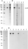
Figure 1
Figure 1
Specificity of the α-actinin–CRP1 interaction under nondenaturing conditions. (A) A Coomassie blue–stained gel showing molecular mass markers M, purified α-actinin (lane 1), and the 27–34% ammonium sulfate precipitate from avian smooth muscle extract (lane 2) that was loaded onto the affinity columns and used in the affinity resin binding assay. (B) Lane 1, Western immunoblot analysis of the 27–34% ammonium sulfate precipitate that was loaded onto the affinity columns using a polyclonal antibody raised against chicken α-actinin; lane 2, silver-stained gel showing the proteins eluted from the CRP1 column; lane 3, Western immunoblot analysis of the proteins shown in lane 2 using a polyclonal antibody raised against α-actinin; lane 4, silver-stained gel showing the material eluted from the BSA column; lane 5, Western immunoblot revealed that no α-actinin bound to the BSA column (α-a, α-actinin). (C) Coomassie blue– stained gel showing the purified GST (lane 1) and GST-CRP1 (lane 2) proteins that were used to generate the affinity resins. (D) Western immunoblot analysis to detect chicken α-actinin. The gel was loaded with α-actinin (lane 1) or a 27–34% ammonium sulfate precipitate from a smooth muscle cell extract (lane 2). Purified α-actinin or proteins found in the 27–34% ammonium sulfate precipitate were incubated with GST agarose (lanes 3 and 5) or GST-CRP1 agarose (lanes 4 and 6). α-Actinin binds to the GST-CRP1 affinity resin. A mock affinity resin binding assay was performed with GST-CRP1 agarose beads in the absence of α-actinin; no immunoreactive product is observed (lane 7). (E) [125I]α-actinin was incubated with GST-CRP1 (left) or GST agarose beads (right) in the absence of competing proteins (+ buffer), in the presence of a 2,000-fold molar excess of unlabeled α-actinin (+ unlabeled α-actinin), or in the presence of an equivalent molar amount of BSA (+ BSA). The counts bound to the agarose beads were analyzed using a γ counter and expressed as a percentage of bound [125I]α-actinin in absence of competing proteins. Mean and SEM from three experiments are shown.
Figure 1
Figure 1
Specificity of the α-actinin–CRP1 interaction under nondenaturing conditions. (A) A Coomassie blue–stained gel showing molecular mass markers M, purified α-actinin (lane 1), and the 27–34% ammonium sulfate precipitate from avian smooth muscle extract (lane 2) that was loaded onto the affinity columns and used in the affinity resin binding assay. (B) Lane 1, Western immunoblot analysis of the 27–34% ammonium sulfate precipitate that was loaded onto the affinity columns using a polyclonal antibody raised against chicken α-actinin; lane 2, silver-stained gel showing the proteins eluted from the CRP1 column; lane 3, Western immunoblot analysis of the proteins shown in lane 2 using a polyclonal antibody raised against α-actinin; lane 4, silver-stained gel showing the material eluted from the BSA column; lane 5, Western immunoblot revealed that no α-actinin bound to the BSA column (α-a, α-actinin). (C) Coomassie blue– stained gel showing the purified GST (lane 1) and GST-CRP1 (lane 2) proteins that were used to generate the affinity resins. (D) Western immunoblot analysis to detect chicken α-actinin. The gel was loaded with α-actinin (lane 1) or a 27–34% ammonium sulfate precipitate from a smooth muscle cell extract (lane 2). Purified α-actinin or proteins found in the 27–34% ammonium sulfate precipitate were incubated with GST agarose (lanes 3 and 5) or GST-CRP1 agarose (lanes 4 and 6). α-Actinin binds to the GST-CRP1 affinity resin. A mock affinity resin binding assay was performed with GST-CRP1 agarose beads in the absence of α-actinin; no immunoreactive product is observed (lane 7). (E) [125I]α-actinin was incubated with GST-CRP1 (left) or GST agarose beads (right) in the absence of competing proteins (+ buffer), in the presence of a 2,000-fold molar excess of unlabeled α-actinin (+ unlabeled α-actinin), or in the presence of an equivalent molar amount of BSA (+ BSA). The counts bound to the agarose beads were analyzed using a γ counter and expressed as a percentage of bound [125I]α-actinin in absence of competing proteins. Mean and SEM from three experiments are shown.
Figure 2
Figure 2
Demonstration of a direct interaction between CRP1 and [125I]α-actinin using a blot overlay assay. (A) Coomassie blue–stained gel showing a 27–34 (lane 1), a 34–43 (lane 2), and a 43–61% (lane 3) ammonium sulfate precipitates from an avian smooth muscle extract. Proteins from a parallel gel were transferred to nitrocellulose and the nitrocellulose strip was probed with [125I]α-actinin. The resulting autoradiograph shown in B illustrates [125I]α-actinin binding to CRP1. (C) Autoradiograph demonstrating the purity of the radioiodinated α-actinin probe. The position of the molecular mass markers is indicated on the left, in kD.
Figure 3
Figure 3
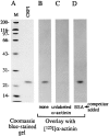
Specificity of the [125I]α-actinin–CRP1 interaction. (A) Coomassie blue–stained gel showing molecular mass markers and the purified recombinant CRP1. Autoradiograph of parallel nitrocellulose strips probed with [125I]α-actinin in the absence of competing protein (B), or in the presence of either a 2,000-fold molar excess of unlabeled α-actinin (C), or a 2,000-fold molar excess of unlabeled BSA (D).
Figure 4
Figure 4
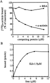
Binding of [125I]α-actinin to CRP1 in a solid-phase binding assay. (A) The specificity of the association between CRP1 and [125I]α-actinin in a solid-phase binding assay was analyzed in a competition experiment. A constant amount of [125I]α-actinin (0.26 pmoles in 120 μl) was incubated in CRP1-coated wells with increasing concentrations of unlabeled α-actinin (+ α-actinin) or BSA (+ BSA). In this experiment, a total of 3,076 cpm were bound specifically to CRP1 when no competing unlabeled α-actinin was added. The data are expressed as a percentage of the maximum binding obtained when the [125I]α-actinin is incubated with the CRP1-coated wells in the absence of competing protein. (B) From the competition experiment shown in A, the moles of α-actinin bound to CRP1 were plotted as a function of the free α-actinin concentration. In this particular experiment, the α-actinin was radioiodinated to a specific activity of 5.8 × 10⁶ cpm/μg; assuming a mol wt of 200,000 g/mol for α-actinin. The calculated dissociation constant (K d) was 1.9 μM. The mean dissociation constant determined from three different experiments using two different probes is 1.8 ± 0.3 μM (mean ± SEM).
Figure 5
Figure 5
Characterization of an anti-peptide antibody (B37) directed against cCRP1. (A) A Coomassie blue–stained gel showing molecular mass markers M and total CEF proteins L. (B) A parallel gel was transferred to nitrocellulose and probed with the anti-CRP1 antibody B37 or its corresponding preimmune serum pre. A single polypeptide of 23 kD is recognized by the antibody. (C) Autoradiograph of a gel loaded with a CEF lysate prepared from [35S]methionine-cysteine–labeled cells L, the proteins immunoprecipitated from this lysate with the polyclonal antibody raised against CRP1 B37, and with its corresponding preimmune serum pre. A single protein of 23 kD is specifically immunoprecipitated with the antibody against CRP1.
Figure 6
Figure 6
CRP1 and α-actinin are extensively codistributed in CEF and in smooth muscle cells. CEF cells (A–C) and smooth muscle cells (D–F), prepared for confocal indirect immunofluorescence microscopy, were double-labeled with a polyclonal antibody raised against CRP1 (A and D), and a monoclonal antibody raised against α-actinin (B and E). C and F are composite images of CRP1 (green) and α-actinin (red) staining; the overlapping regions appear in yellow. Confocal microscopy reveals that CRP1 and α-actinin are extensively colocalized along the actin stress fibers. Both α-actinin and CRP1 are detected at the leading edges of the cells (arrows) and in the adhesion plaques (arrowheads and data not shown). Bars, 30 μm.
Figure 7
Figure 7
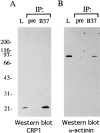
An in vivo interaction between CRP1 and α-actinin in smooth muscle cells. Proteins were immunoprecipitated from a chicken gizzard smooth muscle lysate L with the polyclonal antibody raised against CRP1 B37 and with the corresponding preimmune serum pre. The immunoprecipitated proteins were resolved by SDS-PAGE and were transferred to nitrocellulose and probed with polyclonal antibodies raised against CRP1 (A) or α-actinin (B). α-actinin is immunoprecipitated under nondenaturing conditions with the anti-CRP1 antibody, but not with the preimmune serum. The position of the molecular mass markers is indicated on the left in kD.
Figure 8
Figure 8
CRP1 interacts with the 27-kD actin-binding site of α-actinin. (A) A Coomassie blue–stained gel showing molecular mass markers M, purified α-actinin (lane 1), and the 53- and 27-kD proteolytic products of α-actinin generated by thermolysin cleavage (lane 2). Autoradiograph of overlay assay performed on parallel nitrocellulose strips containing purified α-actinin (lanes 1′ and 1″) and the proteolytic fragments of α-actinin (lanes 2′ and 2″) probed with [32P]GST-CRP1 (B), or [32P]GST (C). Note that in the experiment shown, thermolysin cleavage of α-actinin was not complete, therefore products other than the 53- and 27-kD fragments are also detected. (D) Autoradiograph illustrating the quality of the bacterially expressed, purified, 32P-labeled probes, [32P]GST-CRP1 and [32P]GST.
Figure 9
Figure 9
The binding site for α-actinin on CRP1 is contained within the CRP1-LIM1 fragment. (A) Coomassie blue–stained gel showing the purified CRP1 (lane 1), the purified CRP1-LIM1 fragment (lane 2) and the purified CRP1-LIM2 fragment (lane 3). 100 pmoles of CRP1, 200 pmoles of CRP1-LIM1, and 200 pmoles of CRP1-LIM2 were loaded on the gel. The positions of CRP1, CRP1-LIM1, and CRP1-LIM2 are marked (CRP1, LIM1, and LIM2, respectively). The corresponding blot overlay assay probed with [125I]α-actinin is shown in B. (C) Autoradiograph illustrating the purity of the radioiodinated α-actinin probe. The position of the molecular mass markers is indicated on the left in kD.
Figure 10
Figure 10
CRP1-LIM1 is targeted to actin stress fibers. Expression constructs encoding myc-tagged CRP1 (A and B), CRP1-LIM1 (C and D), or CRP1-LIM2 (E and F) were microinjected into rat embryo fibroblast (REF52) cells. Double-label immunofluorescence was used to compare the subcellular distributions of the expressed CRP1 polypeptides (A, C, and E) and the actin stress fibers (B, D, and F). The expressed proteins were visualized using an anti-myc monoclonal antibody whereas the actin stress fibers were visualized with phalloidin. Bar, 30 μm.
Figure 11
Figure 11
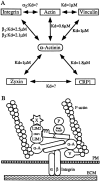
α-Actinin and its binding partners. (A) A summary of the interactions between α-actinin and its binding partners: integrin, zyxin, actin, vinculin, and CRP1. References for the dissociation constant values are as follows: α-actinin–integrin, (Otey et al., 1990); α-actinin– zyxin, (Crawford et al., 1992); α-actinin–vinculin, (Wachsstock et al., 1987); α-actinin–actin, (Wachsstock et al., 1993); α-actinin–CRP1, this report; vinculin–actin, (Menkel et al., 1994). (B) A schematic representation of an adhesion plaque showing a model for the association of α-actinin with its known binding partners within a cell. α-Actinin and CRP1 may cooperate to localize a complex of zyxin, Ena/VASP, and profilin and thus could participate in the regulation of actin assembly dynamics. (Z) zyxin; (P) profilin; (V) vinculin; (α-A) α-actinin; (X) other CRP1-binding partners; (PM) plasma membrane; (ECM) extracellular matrix.

References

    1. Arber S, Caroni P. Specificity of single LIM motifs in targeting and LIM/LIM interactions in situ. Genes Dev. 1996;10:289–300. - PubMed
    1. Arber S, Halder G, Caroni P. Muscle LIM protein, a novel essential regulator of myogenesis, promotes myogenic differentiation. Cell. 1994;79:221–231. - PubMed
    1. Arber S, Hunter JJ, Ross J, Hongo M, Sansig G, Borg J, Perriard J-C, Chien KR, Caroni P. MLP-deficient mice exhibit a disruption of cardiac cytoarchitectural organization, dilated cardiomyopathy, and heart failure. Cell. 1997;88:393–403. - PubMed
    1. Beckerle MC. Identification of a new protein localized at sites of cell-substrate adhesion. J Cell Biol. 1986;103:1679–1687. - PMC - PubMed
    1. Beckerle MC, Porter KR. Analysis of the roles of microtubules and actin in erythrophore intracellular transport. J Cell Biol. 1983;96:354–362. - PMC - PubMed

Publication types

MeSH terms