. 1997 Oct 6;139(1):257-64.
doi: 10.1083/jcb.139.1.257.

Induction of the angiogenic phenotype by Hox D3

Affiliations

Induction of the angiogenic phenotype by Hox D3

N Boudreau et al. J Cell Biol. .

Abstract

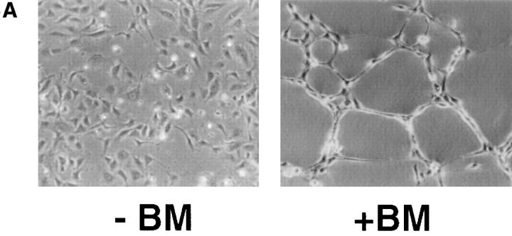
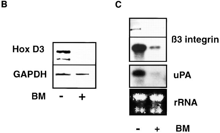
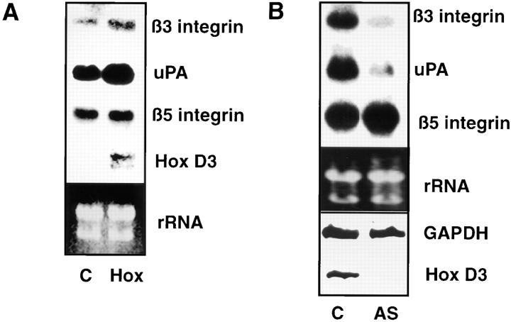
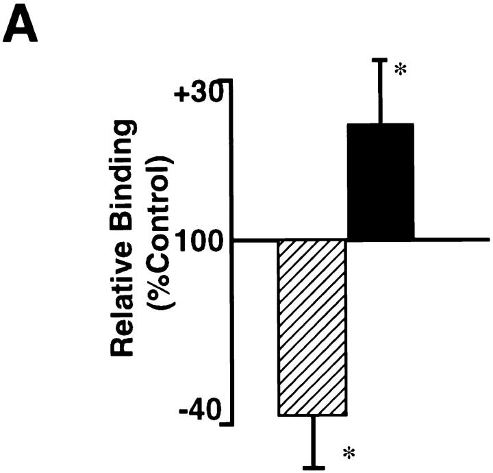
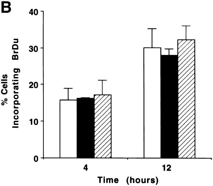
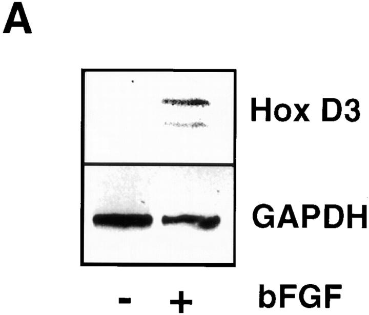
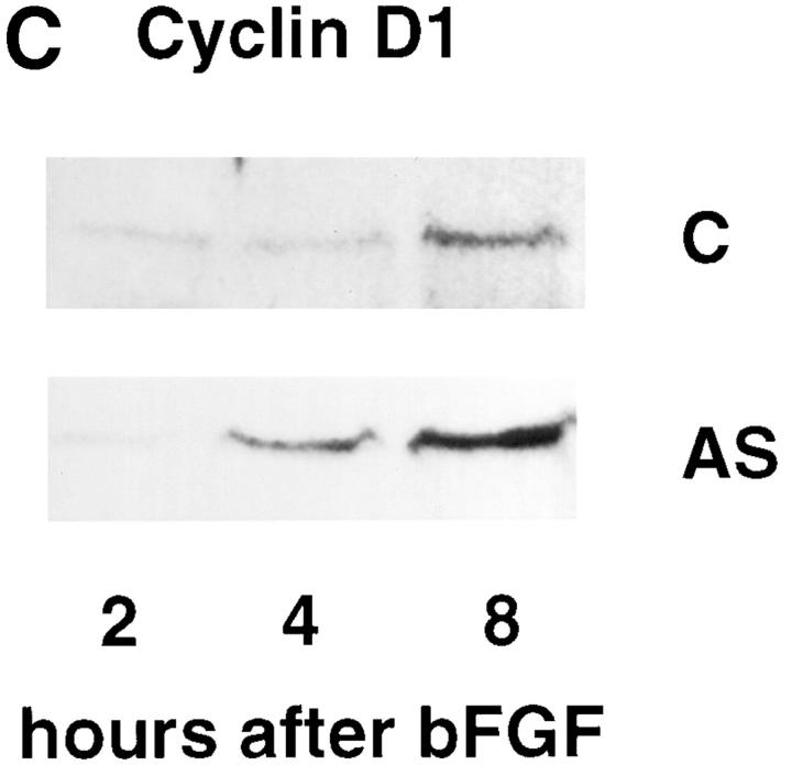
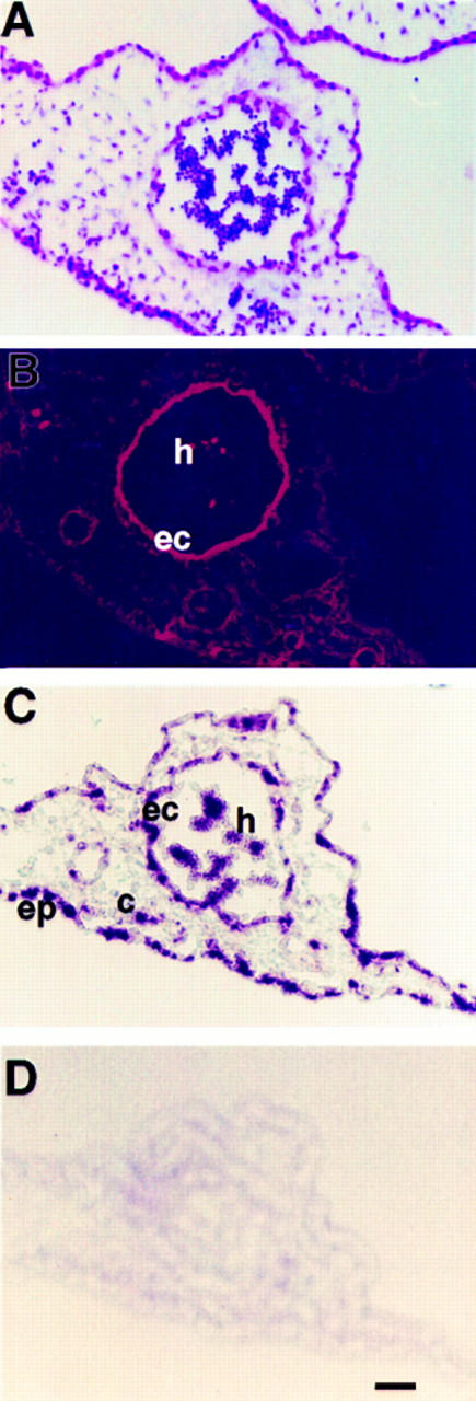
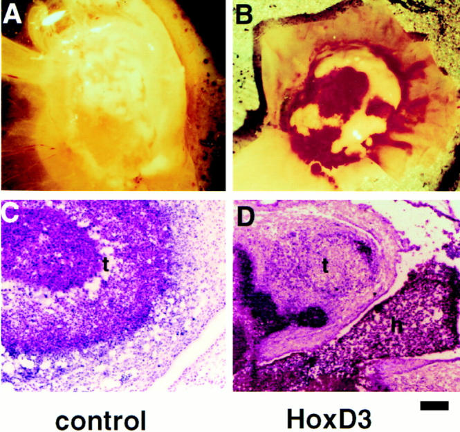
Angiogenesis is characterized by distinct phenotypic changes in vascular endothelial cells (EC). Evidence is provided that the Hox D3 homeobox gene mediates conversion of endothelium from the resting to the angiogenic/invasive state. Stimulation of EC with basic fibroblast growth factor (bFGF) resulted in increased expression of Hox D3, integrin alphavbeta3, and the urokinase plasminogen activator (uPA). Hox D3 antisense blocked the ability of bFGF to induce uPA and integrin alphavbeta3 expression, yet had no effect on EC cell proliferation or bFGF-mediated cyclin D1 expression. Expression of Hox D3, in the absence of bFGF, resulted in enhanced expression of integrin alphavbeta3 and uPA. In fact, sustained expression of Hox D3 in vivo on the chick chorioallantoic membrane retained EC in this invasive state and prevented vessel maturation leading to vascular malformations and endotheliomas. Therefore, Hox D3 regulates EC gene expression associated with the invasive stage of angiogenesis.


Figures

Figure 1
Figure 1
Expression of Hox D3, β3 integrin, and uPA in quiescent and proliferative EC. (A) Morphology of HUVEC cultured in the presence (+) or absence (−) of reconstituted BM after 24 h in M199 containing 5% FCS. (B) RT-PCR of 1 μg mRNA from HUVEC cultured for 48 h with (+) or without (−) reconstituted BM using primers for Hox D3 or GAPDH. (C) Western (top) and Northern (bottom) blot analysis of integrin β3 expression in HUVEC cultured in the presence (+) or absence (−) of BM for 48 h. Northern blots were reprobed for uPA mRNA expression. The bottom panel shows ethidium bromide staining of total RNA loaded in the corresponding gel.
Figure 1
Figure 1
Expression of Hox D3, β3 integrin, and uPA in quiescent and proliferative EC. (A) Morphology of HUVEC cultured in the presence (+) or absence (−) of reconstituted BM after 24 h in M199 containing 5% FCS. (B) RT-PCR of 1 μg mRNA from HUVEC cultured for 48 h with (+) or without (−) reconstituted BM using primers for Hox D3 or GAPDH. (C) Western (top) and Northern (bottom) blot analysis of integrin β3 expression in HUVEC cultured in the presence (+) or absence (−) of BM for 48 h. Northern blots were reprobed for uPA mRNA expression. The bottom panel shows ethidium bromide staining of total RNA loaded in the corresponding gel.
Figure 2
Figure 2
Influence of Hox D3 on β3 integrin and uPA expression in endothelial cells. (A) Northern blot analysis of β3 integrin, β5 integrin, uPA, and Hox D3 mRNA levels from 20 μg total RNA from immortalized HUVEC stably transfected with control (C) or Hox D3 (Hox) expression vectors. The lower panel shows ethidium bromide staining of total RNA loaded for each sample in the corresponding gel. (B) Northern blot analysis of β3 integrin, β5 integrin, and uPA mRNA expression levels from immortalized HUVECS transfected with control (C) or antisense expression vectors for Hox D3 (AS). RNA (20 μg) from each sample was loaded, and total RNA was visualized by ethidium bromide staining of the corresponding gel. The lower box shows RT-PCR of 1 μg total RNA from cells transfected with control (C) or antisense expression vectors against Hox D3 (AS) using primers for Hox D3 or GAPDH.
Figure 3
Figure 3
Effects of Hox D3 sense and antisense expression on EC adhesion and proliferation. (A) Adhesion to microtitre wells coated with 10 μg/ml fibrinogen by EC transfected with Hox D3 sense (▪) or antisense (▨ ▨ ▨ ) expression vectors. Adhesion after 30 min was assessed by absorbance at 600 nm and expressed as a percentage of adhesion displayed by control-transfected EC (n = 3). *P < 0.05. (B) BrdU incorporation in control-transfected EC (□), Hox D3-overexpressing cells (▪), or cells expressing antisense against Hox D3 (▨ ▨ ▨ ). Cells were labeled for 4 or 12 h with 10 mM BrdU. The percentage of cells staining positive for BrdU was assessed by counting at least six different fields containing a total of at least 400 cells each.
Figure 3
Figure 3
Effects of Hox D3 sense and antisense expression on EC adhesion and proliferation. (A) Adhesion to microtitre wells coated with 10 μg/ml fibrinogen by EC transfected with Hox D3 sense (▪) or antisense (▨ ▨ ▨ ) expression vectors. Adhesion after 30 min was assessed by absorbance at 600 nm and expressed as a percentage of adhesion displayed by control-transfected EC (n = 3). *P < 0.05. (B) BrdU incorporation in control-transfected EC (□), Hox D3-overexpressing cells (▪), or cells expressing antisense against Hox D3 (▨ ▨ ▨ ). Cells were labeled for 4 or 12 h with 10 mM BrdU. The percentage of cells staining positive for BrdU was assessed by counting at least six different fields containing a total of at least 400 cells each.
Figure 4
Figure 4
Hox D3 mediates bFGF-induced expression of β3 integrin and uPA. (A) RT-PCR of 1 μg total RNA from HUVEC 24 h after treatment with 20 ng/ml bFGF (+bFGF) using primers for Hox D3 or GAPDH. (B) Northern blot analysis of β3 integrin, β5 integrin, and uPA mRNA levels in HUVEC transfected with control plasmid (C) or antisense against Hox D3 (AS) after 24 h with (+) or without (−) addition of 20 ng/ml bFGF. (C) Western blot of cyclin D1 in immortalized HUVEC transfected with control or antisense against Hox D3. After 24 h in serum-free M199, samples were collected at 2, 4, and 8 h after addition of 20 ng/ml bFGF. 10 μg total cell lysate from each time point was separated on 12% SDS-PAGE, transferred to nylon membranes, and probed with a polyclonal antibody against human cyclin D1.
Figure 4
Figure 4
Hox D3 mediates bFGF-induced expression of β3 integrin and uPA. (A) RT-PCR of 1 μg total RNA from HUVEC 24 h after treatment with 20 ng/ml bFGF (+bFGF) using primers for Hox D3 or GAPDH. (B) Northern blot analysis of β3 integrin, β5 integrin, and uPA mRNA levels in HUVEC transfected with control plasmid (C) or antisense against Hox D3 (AS) after 24 h with (+) or without (−) addition of 20 ng/ml bFGF. (C) Western blot of cyclin D1 in immortalized HUVEC transfected with control or antisense against Hox D3. After 24 h in serum-free M199, samples were collected at 2, 4, and 8 h after addition of 20 ng/ml bFGF. 10 μg total cell lysate from each time point was separated on 12% SDS-PAGE, transferred to nylon membranes, and probed with a polyclonal antibody against human cyclin D1.
Figure 5
Figure 5
Colocalization of Hox D3 and αvβ3 integrin in endotheliomas in vivo. (A) H&E-stained cross-section of tissue infected by Hox D3-expressing retrovirus shows an abnormally large capillary-like structure filled with erythrocytes. (B) Immunofluorescent staining with LM609 against αvβ3 in the corresponding serial section shows strong positive staining in both the endothelial component (ec) of this vascular structure and in hematopoietic cells (h) contained within. (C) In situ hybridization of retrovirally expressed Hox D3 in corresponding serial section showing widespread expression in epithelium (ep), endothelial cells (ec), connective tissue (c), and hematopoietic cells (h). (D) Control in situ hybridization in a serial section performed with a sense riboprobe for Hox D3. Bar, 20 μm.
Figure 6
Figure 6
Effect of retroviral-mediated expression of Hox D3 in chick embryos. (A and B) Morphology of tumors generated 3 d after grafting of QT6 cells producing control or Hox D3-expressing retrovirus onto 10 d chick CAMs. (C and D) H&E staining in tissue cross-sections from tumors (t) and vasculature produced by cells shedding control or Hox D3-expressing retrovirus. Note the large hemorrhagic region containing hematopoietic cells (h) adjacent to tumor tissue in the Hox D3-infected tissue. Bar, 50 μm.

References

    1. Bautch VL, Toda S, Hassell JA, Hanahan D. Endothelial cell tumors develop in transgenic mice carrying polyoma middle T oncogene. Cell. 1987;51:529–538. - PubMed
    1. Botas J. Control of morphogenesis and differentiation by HOM/Hoxgenes. Curr Opin Cell Biol. 1993;5:1015–1022. - PubMed
    1. Brooks PC, Clark RAF, Cheresh DA. Requirement of vascular integrin αvβ3 for angiogenesis. Science (Wash DC) 1994;264:569–571. - PubMed
    1. Brooks PC, Strömblad S, Sanders L, von Schalscha TL, Aimes RT, Stetler-Stevenson WG, Quigley JP, Cheresh DA. Localization of matrix metalloproteinase MMP-2 to the surface of invasive cells by interaction with integrin αvβ3. Cell. 1996;85:683–693. - PubMed
    1. Brown WM, Zhou L, Taylor GR. The nucleotide sequence of the murine Hox D3gene reveals extensive identity with the human protein. Biochim Biophys Acta. 1994;1219:219–222. - PubMed

Publication types

MeSH terms