. 1997 Oct 6;139(1):265-78.
doi: 10.1083/jcb.139.1.265.

Beta 1 integrin is essential for teratoma growth and angiogenesis

Affiliations

Beta 1 integrin is essential for teratoma growth and angiogenesis

W Bloch et al. J Cell Biol. .

Abstract

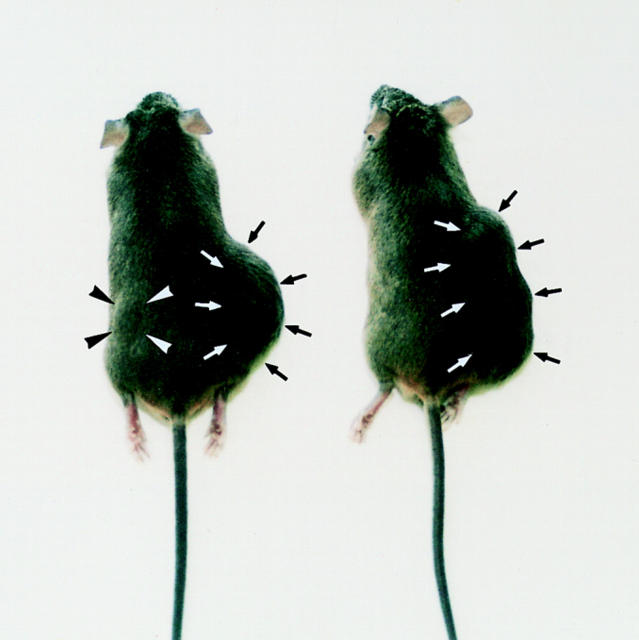
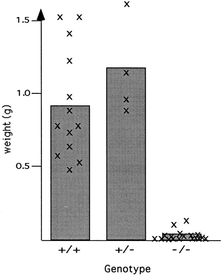
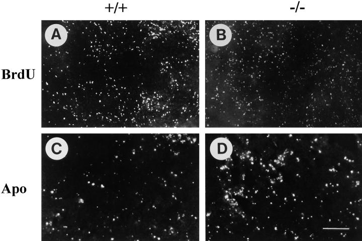
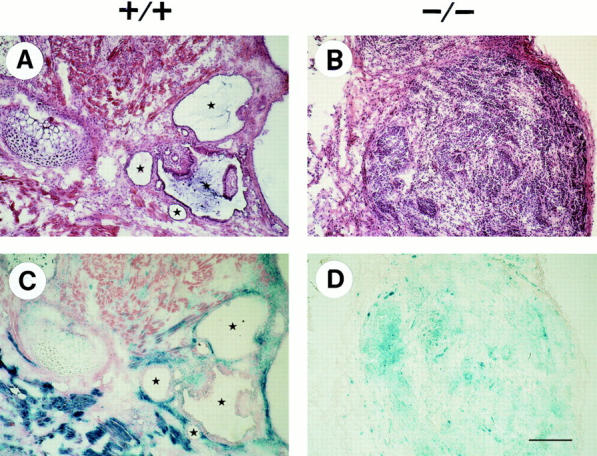
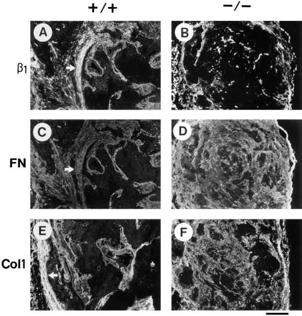
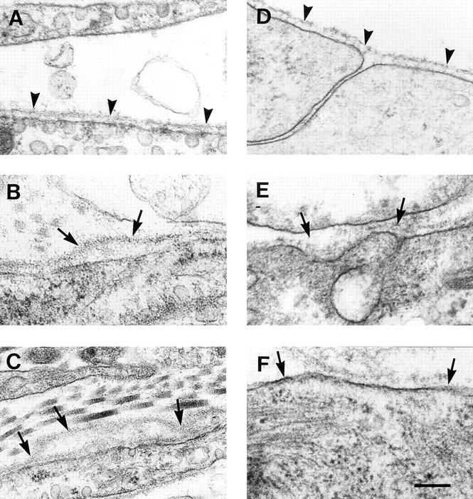
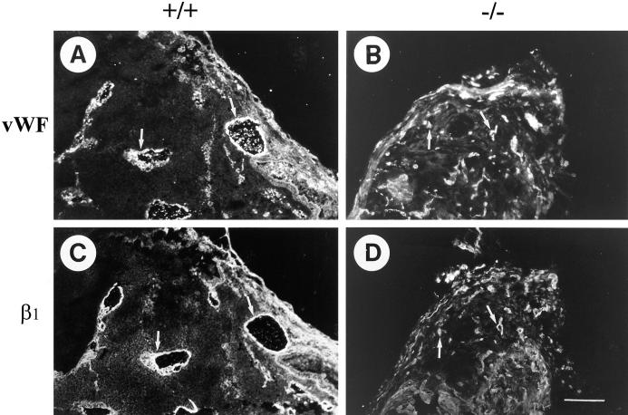
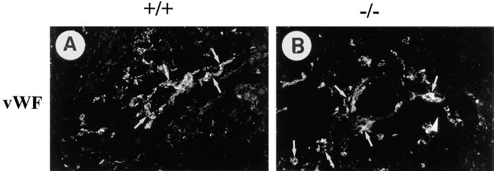
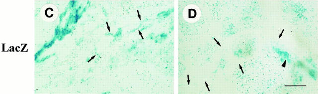
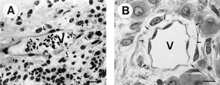
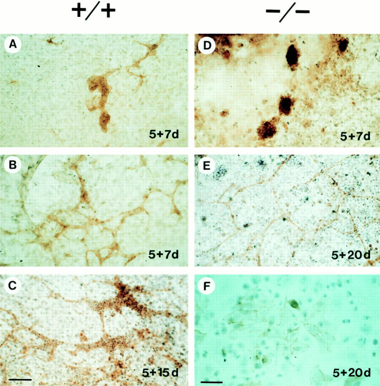
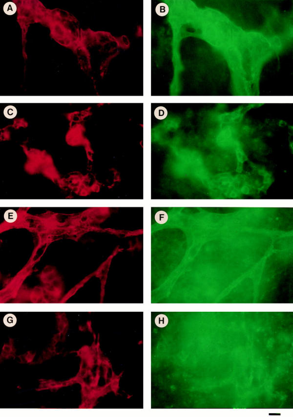
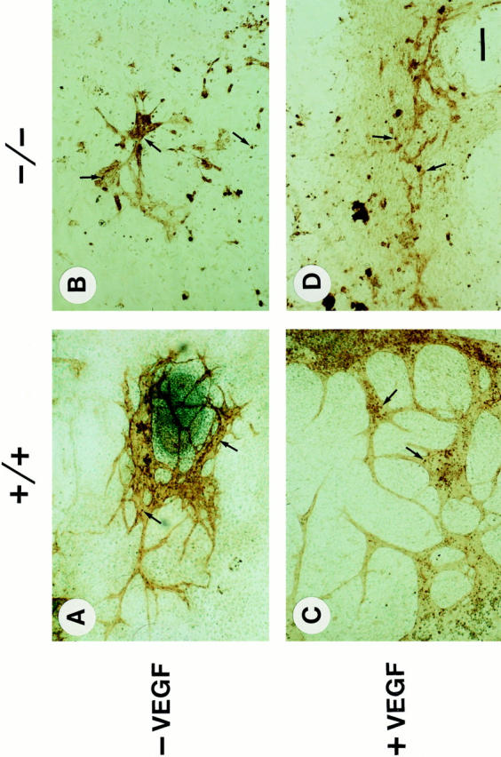
Teratomas are benign tumors that form after ectopic injection of embryonic stem (ES) cells into mice and contain derivatives of all primitive germ layers. To study the role of beta 1 integrin during teratoma formation, we compared teratomas induced by normal and beta1-null ES cells. Injection of normal ES cells gave rise to large teratomas. In contrast, beta 1-null ES cells either did not grow or formed small teratomas with an average weight of <5% of that of normal teratomas. Histological analysis of beta 1-null teratomas revealed the presence of various differentiated cells, however, a much lower number of host-derived stromal cells than in normal teratomas. Fibronectin, collagen I, and nidogen were expressed but, in contrast to normal teratomas, diffusely deposited in beta1-null teratomas. Basement membranes were present but with irregular shape and detached from the cell surface. Normal teratomas had large blood vessels with a smooth inner surface, containing both host- and ES cell-derived endothelial cells. In contrast, beta 1-null teratomas had small vessels that were loosely embedded into the connective tissue. Furthermore, endothelial cells were always of host-derived origin and formed blood vessels with an irregular inner surface. Although beta 1- deficient endothelial cells were absent in teratomas, beta 1-null ES cells could differentiate in vitro into endothelial cells. The formation of a complex vasculature, however, was significantly delayed and of poor quality in beta1-null embryoid bodies. Moreover, while vascular endothelial growth factor induced proliferation of endothelial cells as well as an extensive branching of blood vessels in normal embryoid bodies, it had no effect in beta 1-null embryoid bodies.


Figures

Figure 1
Figure 1
Male 129/Sv mice inoculated with 107 D3 cells (β1+/+; right side of the back) and 107 G 201 cells (β1−/−, left side of the back). The right side of the back was inoculated with normal ES cells. After an incubation period of 3 wk, both mice developed a big teratoma (arrows). The left side of the back was inoculated with β1-null ES cells. Note that a small teratoma formed and is visible in the left mouse (arrowheads) but not in the right mouse.
Figure 2
Figure 2
Weights of teratomas derived from normal (+/+), β1-heterozygous (+/−), and β1-null (−/−) ES cells. The mean values were calculated from 13 +/+, 4 +/−, and 9 −/− teratomas, respectively. Individual values are designated by an x.
Figure 3
Figure 3
Analysis of cell proliferation and apoptosis in a normal (+/+) teratoma and a β1-null (−/−) teratoma. ES cells were injected under the skin of the back and incubated for 21 d. 2 h before tumor isolation BrdU was injected intraperitoneally. Incorporated BrdU was detected using specific antibodies labeled with FITC (A and B). Apoptotic cells were identified by staining for the presence of free 3′-hydroxyl groups in tissue sections of +/+ and −/− teratomas (C and D). Bar, 100 μm.
Figure 4
Figure 4
Histology of normal (+/+) and β1-null (−/−) teratomas. Tumors derived from normal ES cells (β1 +/+) and from β1-null ES cells (β1 −/−) were stained with Hematoxylin/Eosin (A and B) and lacZ/eosin (C and D), respectively. Blue, lacZ-positive cells are ES cell-derived whereas lacZ-negative cells are host-derived stromal cells that have migrated into the tumor tissue. Stars indicate lumina of glandular structures. Bar, 100 μm.
Figure 5
Figure 5
Immunostaining for ECM components in normal (+/+) and in β1-null (−/−) teratomas. The antibodies used were directed against β1 integrin (A and B), fibronectin (C and D), and collagen I (E and F). The staining shown in A and C, and B and D, respectively, are double stainings of the same tissue specimens. Arrows indicate dense cords of connective tissue. Bar, 100 μm.
Figure 6
Figure 6
Electronmicroscopy of normal (A and D) and β1-null teratomas and embryoid bodies (B, C, E, and F). Normal cells in teratomas (A) and embryoid bodies (D) are covered by a smooth basement membrane that is strictly located in close vicinity of the cell surface (arrowhead). In β1-null teratomas as well as β1-null embryoid bodies, the basement membranes are partially detached (B and F, arrows), multilayered (C), or show an increased thickness and a loss of typical structure (E). Bar, 250 nm.
Figure 7
Figure 7
Immunostaining of a normal (+/+) and a β1-null (−/−) teratoma for the expression of vWF and for β1 integrin expression. Teratomas were removed 21 d after inoculation and sectioned. Neighboring sections were stained for vWF and β1 integrin expression, respectively. Whereas normal teratomas have large vessels, β1-null teratomas have small vessels. Note that like in normal tumors vessels of β1-null tumors coexpress vWF and β1 integrin indicating that endothelial cells are host derived. Bar, 100 μm.
Figure 8
Figure 8
Localization of vWF and lacZ expression in normal (+/+) and β1-null (−/−) teratomas. Teratomas were removed 21 d after inoculation of ES cells and sectioned. Sections were stained overnight for lacZ expression and subsequently immunostained for vWF expression. In normal teratomas, vWF-positive areas (A, arrows) are found to be positive and negative for lacZ expression (C, corresponding arrows) indicating that endothelial cells are both host- and ES cell–derived. In contrast, in β1-null teratomas, all vWF-positive areas (B, arrows) are lacZ-negative (D, corresponding arrows) indicating that endothelial cells are solely host derived. Many other areas, however, that are vWF-negative (B, arrowhead) are clearly lacZ-positive (D, corresponding arrowhead) indicating that many other regions in β1-null teratomas are ES cell–derived. Bar, 100 μm.
Figure 8
Figure 8
Localization of vWF and lacZ expression in normal (+/+) and β1-null (−/−) teratomas. Teratomas were removed 21 d after inoculation of ES cells and sectioned. Sections were stained overnight for lacZ expression and subsequently immunostained for vWF expression. In normal teratomas, vWF-positive areas (A, arrows) are found to be positive and negative for lacZ expression (C, corresponding arrows) indicating that endothelial cells are both host- and ES cell–derived. In contrast, in β1-null teratomas, all vWF-positive areas (B, arrows) are lacZ-negative (D, corresponding arrows) indicating that endothelial cells are solely host derived. Many other areas, however, that are vWF-negative (B, arrowhead) are clearly lacZ-positive (D, corresponding arrowhead) indicating that many other regions in β1-null teratomas are ES cell–derived. Bar, 100 μm.
Figure 9
Figure 9
Semithin sections of a normal and a β1-null teratoma stained with methylene blue and immunostained for vWF. Vessels (V) in normal teratomas (A) have a smooth inner surface and are tightly embedded within the surrounding tissue (arrows). Vessels of β1-null teratomas (B) have an irregular surface and have lost contacts to the surrounding tissue (arrows). Bar, 20 μm.
Figure 10
Figure 10
Normal (+/+) and β1-null (−/−) embryoid bodies immunostained for PECAM. ES cells were differentiated in hanging drops for 2 d, cultured in bacteriological dishes for another 3 d (which gives a total culture period of 5 d in suspension), and plated on gelatin-coated cover slips. After various periods on the cover slips (7, 15, 20 d) outgrowth were fixed and stained for PECAM. The culture periods are designated in each picture. Note the presence of blood cells in vessels of normal embryoid body outgrowths (C). Bars: (A–D) 80 μm; (F) 40 μm.
Figure 11
Figure 11
Diameter of vessel lumen in normal (+/+) and β1-null (−/−) embryoid bodies. •, −/− PECAM1; □, +/+ PECAM1. Morphometrical analysis of PECAM-positive vessels after 5 + 20 d in culture. Normal embryoid bodies have variously sized vessels with diam up to 70 μm. In β1-null embryoid bodies most vessels have a diam of 10 μm and only a few are larger.
Figure 12
Figure 12
Double immunostaining of normal (+/+) and β1-null (−/−) embryoid bodies for β3 integrin and PECAM (A–D), and αv integrin and PECAM (E–H). Normal and mutant ES cells were differentiated for 20 d, fixed and stained for PECAM (A and C, red) and β3 (B and D, green), or PECAM (E and G, red) and αv (F and H, green), respectively. In normal embryoid bodies large vessels stain for PECAM (A and C) and β3 (B), or αv integrin (D). In β1-null embryoid bodies the vessel diameters are smaller but the staining for PECAM (E and G), β3 (D), and αv (H) is similar, like in normal bodies. Note the high background for αv staining, which is expressed on many PECAM-negative cells in the embryoid bodies. Bar, 15 μm.
Figure 13
Figure 13
VEGF treatment of normal (+/+) and β1-null (−/−) embryoid bodies. Normal or β1-null embryoid bodies were treated either without (A and B) or with (C and D) 10 ng/ml VEGF during a period of 5 d in suspension and 12 d on a gelatinized glass cover slip. 2 h before fixation, the cultures were treated with BrdU. Sections were first stained for BrdU incorporation (arrows), then for PECAM-1, and finally counterstained with methyl green. Note that VEGF treatment increases the branching of blood vessels (C) and the proliferation rate of PECAM-positive cells in normal (C, arrows) but not in β1-null embryoid bodies (D, arrows). Bar, 100 μm.

References

    1. Alexander, C.M., and Z. Werb. 1991. Degradation of extracellular matrix. In Cell Biology of Extracellular Matrix. E. Hay, editor. Plenum Press, New York. pp. 255–294.
    1. Bagutti C, Wobus AM, Fässler R, Watt FM. Differentiation of embryonal stem cells into keratinocytes: comparison of wild-type and β1 integrin-deficient cells. Dev Biol. 1996;179:184–196. - PubMed
    1. Boudreau N, Sympson CJ, Werb Z, Bissel MJ. Suppression of ICE and apoptosis in mammary epithelial cells by extracellular matrix. Science (Wash DC) 1995;267:891–893. - PMC - PubMed
    1. Brooks PC, Clark RAF, Cheresch DA. Requirement of vascular integrin αvβ3 for angiogenesis. Science (Wash DC) 1994a;264:569–571. - PubMed
    1. Brooks PC, Montgomery AMP, Rosenfeld M, Reisfeld RA, Hu T, Klier G, Cheresh DA. Integrin αvβ3antagonists promote tumor regression by inducing apoptosis of angiogenic blood vessels. Cell. 1994b;79:1157–1164. - PubMed

Publication types

MeSH terms