. 1997 Oct 6;186(7):989-97.
doi: 10.1084/jem.186.7.989.

Reduced incidence and delayed onset of diabetes in perforin-deficient nonobese diabetic mice

Affiliations

Reduced incidence and delayed onset of diabetes in perforin-deficient nonobese diabetic mice

D Kägi et al. J Exp Med. .

Abstract

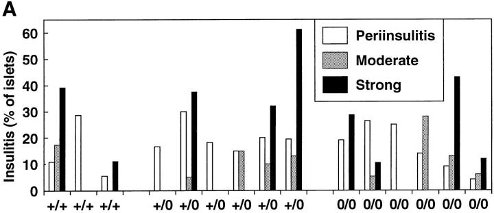
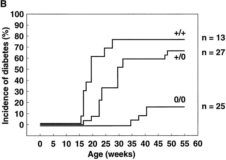
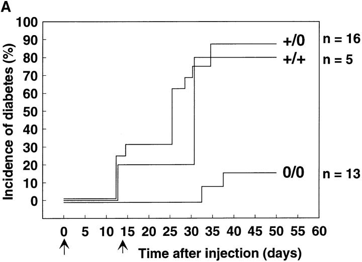
To investigate the role of T cell-mediated, perforin-dependent cytotoxicity in autoimmune diabetes, perforin-deficient mice were backcrossed with the nonobese diabetes mouse strain. It was found that the incidence of spontaneous diabetes over a 1 yr period was reduced from 77% in perforin +/+ control to 16% in perforin-deficient mice. Also, the disease onset was markedly delayed (median onset of 39.5 versus 19 wk) in the latter. Insulitis with infiltration of CD4(+) and CD8(+) T cells occurred similarly in both groups of animals. Lower incidence and delayed disease onset were also evident in perforin-deficient mice when diabetes was induced by cyclophosphamide injection. Thus, perforin-dependent cytotoxicity is a crucial effector mechanism for beta cell elimination by cytotoxic T cells in autoimmune diabetes. However, in the absence of perforin chronic inflammation of the islets can lead to diabetogenic beta cell loss by less efficient secondary effector mechanisms.


Figures

Figure 2
Figure 2
Histological analysis of pancreatic islets. (A) Comparable degree of insulitis in pancreata of perforin-competent (+/0, top) and perforin-deficient (0/0, bottom) NOD mice. Pancreas sections from young (8-wk-old) and adult (55-wk-old) mice were stained with either hematoxylin and eosin (HE) or used for immunohistochemistry with CD4- or CD8-specific antibodies. Antibody binding resulted in red staining. (B) Presence of insulin-containing β cells in the islets of diabetic perforin-deficient mice. Sections from a 30-wk-old heterozygous and a 41-wk-old perforin-deficient diabetic NOD mouse were stained either with hematoxylin and eosin (HE) or with insulin-specific antibodies (red staining). Arrowheads in the left panel indicate reddish, eosinophilic dying islet cells. (C) Pancreatic islets of diabetic heterozygous and perforin-deficient NOD mice (both 7–10 wk old) after injection with cyclophosphamide. Diabetes occurred in the heterozygous mice 13 d after the first injection with 6 mg cyclophosphamide and in the perforin-deficient mouse 16 d after the second injection. Pancreas sections were either stained with hematoxylin and eosin (HE) or antiinsulin antibodies (red staining). Note the presence of insulin-containing β cells in the diabetic perforin-deficient NOD mouse and their lower intensity of insulin staining compared to islets from a healthy, untreated control C57BL/6 mouse.
Figure 2
Figure 2
Histological analysis of pancreatic islets. (A) Comparable degree of insulitis in pancreata of perforin-competent (+/0, top) and perforin-deficient (0/0, bottom) NOD mice. Pancreas sections from young (8-wk-old) and adult (55-wk-old) mice were stained with either hematoxylin and eosin (HE) or used for immunohistochemistry with CD4- or CD8-specific antibodies. Antibody binding resulted in red staining. (B) Presence of insulin-containing β cells in the islets of diabetic perforin-deficient mice. Sections from a 30-wk-old heterozygous and a 41-wk-old perforin-deficient diabetic NOD mouse were stained either with hematoxylin and eosin (HE) or with insulin-specific antibodies (red staining). Arrowheads in the left panel indicate reddish, eosinophilic dying islet cells. (C) Pancreatic islets of diabetic heterozygous and perforin-deficient NOD mice (both 7–10 wk old) after injection with cyclophosphamide. Diabetes occurred in the heterozygous mice 13 d after the first injection with 6 mg cyclophosphamide and in the perforin-deficient mouse 16 d after the second injection. Pancreas sections were either stained with hematoxylin and eosin (HE) or antiinsulin antibodies (red staining). Note the presence of insulin-containing β cells in the diabetic perforin-deficient NOD mouse and their lower intensity of insulin staining compared to islets from a healthy, untreated control C57BL/6 mouse.
Figure 1
Figure 1
Development of insulitis and diabetes. (A) Semiquantitative assessment of insulitis in 9–12-wk-old normal control (+/+), heterozygous (+/0), or perforin-deficient (0/0) NOD mice. 20–40 randomly chosen islets/mouse were assessed on pancreas sections stained with hematoxylin and eosin. Insulitis was classified as either periinsulitis, moderate insulitis, or strong insulitis. Periinsulitis indicates a weak peripheral inflammatory infiltrate that does not penetrate the islet tissue, moderate insulitis indicates an infiltrate <50% of the islet area, and strong insulitis indicates an infiltrate >50% of the islet area. The results are given as percentage of islets per total islets scored for each mouse. Sections from three perforin +/+, six perforin +/0, and six perforin 0/0 mice were assessed. (B) Delayed onset and reduced incidence of spontaneous diabetes in perforin-deficient mice. Normal control (+/+, n = 13), heterozygous (+/0, n = 27), and perforin-deficient (0/0, n = 25) female NOD mice (seventh generation backcross, littermates) were observed over a period of 55 wk. Animals were considered diabetic if two successive blood glucose measurements at least 1 d apart yielded values >17 mM. (C) Analysis of spontaneous diabetes in perforin-deficient mice. Insulin dependence in perforin-expressing and perforin-deficient diabetes mice. Newly diagnosed diabetic mice were injected with 1 IU of human recombinant insulin s.c. into the flank and blood glucose levels were monitored subsequently. Normal NOD and heterozygous mice were 25 and 33 wk old, respectively, whereas diabetic perforin-deficient mice were both 41 wk old. Blood glucose levels from individual mice are shown. The experiment was repeated twice with similar results.
Figure 1
Figure 1
Development of insulitis and diabetes. (A) Semiquantitative assessment of insulitis in 9–12-wk-old normal control (+/+), heterozygous (+/0), or perforin-deficient (0/0) NOD mice. 20–40 randomly chosen islets/mouse were assessed on pancreas sections stained with hematoxylin and eosin. Insulitis was classified as either periinsulitis, moderate insulitis, or strong insulitis. Periinsulitis indicates a weak peripheral inflammatory infiltrate that does not penetrate the islet tissue, moderate insulitis indicates an infiltrate <50% of the islet area, and strong insulitis indicates an infiltrate >50% of the islet area. The results are given as percentage of islets per total islets scored for each mouse. Sections from three perforin +/+, six perforin +/0, and six perforin 0/0 mice were assessed. (B) Delayed onset and reduced incidence of spontaneous diabetes in perforin-deficient mice. Normal control (+/+, n = 13), heterozygous (+/0, n = 27), and perforin-deficient (0/0, n = 25) female NOD mice (seventh generation backcross, littermates) were observed over a period of 55 wk. Animals were considered diabetic if two successive blood glucose measurements at least 1 d apart yielded values >17 mM. (C) Analysis of spontaneous diabetes in perforin-deficient mice. Insulin dependence in perforin-expressing and perforin-deficient diabetes mice. Newly diagnosed diabetic mice were injected with 1 IU of human recombinant insulin s.c. into the flank and blood glucose levels were monitored subsequently. Normal NOD and heterozygous mice were 25 and 33 wk old, respectively, whereas diabetic perforin-deficient mice were both 41 wk old. Blood glucose levels from individual mice are shown. The experiment was repeated twice with similar results.
Figure 3
Figure 3
Induction of diabetes by injection of cyclophosphamide. (A) Incidence of diabetes after i.p. injection of 6 mg of cyclophosphamide on days 0 and 14 into 8–12-wk-old normal control (n = 5), heterozygous (n = 16), and perforin-deficient (n = 13) mice. The experiment was repeated twice with similar results. (B) Blood glucose values in four representative heterozygous or perforin-deficient mice from the experiment shown in A.
Figure 3
Figure 3
Induction of diabetes by injection of cyclophosphamide. (A) Incidence of diabetes after i.p. injection of 6 mg of cyclophosphamide on days 0 and 14 into 8–12-wk-old normal control (n = 5), heterozygous (n = 16), and perforin-deficient (n = 13) mice. The experiment was repeated twice with similar results. (B) Blood glucose values in four representative heterozygous or perforin-deficient mice from the experiment shown in A.

References

    1. Makino S, Kunimoto K, Muraoka Y, Mizushima Y, Katagiri K, Tochino Y. Breeding of a non-obese diabetic strain of mice. Exp Anim (Tokyo) 1980;29:1–13. - PubMed
    1. Bendelac A, Carnaud C, Boitard C, Bach JF. Syngeneic transfer of autoimmune diabetes from diabetic NOD mice to healthy neonates: requirement for both L3T4+ and Lyt-2+T cells. J Exp Med. 1987;166:823–832. - PMC - PubMed
    1. Miller BJ, Appel MC, O'Neil JJ, Wicker LS. Both the Lyt-2+ and L3T4+T cell subsets are required for the transfer of diabetes in nonobese diabetic mice. J Immunol. 1988;140:52–58. - PubMed
    1. Bradley BJ, Haskins K, La FG, Rosa, Lafferty KJ. CD8 T cells are not required for islet destruction induced by a CD4-positive islet-specific T cell clone. Diabetes. 1992;41:1603–1608. - PubMed
    1. Katz JD, Benoist C. T helper cell subsets in insulin-dependent diabetes. Science (Wash DC) 1995;268:1185–1188. - PubMed

Publication types

MeSH terms