. 1997 Oct 6;186(7):999-1014.
doi: 10.1084/jem.186.7.999.

Gastric hyperplasia and increased proliferative responses of lymphocytes in mice lacking the COOH-terminal ankyrin domain of NF-kappaB2

Affiliations

Gastric hyperplasia and increased proliferative responses of lymphocytes in mice lacking the COOH-terminal ankyrin domain of NF-kappaB2

H Ishikawa et al. J Exp Med. .

Abstract

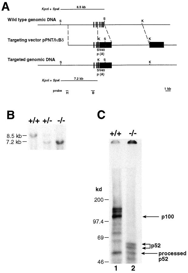
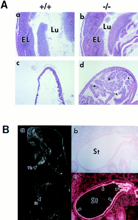
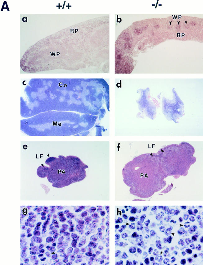
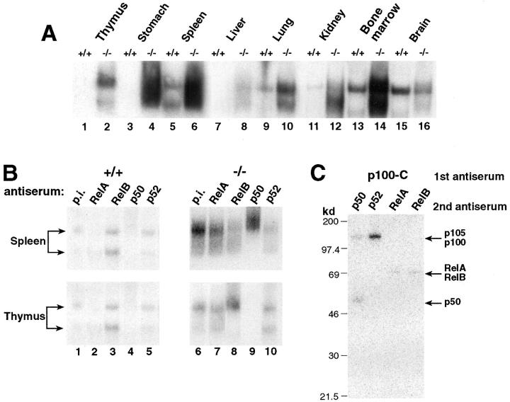
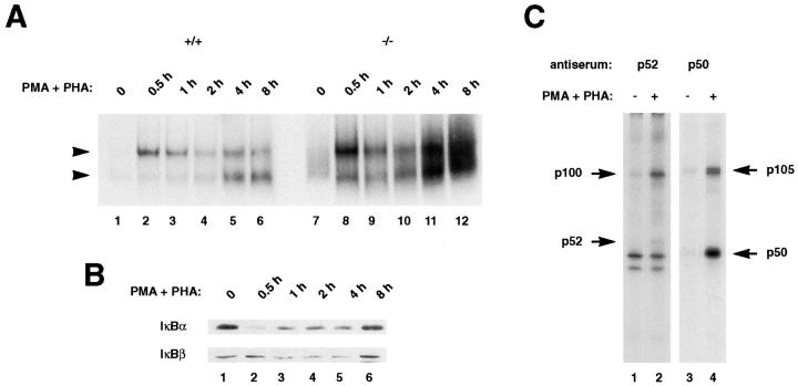
The nfkb2 gene encodes the p100 precursor which produces the p52 protein after proteolytic cleavage of its COOH-terminal domain. Although the p52 product can act as an alternative subunit of NF-kappaB, the p100 precursor is believed to function as an inhibitor of Rel/NF-kappaB activity by cytoplasmic retention of Rel/NF-kappaB complexes, like other members of the IkappaB family. However, the physiological relevance of the p100 precursor as an IkappaB molecule has not been understood. To assess the role of the precursor in vivo, we generated, by gene targeting, mice lacking p100 but still containing a functional p52 protein. Mice with a homozygous deletion of the COOH-terminal ankyrin repeats of NF-kappaB2 (p100(-/-)) had marked gastric hyperplasia, resulting in early postnatal death. p100(-/-) animals also presented histopathological alterations of hematopoietic tissues, enlarged lymph nodes, increased lymphocyte proliferation in response to several stimuli, and enhanced cytokine production in activated T cells. Dramatic induction of nuclear kappaB-binding activity composed of p52-containing complexes was found in all tissues examined and also in stimulated lymphocytes. Thus, the p100 precursor is essential for the proper regulation of p52-containing Rel/NF-kappaB complexes in various cell types and its absence cannot be efficiently compensated for by other IkappaB proteins.


Figures

Figure 1
Figure 1
Generation of mice deficient in the p100 precursor. (A) Targeting strategy of the ankyrin-encoding region of the nfkb2 gene. The relevant part of the mouse nfkb2 gene structure is shown at the top. Exons 13–19, encoding residues 332–690, are indicated by closed boxes. Targeting vector pPNT/IκBδ and the targeted allele are shown at the middle and bottom, respectively. Open boxes indicate the SV40 polyadenylation recognition sequences, PGK-neo and PGK-tk cassettes. The position of KpnI and SpeI sites are indicated by K and S, respectively. The diagnostic restriction fragments used for Southern blot analysis are indicated at the top (wild-type allele) and bottom (targeted allele). The DNA fragments used as 5′ external (H) and internal (B) probes are indicated at the bottom. (B) Genotype analysis of mice generated from p100+/− heterozygote intercrosses. Tail DNAs were digested with KpnI and SpeI, and subjected to Southern blot analysis using the 5′ external probe H indicated in A. The 8.5-kb band indicates the wild-type allele, while the 7.2-kb band represents the targeted allele. (C) Absence of p100 in homozygous mutant mice. Protein extracts from control (+/+) and homozygous (/−) mutant thymocytes labeled with [35S]methionine for 8 h in the presence of PMA (20 ng/ml) and PHA (5 μg/ml) were immunoprecipitated with an anti-p52 antiserum. p100 and p52 proteins are indicated by the arrows.
Figure 2
Figure 2
Postnatal growth and survival of p100-deficient mice. (A) Changes in body weight of p100−/− mice (open triangles) and control littermates (+/+ and +/−, closed circles). (B) Survival of control (+/+ and +/−, closed circles, n = 290) and p100−/− mice (open triangles, n = 100). Survival is shown as a percentage of the total initial number of control (+/+ and +/−) or p100−/− mice.
Figure 3
Figure 3
Histopathology of a p100−/− mouse stomach. (A) Stomach sections of 3-wk-old wild-type (a and c) and p100−/− (b and d) animals stained with hematoxylin and eosin (original magnification: 12.5-fold). As shown in (b) the epithelial layer (EL) was markedly thick, whereas the gastric lumen (Lu) was narrow in p100−/− mice. Also, hyperkeratosis in cardiac portion was evident in the p100−/− stomach (d). (B) A section of a wild-type newborn mouse was probed with nfkb2 cDNA, stained with carmine red (b and c), and photographed under dark (a and c) or light (b) field illumination (original magnification: a, 4-fold; b and c, 25-fold). The nfkb2 transcript is expressed in thymus (Th) and in the surface epithelium (c; arrows) of the stomach (St).
Figure 4
Figure 4
Alterations of hematopoietic tissues in p100−/− mice. (A) Sections of 3-wk-old wild type (a, c, e, and g) and p100−/− (b, d, f, and h) spleen (a and b), thymus (c and d), lymph node (e and f), and bone marrow (g and h) stained with hematoxylin and eosin (original magnification: a, b, e and f, 12.5-fold; c and d, 5-fold; g and h, 375-fold). Both spleen (b) and thymus (d) of p100−/− mice were atrophic, showing poorly demarcated white (WP, arrows) and red pulp, and cortico–medullary junctions, respectively. (f) Lymph nodes were clearly enlarged and the lymphatic follicle (LF, arrows) was not well defined in p100−/− mice. (h) Granulocyte precursors and neutrophils (arrows) markedly accumulated in p100−/− bone marrow. WP, white pulp; RP, red pulp; Co, cortex; Me, medulla; LF, lymphatic follicle; PA, paracortical area. (B) Flow cytometry of thymocytes (Th) stained for CD4 and CD8 (a and b), splenocytes (Sp) stained for B220 and Thy-1.2 (c and d), bone marrow cells (BM) stained for Gr-1 (e and f) or B220 (g and h), and lymph node cells (LN) stained for CD25 (i and j) from 3-wk-old wild-type (+/+, a, c, e, g, and i) and p100−/− (−/−, b, d, f, h, and j) mice. Percentages of positive cells are indicated.
Figure 5
Figure 5
Augmented κB-binding activity in p100−/− mice. (A) Tissues from p100−/− mice present increased Rel/NF-κB activity. The κB-binding activity of nuclear extracts (2 μg) from several tissues was determined by EMSA using a palindromic κB site. (B) The accumulated κB-binding complexes contain p52 in p100−/− mice. Antisera used for determining the composition of the Rel/NF-κB complexes are indicated at the top. p.i., preimmune serum. Different mobilities of κB-binding complexes are indicated by arrows. (C) The p100 precursor interacts with all members of the Rel/NF-κB family in primary murine thymocytes. Whole cell lysates from wild-type thymocytes labeled with [35S]methionine for 8 h in the presence of PMA and PHA were incubated with p100–COOH-terminus antiserum (p100-C). The resulting immune complexes were disrupted and reprecipitated with either p50, p52, RelA, or RelB antiserum as indicated (2nd antiserum). Specific signals for the p105, p100, p50, RelA, and RelB proteins are indicated by arrows.
Figure 6
Figure 6
Induction of κB-binding activity after stimulation of thymocytes. (A) Strong Rel/NF-κB activation in stimulated p100−/− thymocytes. Nuclear extracts (1.5 μg) from wild type (+/+, lanes 1–6) and p100−/− (−/−, lanes 7–12) thymocytes treated with PMA and PHA for the indicated periods were analyzed by EMSA. Two major bands are indicated by arrows. (Note: the exposure time of the autoradiogram is significantly shorter than that of Fig. 5 A). (B) IκBα is responsible for the rapid activation of the p50–RelA complexes in thymocytes. The amounts of the IκBα and IκBβ proteins in wild-type thymocytes stimulated with PMA and PHA for different periods were determined by Western blot analysis using 30 μg of cytoplasmic extracts. (C) The processing of the p105 precursor is enhanced by stimulation of thymocytes. Whole cell lysates from wild-type thymocytes labeled with [35S]methionine for 6 h in the absence (lanes 1 and 3) or presence (lanes 2 and 4) of PMA and PHA were incubated with a p52 antiserum (lanes 1 and 2) and the resulting supernatants were immunoprecipitated with a p50 antiserum (lanes 3 and 4). The exposure time of the autoradiogram of the immunoprecipitation with p52 is 2.5 times longer than that of p50, so as to verify the p100 and p52 proteins. Specific signals for the p100, p52, p105, and p50 proteins are indicated by arrows. The numbers of methionine residues contained in human p100, p52, murine p105, and p50 are 16, 10, 20, and 9, respectively (25, 66).
Figure 7
Figure 7
The expression of Rel/NF-κB–regulated genes is upregulated in p100−/− mice. Total RNA (0.25 μg) isolated from the spleen of 10-d-old wild-type (+/+), heterozygous (+/−), and homozygous (−/−) mutant mice was subjected to RT-PCR analysis using the indicated specific primers.
Figure 8
Figure 8
Accelerated proliferative responses and cytokine production in activated T cells of p100−/− mice. (A) T cell proliferation in vitro. Peripheral T cells isolated from spleen of 10-d-old wild-type (closed boxes) and p100−/− mice (open boxes) were treated with either anti-CD3, anti-CD3 plus anti-CD28, or PMA plus PHA, followed by [3H]thymidine incorporation. Values of [3H]thymidine incorporation are shown by mean ± S.D. (B) The cytokine production from stimulated T cells of p100−/− mice is increased. Splenic T cells isolated from 10-d-old wild-type (closed boxes) and p100−/− (open boxes) mice were treated with (+) or without () anti-CD3 and anti-CD28 antibodies for 72 h. The cytokine levels in the supernatants were determined by ELISA. Levels of IL-2, IL-4, IL-10, GM-CSF, and TNF-α produced in p100−/− T cells relative to control T cells are represented by mean values ± S.D.

Similar articles

Cited by

References

    1. Beg AA, Baldwin AS., Jr The IκB proteins: multifunctional regulators of Rel/NF-κB transcription factors. Genes Dev. 1993;7:2063–2070. - PubMed
    1. Gilmore TD, Morin PJ. The IκB proteins: members of a multifunctional family. Trends Genet. 1993;9:427–433. - PubMed
    1. Liou HC, Baltimore D. Regulation of the NF-κB/rel transcription factor and IκB inhibitor system. Curr Opin Cell Biol. 1993;5:477–487. - PubMed
    1. Siebenlist U, Franzoso G, Brown K. Structure, regulation and function of NF-κB. Annu Rev Cell Biol. 1994;10:405–455. - PubMed
    1. Finco TS, Baldwin AS. Mechanistic aspects of NF-κB regulation: the emerging role of phosphorylation and proteolysis. Immunity. 1995;3:263–272. - PubMed

MeSH terms