. 1997 Oct 6;186(7):1027-39.
doi: 10.1084/jem.186.7.1027.

A critical role for Syk in signal transduction and phagocytosis mediated by Fcgamma receptors on macrophages

Affiliations

A critical role for Syk in signal transduction and phagocytosis mediated by Fcgamma receptors on macrophages

M T Crowley et al. J Exp Med. .

Abstract

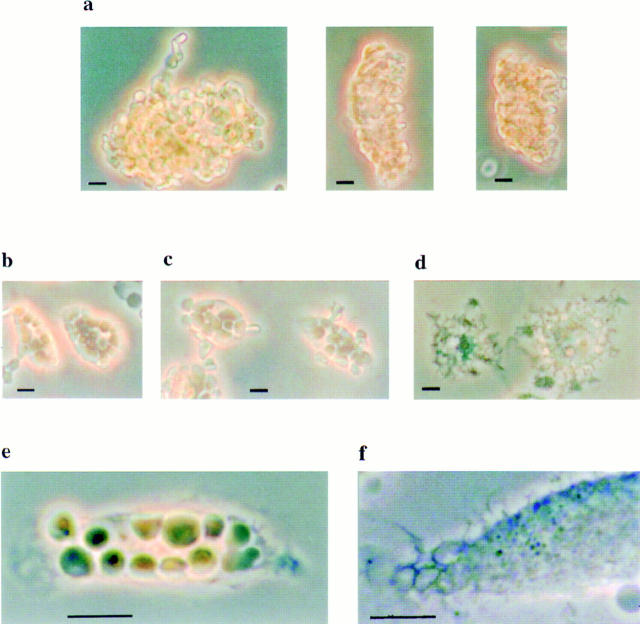
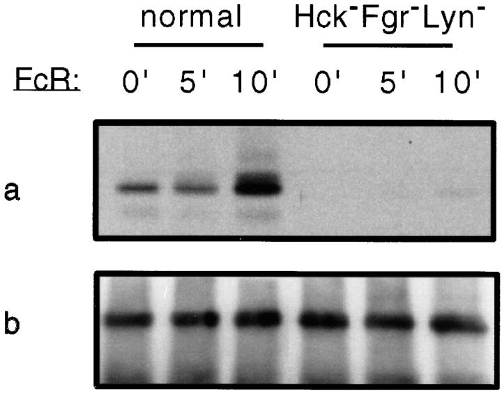
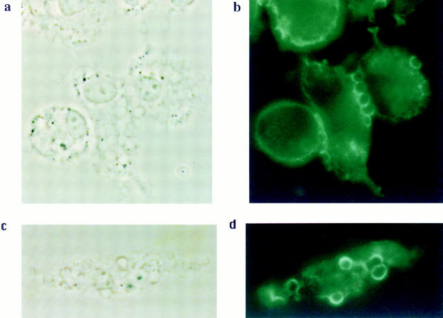
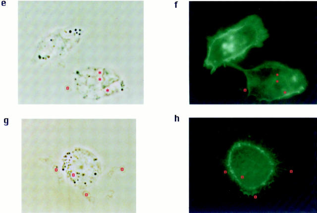
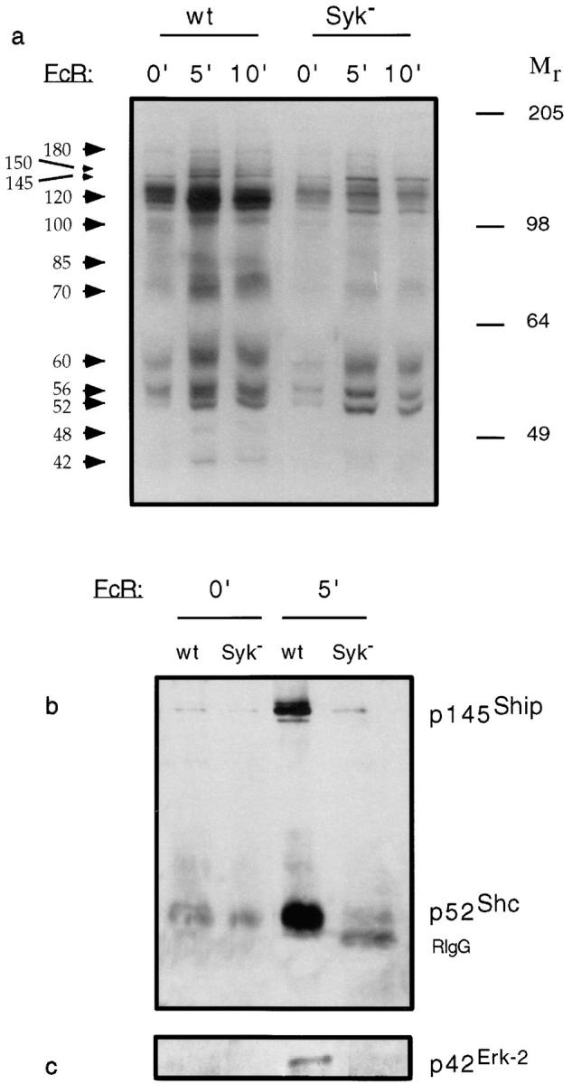
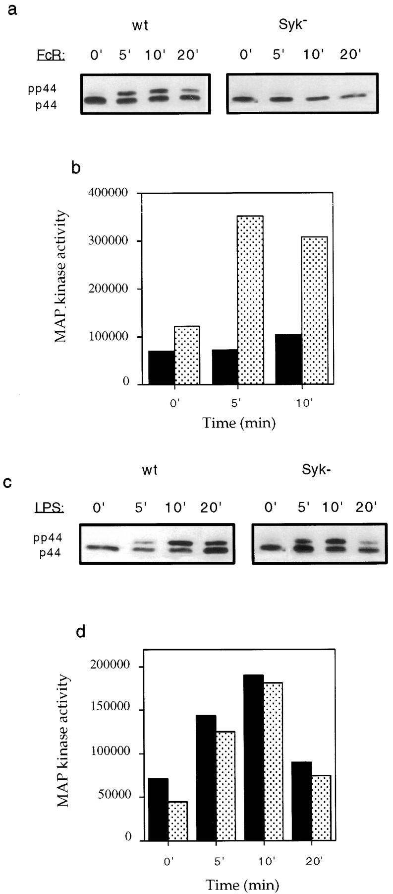
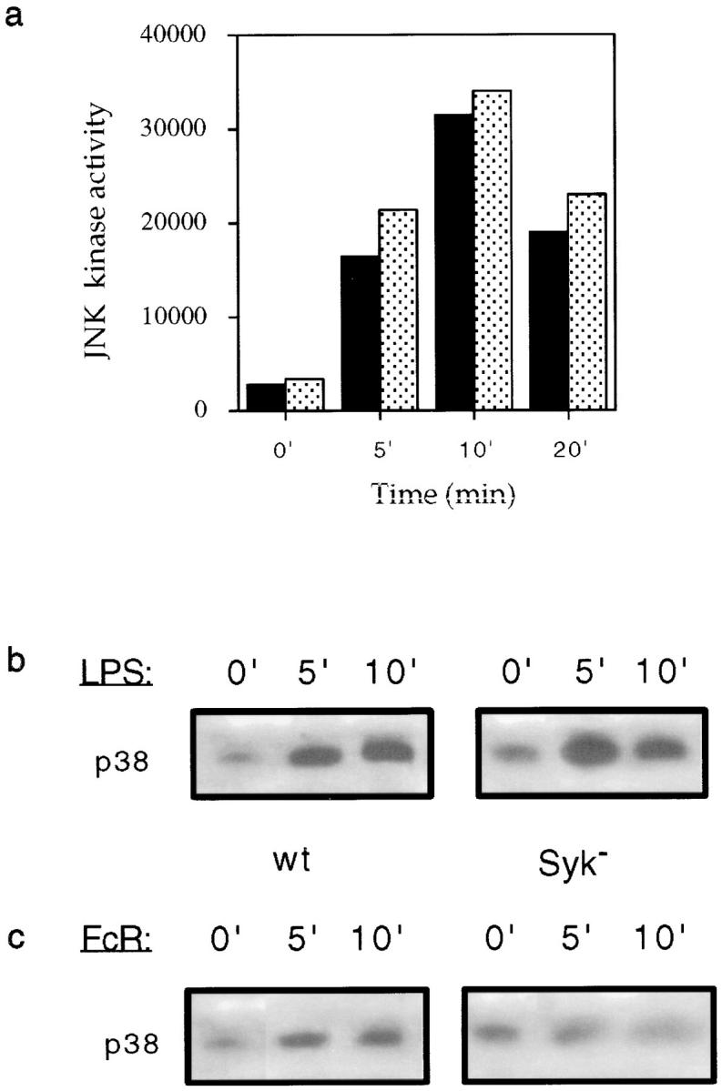
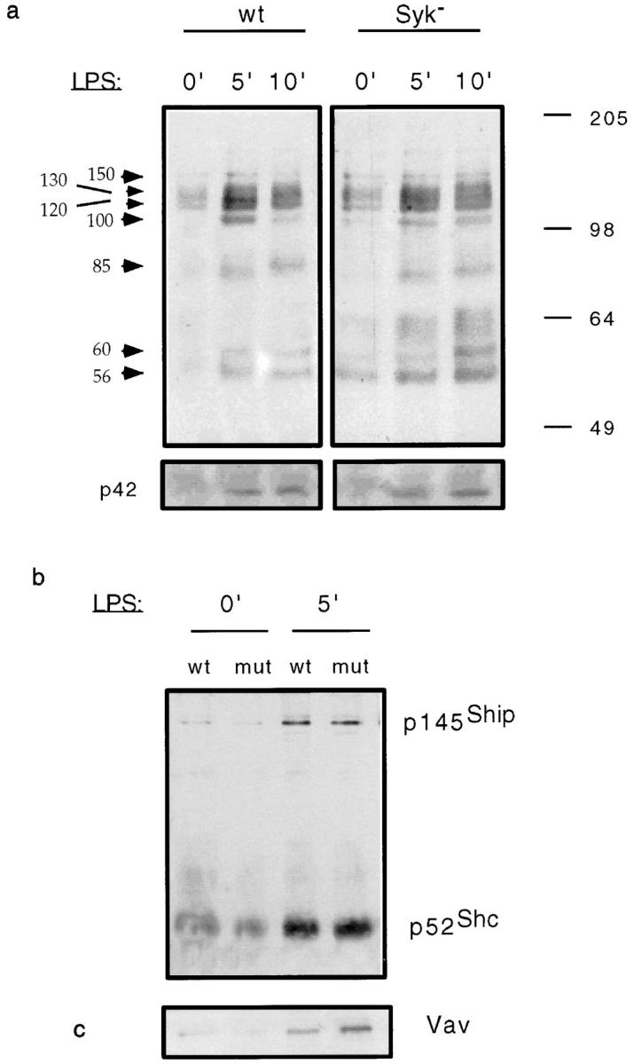
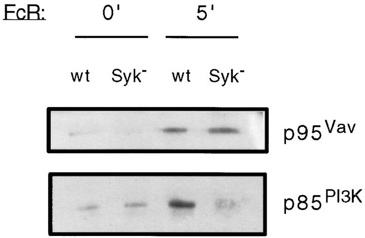
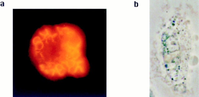
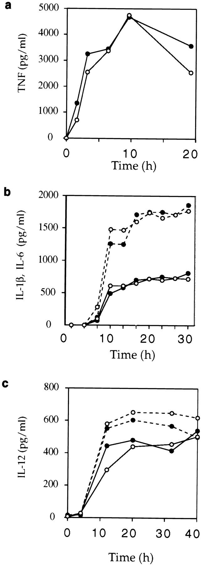
Receptors on macrophages for the Fc region of IgG (FcgammaR) mediate a number of responses important for host immunity. Signaling events necessary for these responses are likely initiated by the activation of Src-family and Syk-family tyrosine kinases after FcgammaR cross-linking. Macrophages derived from Syk-deficient (Syk-) mice were defective in phagocytosis of particles bound by FcgammaRs, as well as in many FcgammaR-induced signaling events, including tyrosine phosphorylation of a number of cellular substrates and activation of MAP kinases. In contrast, Syk- macrophages exhibited normal responses to another potent macrophage stimulus, lipopolysaccharide. Phagocytosis of latex beads and Escherichia coli bacteria was also not affected. Syk- macrophages exhibited formation of polymerized actin structures opposing particles bound to the cells by FcgammaRs (actin cups), but failed to proceed to internalization. Interestingly, inhibitors of phosphatidylinositol 3-kinase also blocked FcgammaR-mediated phagocytosis at this stage. Thus, PI 3-kinase may participate in a Syk-dependent signaling pathway critical for FcgammaR-mediated phagocytosis. Macrophages derived from mice deficient for the three members of the Src-family of kinases expressed in these cells, Hck, Fgr, and Lyn, exhibited poor Syk activation upon FcgammaR engagement, accompanied by a delay in FcgammaR-mediated phagocytosis. These observations demonstrate that Syk is critical for FcgammaR-mediated phagocytosis, as well as for signal transduction in macrophages. Additionally, our findings provide evidence to support a model of sequential tyrosine kinase activation by FcgammaR's analogous to models of signaling by the B and T cell antigen receptors.


Figures

Figure 1
Figure 1
Binding and phagocytosis of antibody-coated EA by wild-type, HckFgrLyn, and Syk macrophages. Adherent wild-type, HckFgrLyn, and Syk macrophages (a, left to right) were incubated with EA (107 cells per ml) for 30 min at 37°C before removal of unbound EA. Uninternalized EA were lysed in b–f. All normal macrophages (b and e) and HckFgrLynmacrophages (c) contained many internalized erythrocytes. The large load of ingested material resulted in a loss of cell body extensions, and a more rounded appearance of the macrophages. None of the Syk-deficient macrophages contained any internalized erythrocytes (d and f   ). These macrophages had a frilled appearance due to the interlacing of actin-rich macrophage cell membrane structures around each bound erythrocyte. Bar, 10 μm.
Figure 2
Figure 2
Greatly decreased FcγR-mediated Syk activation in Hck/ Fgr/Lyn macrophages. Bone marrow–derived macrophages were incubated with growth media (0′) or anti-FcγR mAb 2.4G2 followed by anti–rat IgG cross-linking for the indicated times. The protein kinase activity of Syk was assessed by in vitro autophosphorylation of immunoprecipitated Syk followed by SDS-PAGE and autoradiography (a). Anti-Syk immunoblotting (b) showed that equivalent amounts of Syk protein were present in each reaction.
Figure 3
Figure 3
Formation of actin-rich membrane cups around bound EA in normal and Syk-deficient macrophages. Macrophages were incubated with EA on ice for 20 min. Fixed and permeabilized normal (a and b) and Syk-deficient (c and d) cells were stained with Oregon green–phalloidin to visualize F-actin. The bound erythrocytes are apparent under phase microscopy (a and c). Corresponding actin-lined cups visible by fluorescence microscopy (b and d) are evident. Alternatively, after incubation on ice, cells were washed and further incubated for 20 min at 37°C, during which time there was dissolution of actin cup structures in Syk-deficient macrophages (g and h) as well as internalization of the EA in the case of normal cells (e and f   ). Several internalized EA (*) and bound, noninternalized EA (o) are indicated by symbols inside the erythrocytes.
Figure 3
Figure 3
Formation of actin-rich membrane cups around bound EA in normal and Syk-deficient macrophages. Macrophages were incubated with EA on ice for 20 min. Fixed and permeabilized normal (a and b) and Syk-deficient (c and d) cells were stained with Oregon green–phalloidin to visualize F-actin. The bound erythrocytes are apparent under phase microscopy (a and c). Corresponding actin-lined cups visible by fluorescence microscopy (b and d) are evident. Alternatively, after incubation on ice, cells were washed and further incubated for 20 min at 37°C, during which time there was dissolution of actin cup structures in Syk-deficient macrophages (g and h) as well as internalization of the EA in the case of normal cells (e and f   ). Several internalized EA (*) and bound, noninternalized EA (o) are indicated by symbols inside the erythrocytes.
Figure 4
Figure 4
Impaired FcγR-induced tyrosine phosphorylation of cellular substrates in Syk-deficient macrophages. Cells were incubated with growth media (0′) or anti-FcγR mAb 2.4G2 followed by anti–rat IgG cross-linking for the indicated times. Cells were lysed, and were subjected to SDS-PAGE and immunoblotting with antiphosphotyrosine antibodies (a). The molecular mass (in kD) of markers and the positions of major phosphotyrosine-containing proteins are indicated at the right and left of a, respectively. Alternatively, cell lysates were subjected to immunoprecipitation with anti-Shc antibodies (b) or anti-Erk2 antibodies (c). Erk2, Shc, and Shc-associated proteins were resolved by SDS-PAGE, and their phosphorylation status was determined by immunoblotting with antiphosphotyrosine antibodies.
Figure 5
Figure 5
Impaired FcγR-induced MAPK activation in Syk-deficient macrophages. Wild-type and mutant macrophages were incubated with growth media (0′) or with anti-FcγR mAb 2.4G2, followed by cross-linking with anti–rat IgG for the indicated times. The electrophoretic mobility shift of p44 MAP kinase was assessed by SDS-PAGE, and by immunoblotting with anti-MAP kinase antibodies (a). Alternatively, Erk1 and Erk2 activity was assessed directly by immunoprecipitation with anti-Erk2 antibodies and measurement of the ability to phosphorylate the substrate myelin basic protein in vitro (b and d). Unstimulated or FcγR-stimulated wild-type (stippled bars) or Syk (black bars) macrophages were tested (b). Values shown represent 33PO4 incorporation from labeled ATP into the substrate protein. Seven separate experiments using Syk-deficient cells gave similar results. LPS-induced mobility shift of Erk1 (c) and enzymatic activation of Erk2 (d) was also assessed in wild-type (stippled bars), or Syk (black bars) macrophages exposed to 1 μg/ml LPS for the indicated times. Similar results were obtained in seven separate experiments.
Figure 6
Figure 6
Activation of JNK and tyrosine phosphorylation of p38 in FcγR-stimulated and LPS-activated macrophages. Activation of JNK in wild-type (stippled bars), or Syk (black bars) macrophages exposed to 1 μg/ml LPS for the indicated times was assessed by immunoprecipitation with anti-JNK antibodies, followed by in vitro phosphorylation of a c-Jun– GST fusion protein substrate (a). For analysis of p38 phosphorylation, phosphotyrosine-containing proteins were immunoprecipitated from lysates of unstimulated (0′), LPS-stimulated (b), or FcγR-activated (c) macrophages. Proteins were separated by SDS-PAGE and transferred to nitrocellulose, and the region containing p38 was probed with anti-p38 antibody. Similar results were obtained in three experiments comparing Syk-deficient and normal macrophages.
Figure 7
Figure 7
Lack of defect in LPS-induced tyrosine phosphorylation of cellular substrates in Syk macrophages. Cells were incubated with media or media plus 1 μg/ml LPS for the indicated times. Cells were lysed, and the lysate proteins were subjected to SDS-PAGE and immunoblotting with antiphosphotyrosine antibody (a). Molecular mass markers (in kD) and major phosphotyrosine-containing substrates are indicated on the right and the left of a, respectively. The Erk2 MAPK band, as confirmed by subsequent immunoblotting with anti-Erk2 antibodies (not shown), is indicated as p42. Alternatively, cell lysates were subject to immunoprecipitation with anti-Shc antibodies (b) and immunoblotting with antiphosphotyrosine antibody, or to immunoprecipitation with antiphosphotyrosine antibody and immunoblotting with anti-Vav (c) antibodies. The positions of proteins of interest are indicated.
Figure 8
Figure 8
Altered FcγR-induced signaling events associated with p85 PI 3-kinase, but not with Vav in Syk cells. Fetal liver–derived macrophage cultures were stimulated through their FcγR for 5 min. Cellular lysate proteins were immunoprecipitated with antiphosphotyrosine antibodies, and the immunoprecipitated proteins were subjected to SDS-PAGE and immunoblotting with anti-Vav and anti-p85 PI 3-kinase antibodies as indicated.
Figure 9
Figure 9
Effect of the PI 3-kinase inhibitor wortmannin on FcγR-mediated phagocytosis and actin-based phagocytic cup formation. Macrophages grown on coverslips were pretreated with wortmannin (50 nM), for 20 min, and were then incubated with EA either for 20 min on ice to obtain stable actin-lined phagocytic cups (a), or at 37°C for 40 min, after which time bound, but not internalized erythrocytes were lysed with 1.44% NH4Cl (b). Fixed and permeabilized cells were stained with BODIPY-phalloidin, and were examined by phase microscopy (a) or by fluorescence microscopy to visualize F-actin (b).
Figure 10
Figure 10
Production and release of inflammatory cytokines in Syk-deficient and wild-type macrophages responding to LPS. Syk (•) or wild-type (○) cultured macrophages were stimulated with 50 ng/ml LPS for the time indicated, and concentrations of TNF-α (a), IL-1β (b, solid line), IL-6 (b, dashed line), IL-12 (c, solid line), or IL-12 from γ-IFN (100 U/ml) primed macrophages (c, dashed line) were determined by ELISA.

Similar articles

Cited by

References

    1. Ravetch JV, Kinet JP. Fc receptors. Ann Rev Immunol. 1991;9:457–492. - PubMed
    1. Brooks D, Ravetch JV. Fc receptor signaling. Adv Exp Med Biol. 1994;365:185–195. - PubMed
    1. Takai T, Li M, Sylvestre D, Clynes R, Ravetch JV. FcR gamma chain deletion results in pleiotrophic effector cell defects. Cell. 1994;76:519–529. - PubMed
    1. Takai T, Ono M, Hikida M, Ohmori H, Ravetch JV. Augmented humoral and anaphylactic responses in Fc gamma RII-deficient mice. Nature (Lond) 1996;379:346–349. - PubMed
    1. Ravetch JV. Fc receptors: rubor redux. Cell. 1994;78:553–560. - PubMed

Publication types

MeSH terms