. 1997 Oct 6;186(7):1149-58.
doi: 10.1084/jem.186.7.1149.

How many thymocytes audition for selection?

Affiliations

How many thymocytes audition for selection?

M Merkenschlager et al. J Exp Med. .

Abstract

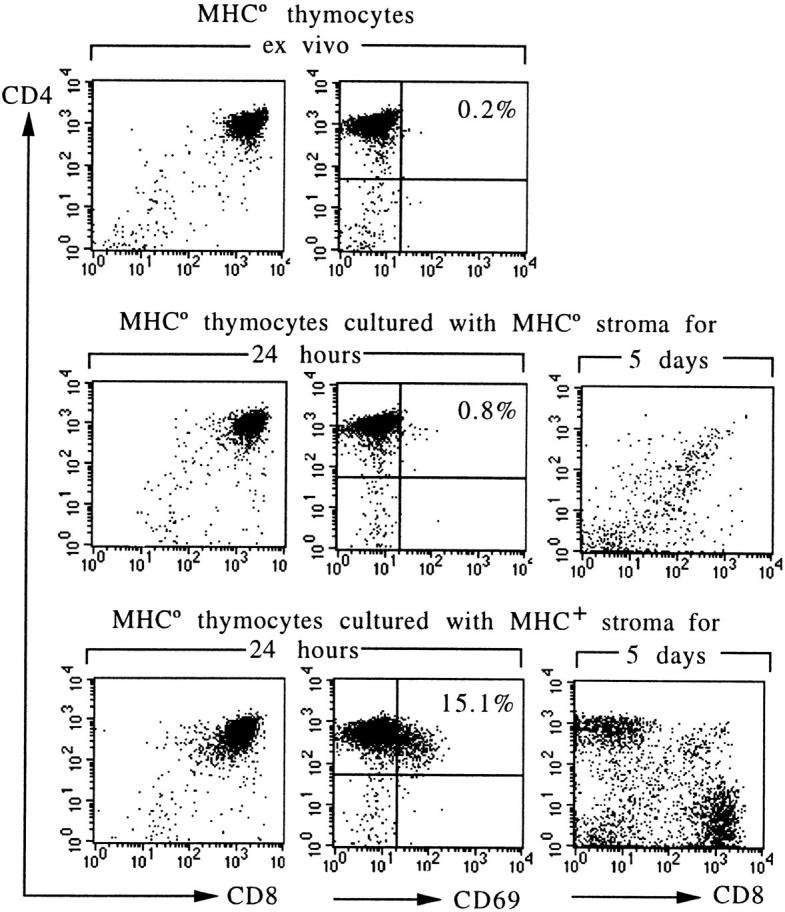
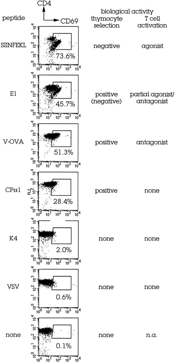
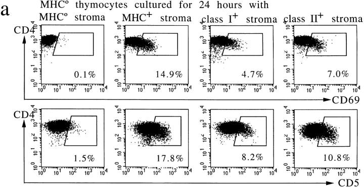
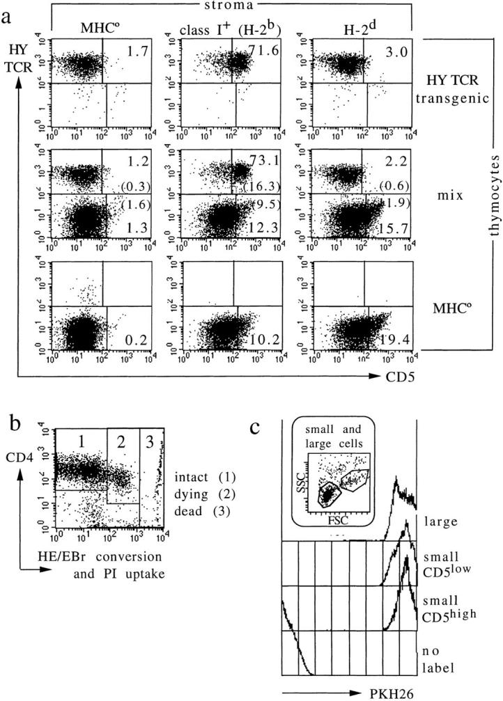
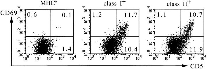
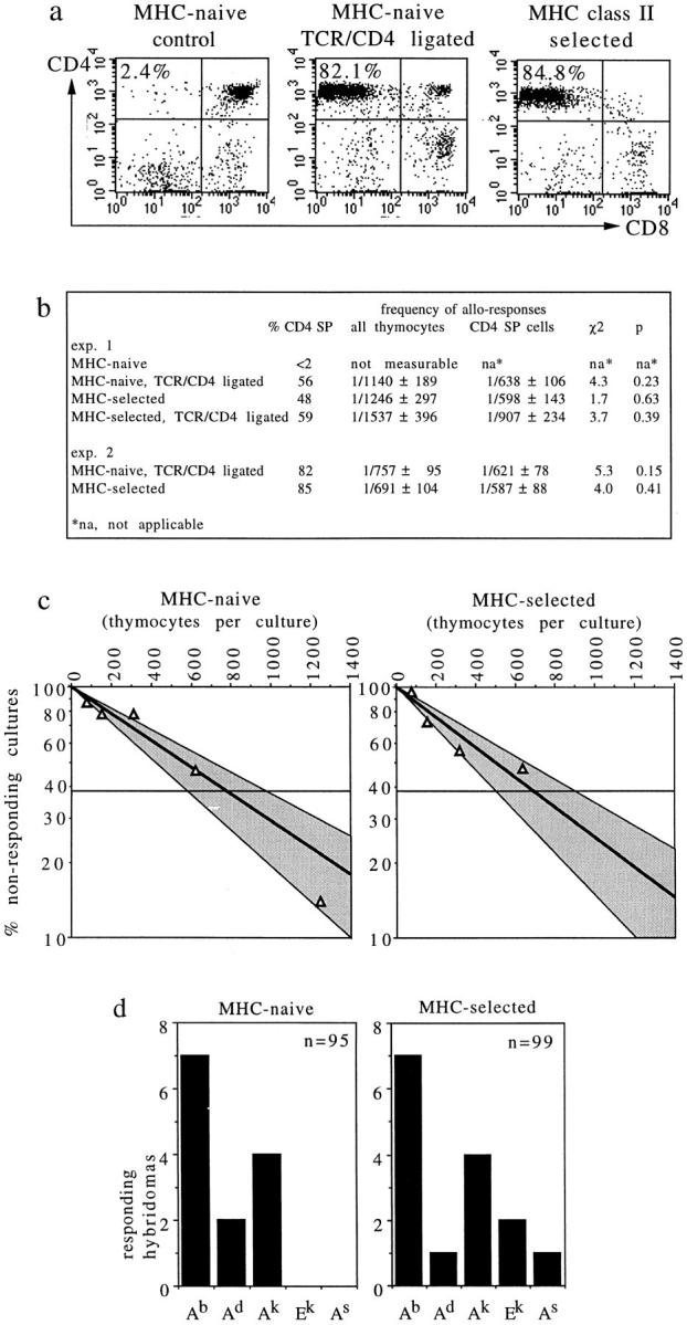
T cell maturation requires the rearrangement of clonotypic T cell receptors (TCR) capable of interacting with major histocompatibility complex (MHC) ligands to initiate positive and negative selection. Only 3-5% of thymocytes mature to join the peripheral T cell pool. To investigate the basis for this low success rate, we have measured the frequency of preselection thymocytes capable of responding to MHC. As many as one in five MHC-naive thymocytes show upregulation of activation markers on exposure to MHC-expressing thymic stroma in short-term reaggregate culture. The majority of these cells display physiological changes consistent with entry into the selection process within 24 h. By exposing TCR transgenic thymocytes to a range of MHC-peptide complexes, we show that CD69 induction is indicative of thymocyte selection, positive or negative. Our data provide evidence that the fraction of thymocytes that qualify to enter the thymic selection process far exceeds the fraction that successfully complete it, and suggest that most MHC-reactive thymocytes are actively eliminated in the course of selection.


Figures

Figure 1
Figure 1
MHC-dependent activation and developmental rescue of MHC-naive thymocytes. Thymocytes from MHC° mice are arrested at the CD4+CD8+ stage and lack selection-associated activation markers. Coculture with MHC+ (H-2b) but not with MHC° thymic stroma results in CD69 expression, and rescues the development of CD4 and CD8 single-positive HSAlow cells (5-d displays are gated on HSAlow cells).
Figure 2
Figure 2
CD69 expression by CD4 CD8 DP thymocytes is triggered by biologically relevant TCR-MHC–peptide contact. Young adult OT-I TCR transgenic/RAG-1°/β2m° thymi were cut into fragments and cultured with or without exogenous peptides (20 μM) and exogenous β2m to allow the reconstitution of Kb–peptide complexes as described (28). CD69 expression was determined 24 h later. Data on thymocyte selection and mature T cell activation are from references , –. Kd (off/on) values are listed in the text.
Figure 3
Figure 3
Preselection thymocyte responses to MHC class I and II are largely additive and, in addition to CD69, involve CD5 upregulation, downregulation of RAG-1 and -2 mRNA, and perturbed CD4 and CD8 expression. (a) Elevated CD5 and CD69 expression by MHC-naive thymocytes 24 h after contact with MHC. Note that responses to class I and II (Ab) are approximately additive. These responses are independent of superantigens, as verified using MHC-expressing cells from mice without mouse mammary tumor virus integrants (provided by Dr. E. Simpson, data not shown). (b) Downregulation of RAG-1 and -2 mRNA 24 h after MHC contact. RT-PCR analysis of thymocytes isolated by fluorescence activated cell sorting from reaggregates with MHC-deficient (lane 1) or MHC+ stroma (lanes 2 and 3). The latter were separated into CD69-negative (lane 2) and -positive cells (lane 3). RAG-1 and -2 signals are barely detectable in CD69-positive thymocytes; CD4 and, to a lesser extent, CD8a mRNA appear downregulated. RT-PCR for HPRT serves as a control; lane 4 contains no cDNA.
Figure 3
Figure 3
Preselection thymocyte responses to MHC class I and II are largely additive and, in addition to CD69, involve CD5 upregulation, downregulation of RAG-1 and -2 mRNA, and perturbed CD4 and CD8 expression. (a) Elevated CD5 and CD69 expression by MHC-naive thymocytes 24 h after contact with MHC. Note that responses to class I and II (Ab) are approximately additive. These responses are independent of superantigens, as verified using MHC-expressing cells from mice without mouse mammary tumor virus integrants (provided by Dr. E. Simpson, data not shown). (b) Downregulation of RAG-1 and -2 mRNA 24 h after MHC contact. RT-PCR analysis of thymocytes isolated by fluorescence activated cell sorting from reaggregates with MHC-deficient (lane 1) or MHC+ stroma (lanes 2 and 3). The latter were separated into CD69-negative (lane 2) and -positive cells (lane 3). RAG-1 and -2 signals are barely detectable in CD69-positive thymocytes; CD4 and, to a lesser extent, CD8a mRNA appear downregulated. RT-PCR for HPRT serves as a control; lane 4 contains no cDNA.
Figure 4
Figure 4
Bystander activation, unscheduled display of activation markers during apoptosis, or preferential expansion do not account for the frequency of MHC responsive thymocytes. (a) CD5 induction by cognate TCR–MHC interactions. HY TCR transgenic thymocytes from RAG-1–deficient mice lacking MHC class I expression respond strongly to selecting (H-2b), but not to nonselecting (H-2d) MHC molecules (top; Note that in contrast to an earlier study [45], our experiment excludes endogenous TCR rearrangements by using RAG-1–deficient thymocytes). Reaggregates containing mixtures of HY TCR transgenic and MHC-naive, TCR nontransgenic thymocytes (middle) show that both populations respond independently, and without significant bystander activation (compare top and bottom to middle). The percentage of cells in each region is indicated. In the middle panel, the percentage of activated cells among TCR transgenic and nontransgenic thymocytes is calculated separately (percentage of total cells is given in brackets). (b) Most cells recovered from short-term aggregate cultures are viable (i.e., exclude propidium iodide; PI) and metabolically intact, as indicated by low oxidative conversion of hydroethidine (HE) to ethidium bromide (EBr ; reference 33). Displays in Fig. 3 a and data in Table 1 are gated on the region marked 1. (c) MHC-responsive thymocytes do not selectively expand in short-term culture. Thymocytes were labeled with the fluorescent membrane dye PKH26 (Sigma Chemical Co.) before coculture with stroma for 24 h. Control traces are of unlabeled and large (cycling) thymocytes; the latter illustrating dye dilution on cell division. PKH26 fluorescence is the same for MHC-responsive and -nonresponsive cells (defined by CD5 expression level).
Figure 5
Figure 5
In vivo expression of selection markers by CD4 CD8 DP thymocytes. Thymocytes taken from young adult mice lacking MHC (MHC°, left), or expressing MHC class I (middle) or class II (Ab; right), respectively, were stained for CD5 and CD69. Displays are gated on CD4+CD8+ double-positive cells as defined by the expression of CD4 (for thymocytes from MHC° and class I+ mice, left and middle) or CD8 (for thymocytes from class II+ mice, right). The percentages of cells expressing elevated levels of CD5, CD69, or both are indicated in the respective quadrants. Although this analysis is representative of H-2b mice, up to 50% of DP thymocytes are apparently engaged in the selection process in MHC haplotypes expressing class II E products (not shown).
Figure 6
Figure 6
MHC-naive CD4 T cells respond to MHC. (a) Treatment of MHC° thymi with CD3/CD4 bispecific antibodies generates CD4 SP cells phenotypically and functionally similar to CD4 SP cells selected by MHC class II. (b) Limiting dilution analysis indicates that MHC-naive and class II–selected CD4 cells mount proliferative responses to allogeneic CBA/Ca splenocytes with similar frequencies, whereas MHC-naive thymocytes not treated with bispecific antibodies mount no detectable responses. (c) Graphic representation of experiment 2. Shaded areas indicate 95% confidence limits. (d) T cell hybridomas were generated from MHC-naive and class II–selected CD4 cells, and tested for reactivity to a panel of MHC class II transfected L cells. 8 of 95 (8.4%) and 13 of 99 (13.1%), respectively, responded to at least one class II transfectant, five hybridomas generated from MHC-naive cells and two hybridomas generated from class II–selected cells responded to two transfectants.

References

    1. Zinkernagel RM, Doherty PC. MHC-restricted cytotoxic T cells: studies on the biological role of polymorphic major transplantation antigens determining T-cell restriction-specificity, function, and responsiveness. Adv Immunol. 1979;27:51–177. - PubMed
    1. Bevan MJ. In a radiation chimaera, host H-2 antigens determine immune responsiveness of donor cytotoxic cells. Nature (Lond) 1977;269:417–418. - PubMed
    1. Jorgensen JL, Reay PA, Ehrich EW, Davis MM. Molecular components of T-cell recognition. Annu Rev Immunol. 1992;10:835–873. - PubMed
    1. Kisielow P, von Boehmer H. Development and selection of T cells: facts and puzzles. Adv Immunol. 1995;58:87–209. - PubMed
    1. Gilfillan S, Benoist C, Mathis D. Mice lacking terminal deoxynucleotidyl transferase: adult mice with a fetal antigen receptor repertoire. Immunol Rev. 1995;148:201–219. - PubMed

Publication types

MeSH terms