. 1997 Oct 15;17(20):7714-24.
doi: 10.1523/JNEUROSCI.17-20-07714.1997.

Late compartments of amyloid precursor protein transport in SY5Y cells are involved in beta-amyloid secretion

Affiliations

Late compartments of amyloid precursor protein transport in SY5Y cells are involved in beta-amyloid secretion

G C Peraus et al. J Neurosci. .

Abstract

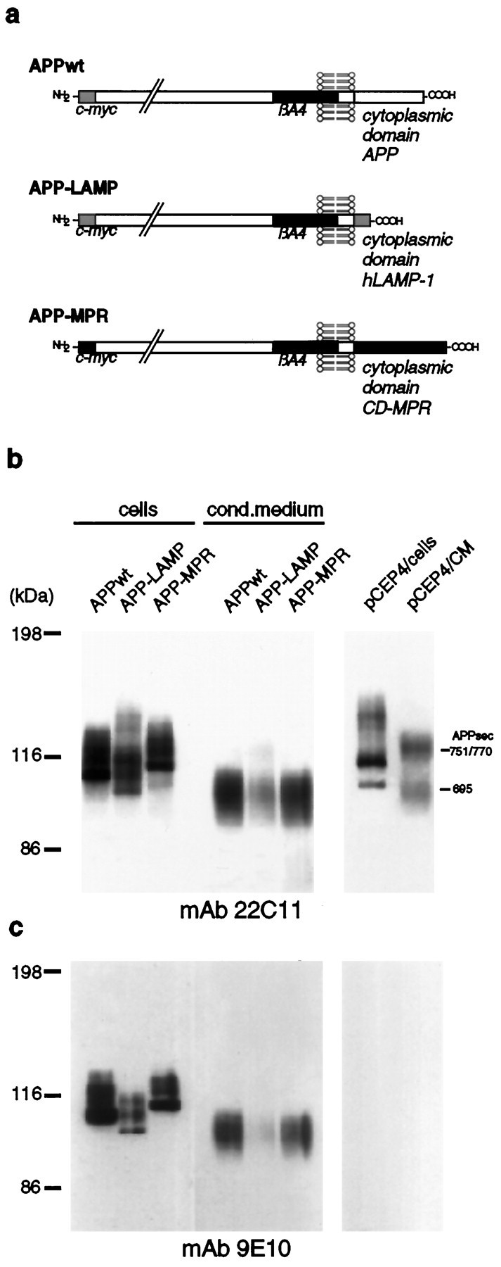
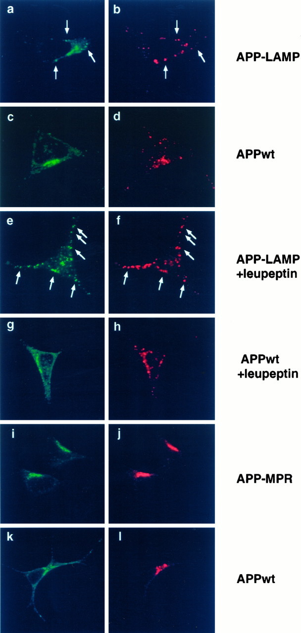
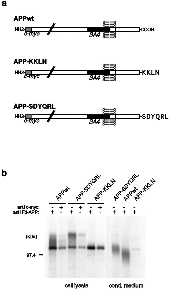
Amyloid plaques, composed mainly of the 39-43 amino acid betaA4 peptide, are a characteristic feature of Alzheimer's disease. Generation of betaA4 by proteolytic processing of the amyloid precursor protein (APP) is thought to occur in a pathway that includes the activity of two as yet unknown proteases, with beta-secretase cleaving at the N terminus and gamma-secretase releasing the C terminus of betaA4. Inhibition studies and the finding that cell surface APP can serve as a direct precursor of betaA4 suggest that the endosomal/lysosomal compartment is involved in the proteolysis of APP into betaA4. In this study we targeted APP695 chimeric proteins directly into the endosomal/lysosomal compartment. This decreased the amount of released betaA4, while the generation of the betaA4 N terminus continued. APP695 proteins were constructed also, which carried sorting signals responsible for recycling between the trans-Golgi network (TGN) and the cell surface. These proteins were processed into secreted betaA4 at even higher levels than wild-type APP695. Moreover, retention of APP695 proteins in the endoplasmic reticulum led to neither betaA4 secretion nor to processing by beta-secretase in human SH-SY5Y neuroblastoma cells. These data suggest that a beta-cleavage activity resides in a late endosomal compartment and that a gamma-cleavage occurs in early endosomes, resulting in the generation of betaA4 peptides with the majority ending at residue 40.


Figures

Fig. 1.
Fig. 1.
Expression of APP-LAMP and APP-MPR by stably transfected SY5Y cells. a, Schematic representation of APP695 (APPwt) and the APP chimeraAPP-LAMP and APP-MPR, which contain the cytoplasmic domain of hLAMP-1 and CD-MPR, respectively. The proteins are N-terminally tagged to a 10 aa c-myc sequence. b, Detection of the chimeric APP proteins by immunoblotting. SY5Y cells stably transfected with APPwt, APP-LAMP, APP-MPR, or thepCEP4 expression vector alone were lysed and cell-associated, and secreted APP proteins were immunoprecipitated with Fd-APP antiserum. Proteins were separated on a 7% SDS-PAGE and analyzed by immunoblotting, using mAb 22C11. c, The same filter was used for a second detection of the APP proteins with mAb 9E10. Because this antibody does not react with endogenous APP, the bands represent the chimeric proteins. CM, Conditioned medium.
Fig. 2.
Fig. 2.
Colocalization of APP-LAMP and APP-MPR with endogenous hLAMP-1 and CI-MPR by immunofluorescence confocal microscopy. SY5Y cells stably transfected with APP-LAMP (a, b, e, f), APP-MPR (i, j), and APPwt (c, d, g, h, k, l) were incubated with polyclonal A-14 (a, c, e, g) or monoclonal mAb 9E10 (i, k) anti-c-myc antibodies and with DTAF-conjugated second antibodies for the detection of the chimeric proteins. Monoclonal antibodies to human LAMP-1 (b, d, f, h) or antiserum to CI-MPR (j, l) and LRSC-conjugated second antibodies were used for the detection of the corresponding endogenous marker proteins. Where indicated, cells were incubated in the presence or absence of 1 mg/ml leupeptin 4 hr before MeOH fixation. In a, b, e, and f, colocalization of APP-LAMP with endogenous hLAMP-1 within vesicular structures is marked byarrows. Leupeptin treatment in e andf resulted in a strong stabilization of APP-LAMP in vesicular structures, most of which colocalized with endogenous hLAMP-1. APPwt mainly colocalized with APP-LAMP in a perinuclear region, which probably is passed by both proteins during their biosynthetic pathways. i and j show colocalization of APP-MPR with CI-MPR, whereas the immunoreactivity of APPwt in k only partially overlaps with that of CI-MPR.
Fig. 3.
Fig. 3.
Cell surface expression and maturation of APP-LAMP and APP-MPR. a, The intact cells, expressing APPwt, APP-LAMP, and APP-MPR, were labeled with sulfo-NHS-biotin, as described in Materials and Methods. Biotinylated cell surface proteins were immunoprecipitated with anti-biotin antibodies, followed by an incubation with Fd-APP antiserum and analyzed by immunoblotting with mAb 22C11. In contrast to APPwt and APP-MPR, biotinylated APP-LAMP was not detectable. b, Pulse chase analysis of the stably transfected cells. After being labeled with [35S]methionine for 8 min, the cells were chased in methionine-enriched growth medium for the times as indicated. Cell-associated and secreted APP proteins were immunoprecipitated with Fd-APP antiserum and analyzed by SDS-PAGE and autoradiography. In comparison to APPwt and APP-MPR, a reduced secretion of APP-LAMP was observed. APPs, Secreted APP (arrow).
Fig. 4.
Fig. 4.
Processing of APPwt, APP-LAMP, and APP-MPR into amyloidogenic fragments. a, Schematic representation of APP, A4CT, and p3CT, generated by α- and β-cleavage and the recognition sites of mAb 4G8 (anti-βA4 17–24) and mAbWO2 (anti-βA4 1–16). b, C-terminal cleavage products of the chimeric proteins arising by the activity of α- and β-secretase. SY5Y cells stably expressing APPwt, APP-LAMP, and APP-MPR were labeled metabolically with [35S]methionine for 4 hr. Immunoprecipitation of the cell lysates was performed with mAb 4G8 and mAbWO2. Precipitates were analyzed by SDS-PAGE and autoradiography. Bands marked by an asterisk (*) correspond to A4CT fragments of APPwt (12 kDa), APP-LAMP (8.4 kDa), and APP-MPR (14.2 kDa) and those labeled by an arrowhead(<) correspond to APPwt (10 kDa), APP-LAMP (6.4 kDa), and APP-MPR (12.2 kDa) p3CT cleavage products. In comparison to APPwt, A4CT and p3CT equivalents of APP-MPR transfected cells were highly enriched, whereas in APP-LAMP-expressing cells the amount of A4CT was reduced and that of p3CT fragments was strongly decreased. c, Stably transfected cells were labeled metabolically with [35S]methionine for 10 min and lysed immediately. According to their expression levels, corresponding amounts of cells were used for immunoprecipitation with Fd-APP antiserum. Precipitates were analyzed by SDS-PAGE and autoradiography. The autoradiogram shows that comparable amounts of APPwt, APP-LAMP, and APP-MPR chimeric proteins were used. d, Detection of secretedβA4 and p3 (arrows). Stably transfected cells were labeled metabolically with [35S]methionine for 16 hr, and the conditioned medium corresponding to their expression level was incubated with βA41–40 antiserum. Immunoprecipitates were analyzed by SDS-PAGE and autoradiography. In APP-MPR transfected cells the secretion of βA4 and p3 was decreased to ∼50%, as compared with APPwt. In the medium of APP-LAMP-expressing cells neither βA4 norp3 was detectable. e, To distinguish between βA41–40 and βA41–42, we radioactive-labeled the APPwt and APP-MPR transfected cells, as described in d, and equal volumes of conditioned media were used for immunoprecipitation with G2-10, specifically recognizing βA41–40, orG2-11, specific for βA41–42. βA4 and p3 were precipitated by G2-10, but not byG2-11.
Fig. 5.
Fig. 5.
Expression of APP-SDYQRL and APP-KKLN chimeras by SY5Y cells. a, Schematic representation ofAPP-SDYQRL and APP-KKLN. Both proteins were N-terminally c-myc-tagged. b, For detection ofAPP-SDYQRL and APP-KKLN, stably transfected SY5Y cells were labeled with [35S]methionine, and immunoprecipitation of equal amounts of the cell lysates was performed with Fd-APP and c-myc antisera. Conditioned medium of the transfected cells was incubated with Fd-APP antiserum. Immunoprecipitates were analyzed by SDS-PAGE and autoradiography. Similar amounts of cell-associatedAPPwt, APP-SDYQRL, andAPP-KKLN proteins were detected. In contrast toAPPwt and APP-SDYQRL, secreted protein was hardly detectable in APP-KKLN-expressing cells.
Fig. 6.
Fig. 6.
Colocalization of APP-SDYQRL and APP-KKLN with TGN38 and the ER resident sec61, respectively, was analyzed by immunofluorescence confocal microscopy. SY5Y cells stably expressing APPwt (c, d, g, h), APP-SDYQRL (a, b), and APP-KKLN (e, f) were incubated with mAb 9E10 (a, c, e, g) for the detection of the c-myc-tagged APP proteins, visualized by using DTAF-coupled second antibodies and TGN38 (b, d) or sec61 antiserum (f, h), followed by a second antibody conjugated with LRSC. Most of APP-SDYQRL colocalized with endogenous TGN38 in a perinuclear region (a, b). In contrast, APPwt also was found outside this juxtanuclear area (c). e and fshow that APP-KKLN colocalized with endogenous sec61, whereas additional staining was observed for the APPwt protein (g).
Fig. 7.
Fig. 7.
Analysis of the maturation and cell surface expression of APP-SDYQRL and APP-KKLN in transfected SY5Y cells.a, Plasma membrane proteins were labeled with sulfo-NHS-biotin on ice, and the biotinylated proteins were immunoprecipitated with biotin antiserum. A second immunoprecipitation with the same cell lysates was performed by using Fd-APP antiserum. The precipitated APP proteins were analyzed by SDS-PAGE and by immunoblotting with mAb 22C11. APPwt and APP-SDYQRL were found to become biotinylated; biotinylated APP-KKLN was not observed.b, Pulse chase analysis of APPwt-, APP-KKLN-, and APP-SDYQRL-expressing cells was performed as described in Figure3b. The APP-KKLN and APP-SDYQRL proteins were immunoprecipitated with c-myc and Fd-APP antiserum, respectively. In contrast to APP-SDYQRL, in cells expressing APP-KKLN no carbohydrate processing and no secretion of APP-KKLN were observed.
Fig. 8.
Fig. 8.
Amyloidogenic processing of APP-SDYQRL and APP-KKLN. SY5Y cells stably expressing APPwt, APP-SDYQRL, and APP-KKLN were labeled metabolically with [35S]methionine.a, A4CT and p3CT cleavage products of the cell lysates were immunoprecipitated with mAb W02 and mAb4G8, respectively, and analyzed by SDS-PAGE and autoradiography. Bands marked by an asterisk (*) correspond to A4CT equivalents of APPwt (12 kDa) and APP-SDYQRL (12.7 kDa). The arrowheads (<) point to bands corresponding to p3CT cleavage products of APPwt (10 kDa) and APP-SDYQRL (10.7 kDa). In APP-KKLN-expressing cells, neither A4CT nor p3CT equivalents were detectable. b, Immunoprecipitation of βA4 and p3 was performed by incubation of the same volumes of conditioned medium with βA41–40 antiserum. The samples were analyzed by SDS-PAGE and autoradiography. In comparison to APPwt, even higher amounts of βA4 were detectable in the medium of APP-SDYQRL transfected cells, whereas neither βA4 nor p3 was found in the medium of APP-KKLN-expressing cells. c, For specific detection of βA41–40 and βA41–42, the same volumes of APP-SDYQRL-conditioned medium were treated with G2-10 orG2-11 in an immunoprecipitation experiment. The precipitates were analyzed by SDS-PAGE and autoradiography. Secreted βA4 and p3 were precipitated by G2-10, but not byG2-11.

References

    1. Busciglio J, Gabuzda DH, Matsudaira P, Yankner BA. Generation of beta-amyloid in the secretory pathway in neuronal and nonneuronal cells. Proc Natl Acad Sci USA. 1993;90:2092–2096. - PMC - PubMed
    1. Caporaso GL, Gandy SE, Buxbaum JD, Greengard P. Chloroquine inhibits intracellular degradation but not secretion of Alzheimer beta/A4 amyloid precursor protein. Proc Natl Acad Sci USA. 1992;89:2252–2256. - PMC - PubMed
    1. Chen JW, Murphy TL, Willingham MC, Pastan I, August JT. Identification of two lysosomal membrane glycoproteins. J Cell Biol. 1985;101:85–95. - PMC - PubMed
    1. Cole GM, Huynh TV, Saitoh T. Evidence for lysosomal processing of amyloid beta-protein precursor in cultured cells. Neurochem Res. 1989;14:933–939. - PubMed
    1. Cosson P, Letourneur F. Coatomer interaction with di-lysine endoplasmic reticulum retention motifs. Science. 1994;263:1629–1631. - PubMed

Publication types

MeSH terms

Substances

LinkOut - more resources