. 1997 Oct 15;17(20):7736-45.
doi: 10.1523/JNEUROSCI.17-20-07736.1997.

Aggregated amyloid-beta protein induces cortical neuronal apoptosis and concomitant "apoptotic" pattern of gene induction

Affiliations

Aggregated amyloid-beta protein induces cortical neuronal apoptosis and concomitant "apoptotic" pattern of gene induction

S Estus et al. J Neurosci. .

Abstract

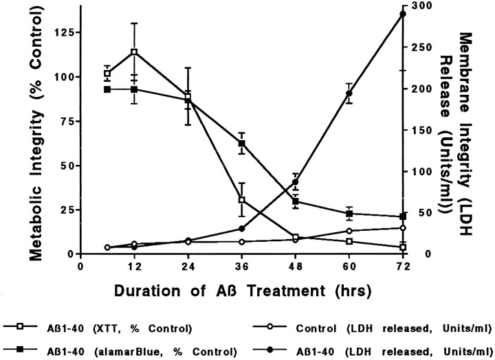
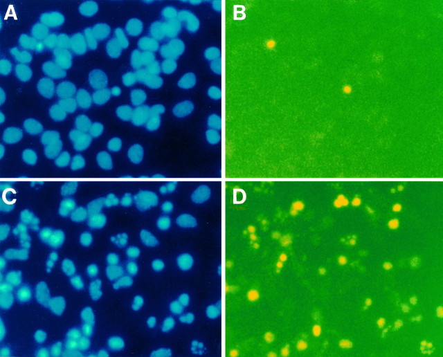
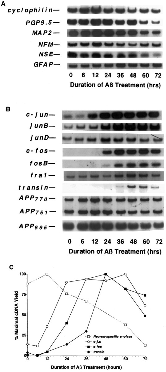
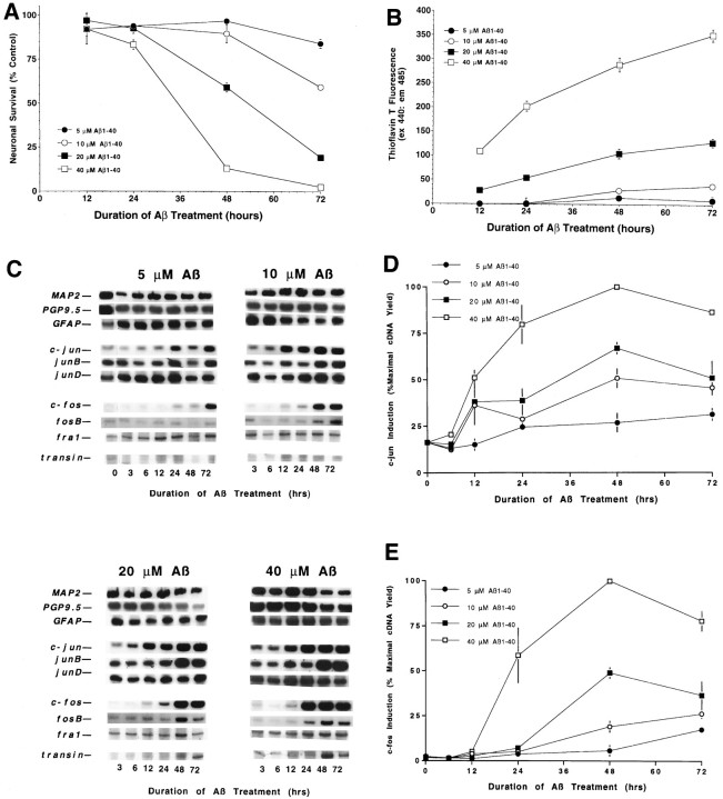
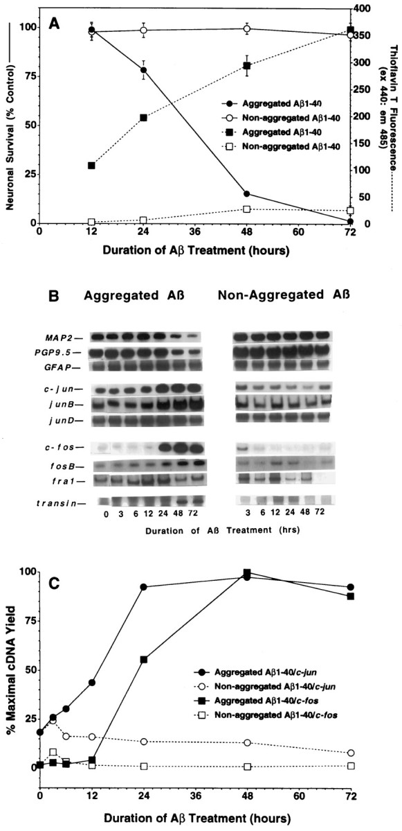
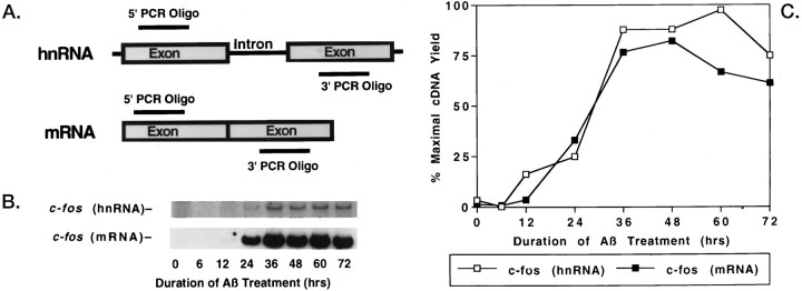
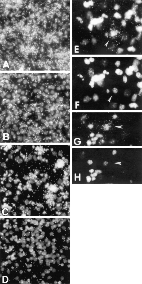
To gain a molecular understanding of neuronal responses to amyloid-beta peptide (Abeta), we have analyzed the effects of Abeta treatment on neuronal gene expression in vitro by quantitative reverse transcription-PCR and in situ hybridization. Treatment of cultured rat cortical neurons with Abeta1-40 results in a widespread apoptotic neuronal death. Associated with death is an induction of several members of the immediate early gene family. Specifically, we (1) report the time-dependent and robust induction of c-jun, junB, c-fos, and fosB, as well as transin, which is induced by c-Jun/c-Fos heterodimers and encodes an extracellular matrix protease; these gene inductions appear to be selective because other Jun and Fos family members, i.e., junD and fra-1, are induced only marginally; (2) show that the c-jun induction is widespread, whereas c-fos expression is restricted to a subset of neurons, typically those with condensed chromatin, which is a hallmark of apoptosis; (3) correlate gene induction and neuronal death by showing that each has a similar dose-response to Abeta; and (4) demonstrate that both cell death and immediate early gene induction are dependent on Abeta aggregation state. This overall gene expression pattern during this "physiologically inappropriate" apoptotic stimulus is markedly similar to the pattern we previously identified after a "physiologically appropriate" stimulus, i.e., the NGF deprivation-induced death of sympathetic neurons. Hence, the parallels identified here further our understanding of the genetic alterations that may lead neurons to apoptosis in response to markedly different insults.


Figures

Fig. 1.
Fig. 1.
Time course of Aβ neurotoxicity. Cultured rat cortical neurons were treated with Aβ1–40 (20 μm, Lot ZK840) for the indicated intervals, and cell viability was assayed by metabolic integrity (XTT reduction and alamarBlue reduction) and plasma membrane integrity (LDH release). Metabolic parameters decrease well before the loss of membrane integrity. Data are mean ± SD (error bars) values from triplicate wells and represent typical results obtained with these cells on a routine basis.
Fig. 2.
Fig. 2.
Aβ toxicity manifests the hallmarks of apoptosis. Cortical neuron preparations were treated with medium change alone (A, B) or Aβ1–40 (40 μm, lot ZM482) for 24 hr (C, D) and then assessed for chromatin integrity as discerned by Hoechst 33258 staining (A, C) and by DNA end labeling (B, D). The incidence of neurons manifesting punctate and fragmented chromatin is much higher in Aβ-treated neurons; note that neurons that manifest punctate chromatin also display an increased amount of fragmented DNA, as assessed by DNA end labeling.
Fig. 3.
Fig. 3.
Time course of mRNA expression in rat cortical cultures undergoing Aβ-mediated neuronal apoptosis.A, Cellular marker genes. B, Jun and Fos family members and related genes. C, Quantification of changes in NSE, c-jun,c-fos, and transin expression. To assess changes in mRNA levels, we maintained primary rat cortical cultures (∼125,000 neurons/well) for 3–4 d and then treated them with Aβ1–40 (40 μm, lot ZM482), as described in Materials and Methods. After various times of Aβ treatment, total RNA was isolated, aliquots were converted to cDNA, and then 3% of the resultant cDNA was analyzed in each PCR sample. The data presented are from a single preparation of neuronal cultures, which were maintained and treated in parallel with those described in Figure 1. Each gene induction was confirmed in at least two independent neuronal culture preparations.
Fig. 4.
Fig. 4.
Gene induction and death are dependent on Aβ concentration. Cortical neuronal cultures were treated with Aβ1–40 (lot ZM605) for the indicated concentrations and times. Viability was determined by measuring alamarBlue reduction (A) and Aβ aggregation assessed by changes in ThT fluorescence (B). Changes in gene expression were assessed by RT-PCR (C), with quantification for c-jun (D) andc-fos (E). Values for the alamarBlue and ThT assays are expressed as mean ± SD (error bars) from triplicate wells, whereas those for c-jun andc-fos inductions are the mean ± SE (error bars) from triplicate determinations. The induction of c-jun,junB, c-fos, fosB,transin, and death shows a strong dependence on Aβ concentration and aggregation. The induction of c-junwas significant (p < 0.05) for 5 μm Aβ at the 48 and 72 hr time points and at every time point after 6 hr for the higher Aβ concentrations. The induction ofc-fos was significant at 72 hr for 5 μmAβ, at 48 and 72 hr for 10 and 20 μm Aβ, and at 24, 48, and 72 hr for 40 μm Aβ [ANOVA comparison of Aβ-treated vs control samples (n = 3), withpost hoc Fisher PLSD test].
Fig. 5.
Fig. 5.
mRNA induction correlates with Aβ aggregation and Aβ neurotoxicity. Changes in neuronal viability and Aβ aggregation (A) and gene expression (B, C) were assessed in neurons treated with Aβ1–40(40 μm, lot ZM605) solvated initially in either water or DMSO. Values for the alamarBlue and ThT assays are expressed as mean ± SD (error bars) from triplicate wells. Similar results were observed in at least two independent neuronal culture preparations.
Fig. 6.
Fig. 6.
Enhanced immediate early gene levels appear to be derived from increased transcription. To assess whether induction of a prototype immediate early gene, c-fos, resulted from increased transcription or mRNA stabilization, we designedc-fos oligos such that the PCR product spanned an intron in hnRNA (A). After Aβ1–40treatment (40 μm, lot ZM605), c-fosheteronuclear RNA (hnRNA) and c-fosmature mRNA were induced in parallel, as revealed by the autoradiograms (B) and PhosphorImager quantitation (C). That the hnRNA-associated PCR product was not a result of contaminating genomic DNA was demonstrated by showing that no product was detected in the absence of reverse transcription (data not shown). The parallel nature of the hnRNA and mRNA inductions indicates that enhanced transcription likely contributes toc-fos induction.
Fig. 7.
Fig. 7.
Patterns of c-jun andc-fos induction in situ. Cultures were analyzed for c-jun (A, B) orc-fos (C–H) expression by performing in situ hybridization and for chromatin integrity by staining with Hoechst 33258. Cultures were treated with Aβ1–40 (40 μm, lot ZM605) for 48 hr (A, C, E–H) or treated with medium change only (B, D). The depicted images represent dark-field-and-fluorescence (A–E, G) or fluorescence (F, H) microscopy. The neurons depicted inE and G are shown again inF and H, respectively; thearrows indicate c-fos-positive neurons and their associated chromatin. A–D originally were magnified 200×, whereas E and F were magnified 400×. Sense cRNA probes did not label above background. These results were replicated in at least two separate neuronal preparations. These genes also were induced at 24 hr after Aβ treatment (data not shown).

References

    1. Anderson AJ, Cummings BJ, Cotman CW. Increased immunoreactivity for Jun- and Fos-related proteins in Alzheimer’s disease: association with pathology. Exp Neurol. 1994;125:286–295. - PubMed
    1. Anderson AJ, Pike CJ, Cotman CW. Differential induction of immediate early gene proteins in cultured neurons by β-amyloid (Aβ): association of c-jun with Aβ-induced apoptosis. J Neurochem. 1995;65:1487–1498. - PubMed
    1. Angel P, Karin M. The role of Jun, Fos, and the Ap-1 complex in cell proliferation and transformation. Biochim Biophys Acta. 1991;1072:129–157. - PubMed
    1. Behl C, Davis JB, Klier FG, Schubert D. Amyloid beta peptide induces necrosis rather than apoptosis. Brain Res. 1994a;645:254–264. - PubMed
    1. Behl C, Davis JB, Lesley R, Schubert D. Hydrogen peroxide mediates amyloid beta protein toxicity. Cell. 1994b;77:817–827. - PubMed

Publication types

LinkOut - more resources